
| 规格 | 价格 | 库存 | 数量 |
|---|---|---|---|
| 1mg |
|
||
| 5mg |
|
||
| 10mg |
|
||
| 25mg |
|
||
| 50mg |
|
||
| 100mg |
|
||
| 250mg |
|
||
| 500mg |
|
||
| Other Sizes |
|

| 靶点 |
Natural product; UGTs/UDP-glucuronosyltransferases
Exerts biological effects via regulating downstream proteins: C/EBP homologous protein (CHOP) in non-small cell lung cancer (NSCLC) cells [1], calcium signaling in erythrocytes [2], and proliferation-related proteins (e.g., Ki-67) in colon carcinoma [3] |
|---|---|
| 体外研究 (In Vitro) |
Licochalcone A (LCA) 对 UGT1A1、1A3、1A4、1A6、1A7、1A9 和 2B7 有显着的抑制作用(IC50 和 Ki 值均低于 5 μM)[2]。
1. NSCLC细胞中的凋亡与自噬诱导([1]): 用甘草查尔酮A(10、20、40 μM)处理A549和H1299(NSCLC)细胞24–48小时,以浓度依赖方式抑制细胞增殖。40 μM处理48小时时,A549细胞凋亡率达60%(Annexin V/PI流式细胞术),H1299细胞达55%。实时PCR显示,40 μM时CHOP mRNA表达在A549中升高3.5倍,H1299中升高3倍;蛋白质印迹法显示CHOP蛋白水平升高4倍,自噬标志物LC3-II/LC3-I比值在24小时时升高2.5倍 [1] 2. 人红细胞中的自杀性死亡诱导([2]): 人红细胞与甘草查尔酮A(10、20、40、80 μM)在37°C孵育24小时,呈浓度依赖诱导自杀性死亡。80 μM时,溶血率达35%(对照组为5%);磷脂酰丝氨酸外翻率(流式细胞术Annexin V结合)升高50%,细胞内Ca²⁺浓度(荧光探针Fura-2 AM)升高2.2倍。10 μM时无显著变化 [2] 3. 结肠癌细胞中的抗增殖活性([3]): 用甘草查尔酮A(5–40 μM)处理HT-29和HCT-116(结肠癌细胞)72小时,表现出抗增殖作用,MTT实验显示IC50值为HT-29(25 μM)、HCT-116(30 μM)。与顺铂(5 μM)联用时,20 μM 甘草查尔酮A增强顺铂诱导的细胞活力降低(HT-29中从45%降至65%),且对正常肠上皮细胞(IEC-6)毒性低(40 μM时活力>80%) [3] |
| 体内研究 (In Vivo) |
本研究的目的是确定licochalcone A (LCA)是否有可能在顺铂化疗期间作为有益的补充。我们发现单独给药LCA显著抑制了CT-26细胞接种的Balb/c小鼠实体瘤的大小,没有任何可检测到的肾毒性、肝毒性和氧化应激诱导。LCA还以剂量依赖的方式通过减少CT-26小鼠结肠癌细胞的DNA合成来抑制细胞增殖。LCA不影响顺铂的治疗效果。此外,LCA抑制顺铂诱导的以血清肌酐和血尿素氮升高为特征的肾损害,以及以血清丙氨酸转氨酶和天冬氨酸转氨酶升高为特征的顺铂诱导的肝损害。顺铂治疗前反复口服LCA对顺铂介导的血清一氧化氮和组织脂质过氧化水平升高有预防作用,恢复组织中耗尽的还原性谷胱甘肽水平。这些结果表明,在癌症患者中补充LCA可能有助于抵消顺铂治疗的副作用。[3]
在感染 L. Major 的小鼠中,甘草查尔酮 A(5 mg/kg,腹膜内注射)可完全阻止病变发展。在感染杜氏乳杆菌的小鼠中,甘草查耳酮 A(150 毫克/千克,口服)可使肝脏和脾脏中的寄生虫负荷分别减少 65% 和 85%。在 CT-26 细胞接种的 Balb/c 小鼠中,甘草查尔酮 A(1 mg/kg,口服)可抑制肿瘤生长,并减轻顺铂诱导的肾毒性和肝毒性。[3] 小鼠结肠癌模型中的抗肿瘤疗效与顺铂毒性减轻([3]): 6–8周龄雌性BALB/c裸鼠皮下接种5×10⁶ HT-29细胞,肿瘤体积达100 mm³时随机分为4组:(1)溶剂对照;(2)甘草查尔酮A单独(20 mg/kg,腹腔注射,每周2次);(3)顺铂单独(5 mg/kg,腹腔注射,每周1次);(4)甘草查尔酮A+顺铂(剂量同上)。21天后: - 联合组肿瘤生长抑制率达50%(甘草查尔酮A单独组30%,顺铂单独组35%); - 顺铂单独组的体重下降(15%)在联合组降至5%; - 顺铂单独组升高的血清ALT(2.5倍)、AST(2倍)在联合组恢复正常,BUN(1.8倍)降低40%; - 肿瘤组织免疫组化显示,联合组增殖标志物Ki-67阳性率降低55% [3] |
| 酶活实验 |
脂质过氧化的测定。[3]
MDA的含量作为脂质过氧化程度的指标,以硫代巴比妥酸反应物质的形式进行测定,如前所述[31]。反应混合物(4ml)由0.2 ml 8.1%十二烷基硫酸钠、1.5 ml 20%醋酸(pH 3.5)、1.5 ml 0.8%硫代巴比妥酸、0.2 ml匀浆和蒸馏水组成。将混合物在95℃下孵育1小时,用自来水冷却5分钟,与5 ml正丁醇-吡啶(15-1,v/v)混合物强烈混合,并在1200 ×g下离心10分钟。在532 nm处测定有机层(上正丁醇相)的吸光度。用1,1,3,3-四甲基氧丙烷建立标准曲线,最终MDA浓度以nmol / mg蛋白表示[3]。 谷胱甘肽水平测定。[3] 根据Higach描述的方法测定谷胱甘肽含量,将0.1 ml组织匀浆加入等体积的10%三氯乙酸溶液中,在1200 ×g离心20分钟。将0.1 ml上清加入0.5 ml含1 mM NaNO2的0.2 N H2SO4溶液中,在室温下孵育5 min,然后加入0.2 ml 0.5%氨基磺酸铵溶液、1 ml含0.1% HgCl2和3%磺胺的0.4 N HCl溶液和1 ml含0.1% N-(1-萘基)乙二胺的0.4 N HCl溶液。5分钟后,在540nm处测定吸光度。用谷胱甘肽标准校准曲线表示谷胱甘肽含量为nmol / mg蛋白。 |
| 细胞实验 |
甘草查尔酮A (Licochalcone A, LCA)是从著名中草药甘草中分离得到的一种黄酮类化合物,具有明显的抗癌作用。本研究探讨了LCA对非小细胞肺癌(NSCLC)细胞的抗癌作用及其可能机制。LCA在非小细胞肺癌细胞中以浓度依赖性的方式降低细胞活力,增加乳酸脱氢酶的释放,并诱导细胞凋亡,而在人胚胎肺成纤维细胞中则没有。经LCA处理后,磷脂酰乙醇胺修饰的微管相关蛋白轻链3 (LC3-II)的表达和两种自噬标志物GFP-LC3 punta的形成均增加。与氯喹(chloroquine, CQ)联用时,LCA诱导的LC3-II表达升高,而自噬相关蛋白(autophagy related protein, ATG) 7或ATG5的敲低可逆转LCA诱导的LC3-II表达和GFP-LC3 punta的形成,提示LCA诱导了NSCLC细胞的自噬。抑制自噬不能逆转lca诱导的细胞活力下降和凋亡。此外,LCA增加了内质网应激相关蛋白的表达,如结合免疫球蛋白蛋白和C/EBP同源蛋白(CHOP)。CHOP的下调逆转了lca诱导的细胞活力降低、凋亡和自噬。综上所述,lca诱导的自噬效应是NSCLC细胞的一种伴随现象,CHOP在lca诱导的细胞活力降低、细胞凋亡和自噬过程中起着关键作用[1]。
从健康个体中提取的人红细胞暴露于1-10µg/ml licochalcone a中24小时,随后使用流式细胞术通过膜联蛋白V结合来估计细胞表面的磷脂酰丝氨酸暴露,通过前向散射来估计细胞体积,通过fluo3荧光来估计[Ca2+]i,并利用特异性抗体来估计神经酰胺。此外,通过血红蛋白释放量来定量溶血[2]。 1. NSCLC细胞实验([1]): - 细胞培养:A549和H1299细胞用含10%胎牛血清的RPMI 1640培养,接种于96孔板(5×10³细胞/孔)或6孔板(2×10⁵细胞/孔)。 - 药物处理:贴壁24小时后,用甘草查尔酮A(10–40 μM)处理24–48小时;溶剂组加入0.1% DMSO。 - 检测: 1. 增殖:96孔板加MTT试剂,570 nm处测吸光度计算活力; 2. 凋亡:6孔板细胞用Annexin V-FITC/PI染色,流式细胞术分析; 3. 蛋白/基因:提取总蛋白行蛋白质印迹法(检测CHOP、LC3-I/II、β-肌动蛋白);提取总RNA行实时PCR(检测CHOP mRNA,GAPDH为内参)[1] 2. 红细胞实验([2]): - 红细胞分离:人静脉血离心(1000×g,10分钟)分离红细胞,用PBS洗涤3次。 - 药物处理:红细胞(10⁶细胞/mL)与甘草查尔酮A(10–80 μM)在PBS中37°C孵育24小时。 - 检测: 1. 溶血:测上清405 nm吸光度计算溶血率; 2. 磷脂酰丝氨酸外翻:Annexin V-FITC染色,流式细胞术定量; 3. 细胞内Ca²⁺:加载Fura-2 AM,检测340/380 nm荧光强度[2] 3. 结肠癌细胞实验([3]): - 细胞培养:HT-29、HCT-116和IEC-6细胞用含10%胎牛血清的DMEM培养,接种于96孔板(3×10³细胞/孔)或6孔板(1×10⁵细胞/孔)。 - 药物处理:细胞用甘草查尔酮A(5–40 μM)单独或与顺铂(5 μM)联合处理72小时。 - 检测: 1. 活力:MTT实验(570 nm吸光度)计算IC50及联合作用; 2. 集落形成:6孔板细胞处理24小时后继续培养14天,结晶紫染色计数集落[3] |
| 动物实验 |
将化合物溶于 20 μL 99% (v/v) 乙醇中,并悬浮于 1% 羧甲基纤维素 (CMC) 溶液中;5 mg/kg;灌胃或腹腔注射。
感染利什曼原虫的小鼠异种移植模型。为了评估利什曼原虫查尔酮 A (LCA) 对肿瘤生长的抑制作用及其对顺铂诱导的肾毒性和肝毒性的保护作用,将小鼠分为五组,每组八只:PBS 处理组、CT-26 细胞接种组、CT-26 细胞接种+顺铂组、CT-26 细胞接种+ LCA 组、CT-26 细胞接种+ LCA 和顺铂组。将培养于含10% FBS的DMEM培养基中的CT-26小鼠结肠癌细胞(2 × 10⁶个细胞,溶于0.1 ml PBS)皮下注射至Balb/c小鼠右侧腹部。24小时后,通过灌胃给予Balb/c小鼠LCA(1 mg/kg体重,溶于PBS)。LCA给药2小时后,腹腔注射顺铂(5 mg/kg体重,溶于PBS)。LCA和顺铂每日给药一次,持续15天。对照组仅注射PBS,不注射LCA和顺铂。第15天,用游标卡尺测量肿瘤大小,并根据以下公式计算肿瘤体积:(长 × 宽²)/2。末次顺铂注射后16小时,麻醉处死小鼠。从每只小鼠采集血液后立即取出肝脏和肾脏,然后进行匀浆处理,用于后续实验。[3] 小鼠结肠癌异种移植方案 ([3]): 1. 细胞接种:将 5×10⁶ 个 HT-29 细胞(悬浮于 0.2 mL PBS + 50% Matrigel 中)皮下注射到 6-8 周龄雌性 BALB/c 裸鼠的右侧腹部。 2. 药物配制:将甘草查尔酮 A溶解于 DMSO (5%, v/v) + 生理盐水 (95%, v/v) 中;将顺铂溶解于生理盐水中。 3. 给药:当肿瘤体积达到 100 mm³ 时,对小鼠进行如下治疗,持续 21 天: - 对照组:DMSO + 生理盐水(腹腔注射,每周两次); - 单独使用利可查尔酮A:20 mg/kg(腹腔注射,每周两次); - 单独使用顺铂:5 mg/kg(腹腔注射,每周一次); - 联合用药:利可查尔酮A + 顺铂(剂量同上)。 4. 样本采集与检测:每周两次测量肿瘤体积(公式:长×宽²/2);于第21天处死小鼠,采集血清用于ALT/AST/BUN检测,采集肿瘤组织用于Ki-67免疫组化[3] |
| 毒性/毒理 (Toxicokinetics/TK) |
1. 体外毒性:
- 甘草查尔酮 A (≤40 μM) 对正常人肺成纤维细胞 (MRC-5) 无显著细胞毒性(细胞活力 >85% vs. 对照组)[1] - 10 μM 的甘草查尔酮 A 未诱导红细胞凋亡(溶血率 <8%,磷脂酰丝氨酸外翻 <10%)[2] - 40 μM 的甘草查尔酮 A 可使正常肠上皮细胞 (IEC-6) 保持 >80% 的活力,表明其对正常组织毒性较低 [3] 2. 体内毒性: - 小鼠经 20 mg/kg 甘草查尔酮 A (21 天) 处理后,未出现体重减轻、肝损伤(ALT/AST 正常)或肾损伤。 (BUN正常)[3] - 与单独使用顺铂相比,利可查尔酮A联合顺铂可降低顺铂引起的肝毒性(ALT/AST正常化)和肾毒性(BUN降低40%)[3] |
| 参考文献 |
[1]. Induction of C/EBP homologous protein-mediated apoptosis and autophagy by licochalcone A in non-small cell lung cancer cells. Sci Rep. 2016 May 17;6:26241.
[2]. Licochalcone A Induced Suicidal Death of Human Erythrocytes. Cell Physiol Biochem. 2015;37(5):2060-70. [3]. Licochalcone A inhibits the growth of colon carcinoma and attenuates cisplatin-induced toxicity without a loss of chemotherapeutic efficacy in mice. Basic Clin Pharmacol Toxicol . 2008 Jul;103(1):48-54. |
| 其他信息 |
甘草查尔酮A是查尔酮类化合物。
据报道,甘草查尔酮A存在于甘草(Glycyrrhiza uralensis)、大戟(Euphorbia helioscopia)以及其他有相关数据的生物体中。 甘草查尔酮A是酚类查尔酮的衍生物,存在于甘草属植物(如光果甘草(Glycyrrhiza glabra)和膨胀甘草(Glycyrrhiza inflata))的根部,并可从中提取。它具有潜在的抗炎、抗菌和抗癌活性。甘草查尔酮A给药后,可抑制磷脂酰肌醇-3-激酶/Akt/哺乳动物雷帕霉素靶蛋白(PI3K/Akt/mTOR)信号通路,并抑制c-Jun N端激酶1(JNK-1)的活性。JNK-1是丝裂原活化蛋白激酶(MAPK)家族的成员,在MAPK介导的信号通路中发挥作用。抑制 PI3K/Akt/mTOR 和 MAPK 信号通路可诱导细胞周期停滞和细胞凋亡,降低癌细胞的迁移和侵袭能力,并抑制肿瘤细胞增殖。甘草查尔酮A还能抑制活性氧(ROS)的产生,并通过核因子E2相关因子2(Nrf2)通路降低氧化应激。 1. 药物背景([1][2][3]): 甘草查尔酮A是一种从光果甘草(Glycyrrhiza glabra)根中分离得到的天然查尔酮化合物,具有固有的抗炎、抗氧化和抗癌特性[1][2][3]。 2. 作用机制([1][2][3]): - 在非小细胞肺癌(NSCLC)细胞中:诱导内质网(ER)应激,上调CHOP表达,并激活CHOP介导的细胞凋亡和自噬通路[1]。 - 在红细胞中:通过增加细胞内Ca²⁺浓度和促进磷脂酰丝氨酸外翻来触发细胞凋亡。 [2] - 在结肠癌中:抑制细胞增殖并减轻顺铂诱导的氧化应激(减少活性氧的产生),从而缓解顺铂的毒性[3] 3. 治疗潜力 ([1][3]): 甘草查尔酮A 显示出作为非小细胞肺癌和结肠癌抗癌药物的潜力;它能够在不影响化疗疗效的情况下降低顺铂的毒性,使其成为铂类化疗的一种很有前景的辅助药物[1][3] |
| 分子式 |
C21H22O4
|
|
|---|---|---|
| 分子量 |
338.3970
|
|
| 精确质量 |
338.151
|
|
| 元素分析 |
C, 74.54; H, 6.55; O, 18.91
|
|
| CAS号 |
58749-22-7
|
|
| 相关CAS号 |
|
|
| PubChem CID |
5318998
|
|
| 外观&性状 |
Yellow to orange solid powder
|
|
| 密度 |
1.2±0.1 g/cm3
|
|
| 沸点 |
532.6±50.0 °C at 760 mmHg
|
|
| 熔点 |
100°
|
|
| 闪点 |
186.9±23.6 °C
|
|
| 蒸汽压 |
0.0±1.5 mmHg at 25°C
|
|
| 折射率 |
1.611
|
|
| LogP |
4.95
|
|
| tPSA |
66.76
|
|
| 氢键供体(HBD)数目 |
2
|
|
| 氢键受体(HBA)数目 |
4
|
|
| 可旋转键数目(RBC) |
6
|
|
| 重原子数目 |
25
|
|
| 分子复杂度/Complexity |
488
|
|
| 定义原子立体中心数目 |
0
|
|
| SMILES |
O([H])C1C([H])=C(C(/C(/[H])=C(\[H])/C(C2C([H])=C([H])C(=C([H])C=2[H])O[H])=O)=C([H])C=1C(C([H])=C([H])[H])(C([H])([H])[H])C([H])([H])[H])OC([H])([H])[H]
|
|
| InChi Key |
KAZSKMJFUPEHHW-DHZHZOJOSA-N
|
|
| InChi Code |
InChI=1S/C21H22O4/c1-5-21(2,3)17-12-15(20(25-4)13-19(17)24)8-11-18(23)14-6-9-16(22)10-7-14/h5-13,22,24H,1H2,2-4H3/b11-8+
|
|
| 化学名 |
(E)-3-(4-hydroxy-2-methoxy-5-(2-methylbut-3-en-2-yl)phenyl)-1-(4-hydroxyphenyl)prop-2-en-1-one
|
|
| 别名 |
|
|
| HS Tariff Code |
2934.99.9001
|
|
| 存储方式 |
Powder -20°C 3 years 4°C 2 years In solvent -80°C 6 months -20°C 1 month |
|
| 运输条件 |
Room temperature (This product is stable at ambient temperature for a few days during ordinary shipping and time spent in Customs)
|
| 溶解度 (体外实验) |
|
|||
|---|---|---|---|---|
| 溶解度 (体内实验) |
配方 1 中的溶解度: ≥ 2.5 mg/mL (7.39 mM) (饱和度未知) in 10% DMSO + 90% Corn Oil (这些助溶剂从左到右依次添加,逐一添加), 澄清溶液。
例如,若需制备1 mL的工作液,可将100 μL 25.0 mg/mL 澄清 DMSO 储备液加入900 μL 玉米油中,混合均匀。 配方 2 中的溶解度: 2.08 mg/mL (6.15 mM) in 10% DMSO + 40% PEG300 + 5% Tween80 + 45% Saline (这些助溶剂从左到右依次添加,逐一添加), 悬浊液; 超声助溶。 例如,若需制备1 mL的工作液,可将 100 μL 20.8 mg/mL澄清的DMSO储备液加入到400 μL PEG300中,混匀;再向上述溶液中加入50 μL Tween-80,混匀;然后加入450 μL生理盐水定容至1 mL。 *生理盐水的制备:将 0.9 g 氯化钠溶解在 100 mL ddH₂O中,得到澄清溶液。 View More
配方 3 中的溶解度: 2.08 mg/mL (6.15 mM) in 10% DMSO + 90% (20% SBE-β-CD in Saline) (这些助溶剂从左到右依次添加,逐一添加), 悬浊液; 超声助溶。 1、请先配制澄清的储备液(如:用DMSO配置50 或 100 mg/mL母液(储备液)); 2、取适量母液,按从左到右的顺序依次添加助溶剂,澄清后再加入下一助溶剂。以 下列配方为例说明 (注意此配方只用于说明,并不一定代表此产品 的实际溶解配方): 10% DMSO → 40% PEG300 → 5% Tween-80 → 45% ddH2O (或 saline); 假设最终工作液的体积为 1 mL, 浓度为5 mg/mL: 取 100 μL 50 mg/mL 的澄清 DMSO 储备液加到 400 μL PEG300 中,混合均匀/澄清;向上述体系中加入50 μL Tween-80,混合均匀/澄清;然后继续加入450 μL ddH2O (或 saline)定容至 1 mL; 3、溶剂前显示的百分比是指该溶剂在最终溶液/工作液中的体积所占比例; 4、 如产品在配制过程中出现沉淀/析出,可通过加热(≤50℃)或超声的方式助溶; 5、为保证最佳实验结果,工作液请现配现用! 6、如不确定怎么将母液配置成体内动物实验的工作液,请查看说明书或联系我们; 7、 以上所有助溶剂都可在 Invivochem.cn网站购买。 |
| 制备储备液 | 1 mg | 5 mg | 10 mg | |
| 1 mM | 2.9551 mL | 14.7754 mL | 29.5508 mL | |
| 5 mM | 0.5910 mL | 2.9551 mL | 5.9102 mL | |
| 10 mM | 0.2955 mL | 1.4775 mL | 2.9551 mL |
1、根据实验需要选择合适的溶剂配制储备液 (母液):对于大多数产品,InvivoChem推荐用DMSO配置母液 (比如:5、10、20mM或者10、20、50 mg/mL浓度),个别水溶性高的产品可直接溶于水。产品在DMSO 、水或其他溶剂中的具体溶解度详见上”溶解度 (体外)”部分;
2、如果您找不到您想要的溶解度信息,或者很难将产品溶解在溶液中,请联系我们;
3、建议使用下列计算器进行相关计算(摩尔浓度计算器、稀释计算器、分子量计算器、重组计算器等);
4、母液配好之后,将其分装到常规用量,并储存在-20°C或-80°C,尽量减少反复冻融循环。
计算结果:
工作液浓度: mg/mL;
DMSO母液配制方法: mg 药物溶于 μL DMSO溶液(母液浓度 mg/mL)。如该浓度超过该批次药物DMSO溶解度,请首先与我们联系。
体内配方配制方法:取 μL DMSO母液,加入 μL PEG300,混匀澄清后加入μL Tween 80,混匀澄清后加入 μL ddH2O,混匀澄清。
(1) 请确保溶液澄清之后,再加入下一种溶剂 (助溶剂) 。可利用涡旋、超声或水浴加热等方法助溶;
(2) 一定要按顺序加入溶剂 (助溶剂) 。
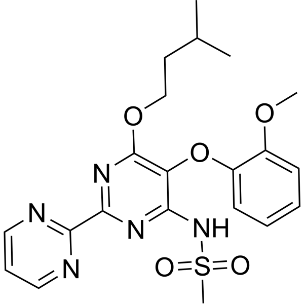 ET receptor antagonist 1
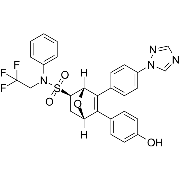 ERα degrader 6
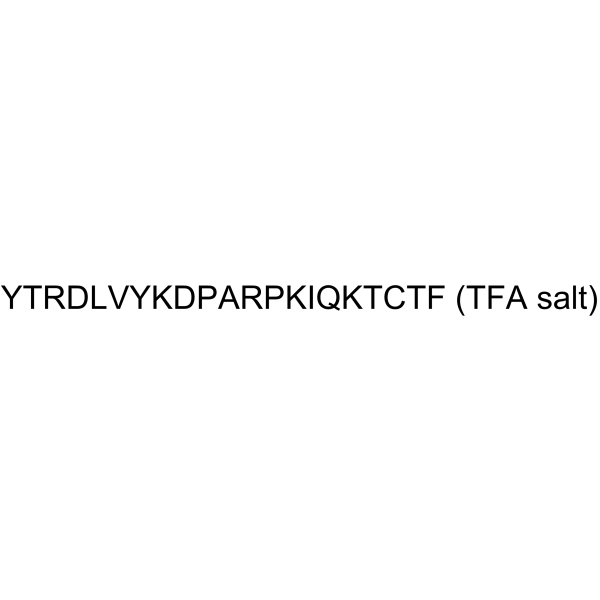 hFSH-β-(33-53) (TFA)
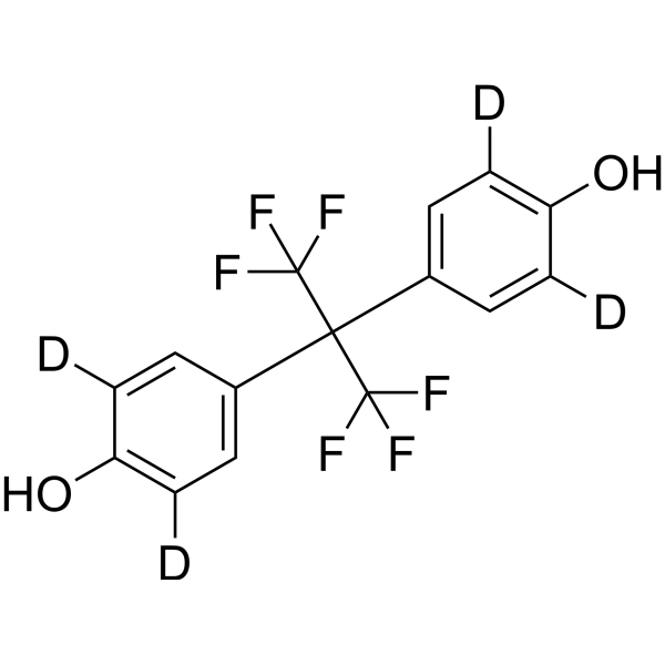 Bisphenol AF-d4 (BPAF-d4; 4,4'-(Perfluoropropane-2,2-diyl)diphenol-d4)
InvivoChem的所有产品仅用于作科学研究,不面向患者销售
Copyright 2020 InvivoChem LLC | All Rights Reserved 粤ICP备20063088号-1


 COA
COA
 463611831
