| 规格 | 价格 | 库存 | 数量 |
|---|---|---|---|
| 1mg |
|
||
| 5mg |
|
||
| 10mg |
|
||
| Other Sizes |
|

| 靶点 |
Cholesterol-desaturating agent; Secondary metabolite; microbial metabolite; secondary bile acid; endogenous metabolite
|
|---|---|
| 体外研究 (In Vitro) |
β-鼠胆酸(100µM;48小时)抑制原代小鼠肝细胞中的脂质积聚[1]。原代肝细胞中的 β-鼠胆酸荧光探针稀释液(100 μM;48 小时)[1]。
|
| 体内研究 (In Vivo) |
β-鼠胆酸(给予 0.5% β-鼠胆酸 8 周)可抑制大鼠饮食诱导或实验性胆酸 [2]。
束缚应激提高了NASH小鼠的肝脏和血清BA水平[1] 接下来,测量肝脏和血清BA水平,以研究血清中皮质酮和BA之间的相关性是否是应激挑战的结果。在MCD饮食下,应激显著增加了血清和肝脏BA水平[23.6-38.8μM(1.64倍)和0.399-0.673μmol/g肝脏(1.69倍)](图2A)。在MCS饮食条件下,应激倾向于提高肝脏BA水平[对照组和应激组分别为0.026和0.137μmol/g肝脏(5.26倍)](图2A)。此外,还研究了肝脏BA成分。MCD诱导的NASH中,肝脏牛磺β-胞浆酸(TβMCA)和牛磺胆酸(TCA)水平显著升高。然而,这些牛磺酸结合的BA在应激后没有变化(图2B)。MCD诱导的NASH中,肝牛磺脱氧胆酸(TCDCA)和牛磺脱氧烟酸(TDCA)水平没有变化(图2B)。有趣的是,非共轭BA和βMCA在应激后显著升高[1.14-3.46 nmol/g肝脏(3.03倍)],胆酸(CA)趋于升高[0.84-2.24 nmol/g肝(2.67倍)](图2B)。在测试的BA相关基因中,MCD饮食增加了细胞色素P450家族(CYP)7A1(CYP7A1)、溶质载体家族(SLC)51β(SLC51B,也称为有机溶质转运蛋白亚基β)和ATP结合盒(ABC)C4基因的肝脏表达水平(分别为3.56倍、11.73倍和7.09倍)(图2C)。在MCD饮食下,应激增强了SCL51B和ABCC4的表达(分别为11.73-15.55倍和7.09-14.47倍)(图2C)。有趣的是,在MCS饮食条件下,应激诱导了肝脏CYP7A1的表达(4.03倍)。研究结果可能表明,应激直接刺激肝脏Cyp7a1的诱导。其他BA相关因子的肝脏表达,即肝核因子4α(HNF4A)、富肝同源物1(LRH1)、法尼醇X受体(FXR)、小异二聚体伴侣(SHP)、CYP7B1、CYP8B1、CYP27A1、胆汁酸辅酶A:氨基酸N-酰基转移酶(BAAT)、ABCB11、ABCC2、SLC51A、ABCC3、ABCC5、SLC10A1和溶质载体有机阴离子1a1(SLCO1A1),在应激后均未在MCS或MCD组中发生变化(图2C)。在MCD应激组中观察到最高的肝脏CYP7A1蛋白水平(图2D)。有趣的是,血清BA和皮质酮水平与肝脏CYP7A1蛋白水平呈正相关,但与肝脏CYP5A1 mRNA水平无关(图2E)。这些结果表明,应激后NASH肝脏中存在皮质酮-CYP7A1蛋白-BA级联反应。 束缚应激通过升高肝脏βMCA水平降低NASH小鼠的肝脏脂质水平[1] 进行油红O染色,以研究应激是否会影响NASH发展过程中的肝脏脂质稳态。油红O染色显示,MCD应激组肝脏中的脂滴尺寸小于MCD非应激组肝脏(图4A)。此外,在应激挑战后,测量了血清和肝脏中的甘油三酯(TG)、总胆固醇(TChol)和NEFA水平。应激显著降低了MCD组的肝脏TG和NEFA水平[624-353 mg/g肝脏(0.57倍)和0.336-0.242 Eq/g肝脏(0.72倍)](图4B),但不影响肝脏中脂肪酸相关基因的表达(图4C)。然而,应激对肝脏TChol(图4B)和肝脏胆固醇相关基因表达水平(图S1B)没有影响。有趣的是,βMCA在暴露于棕榈酸(PA)/油酸(OA)后抑制了原代培养的小鼠肝细胞中的脂质积累,而CA则没有(图5A,B)。然而,经βMCA处理后,肝细胞中脂质相关基因的表达水平没有改变(图5C)。另一方面,PA/OA降低了CYP7A1 mRNA,在用βMCA治疗后,蛋白质水平趋于恢复(图5D,E)。这些结果表明,βMCA可以抑制NASH小鼠的肝脏脂质积聚。 |
| 细胞实验 |
原代肝细胞培养[1]
如前所述制备原代肝细胞。在皮质酮治疗中,用FBS阴性Williams培养基E饥饿2小时后,将肝细胞暴露于0.1%二甲亚砜作为载体和30-1000nM皮质酮中。六小时后,收集细胞并进行定量PCR和蛋白质印迹分析。对于脂质积聚,肝细胞暴露于0.2%异丙醇/1%乙醇/1%FBS溶液作为载体、100µM棕榈酸或100µM棕榈酸和100µM胆汁酸(βMCA或CA)。48小时后,收集细胞并进行定量PCR和蛋白质印迹分析。另一组细胞进行油红O染色。使用60%异丙醇溶液和油红O染料进行油红O染色。然后,用Mayer的苏木精溶液和纯伊红溶液进行苏木精和伊红(HE)染色。为了定量脂质积累,在油红O染色后,用40 mM HCl/异丙醇溶液从染色细胞中提取染料。通过测量500nm处的吸光度来确定染料浓度,并将其用作相对脂质量。 |
| 动物实验 |
动物/疾病模型: 6-8 周龄雄性 C57L/J 小鼠(含 2% 胆固醇和 0.5% 胆酸的致胆饮食)[2]
剂量: 0.5% β-鼠胆酸 给药途径: 使用 0.5% β-鼠胆酸。8 周鼠胆酸喂养饮食的结果:胆结石患病率降低至 20%,其机制是通过显著降低胆汁胆固醇分泌率、饱和指数和肠道吸收,以及诱导相界移动和扩大 E 区,从而阻止胆固醇从液晶相转变为固态晶体和结石。 |
| 参考文献 |
|
| 其他信息 |
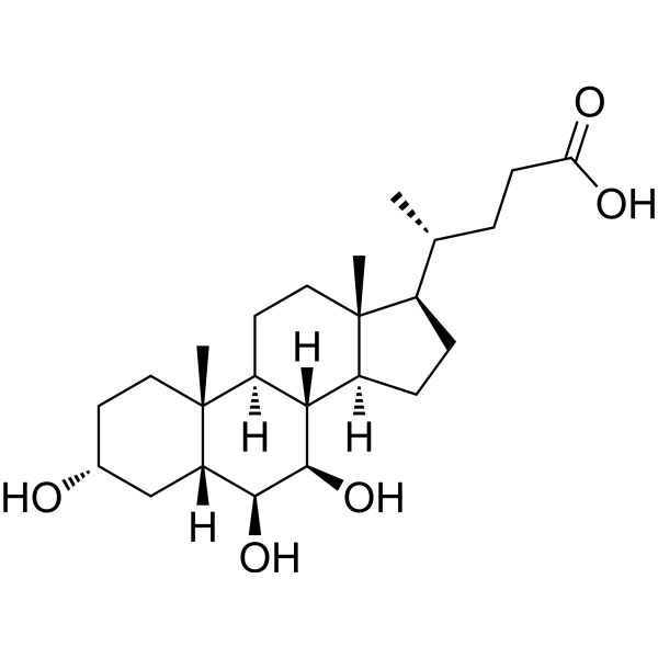
β-鼠胆酸是鼠胆酸类化合物中的一种,其6位和7位羟基均为β构型。它属于鼠胆酸类化合物、6β-羟基甾醇和7β-羟基甾醇。它是β-鼠胆酸的共轭酸。
另见:β-鼠胆酸(注释已移至)。 据报道,在人类中,非酒精性脂肪性肝炎(NASH)会升高乙二醇-CA和TCA水平,并降低二级胆汁酸/一级胆汁酸的比值。在本研究中,MCD饮食降低了小鼠肝脏中TCDCA和TDCA水平与TβMCA和TCA水平的比值。由于胆汁酸的亲水性顺序为:共轭胆汁酸 > 非共轭胆汁酸,且βMCA > CA > CDCA > DCA,这些结果可能表明,NASH中亲水性胆汁酸水平上调。此外,在非酒精性脂肪性肝炎(NASH)中,应激可增加肝脏β-甲基胆碱(βMCA)和胆酸(CA)水平,而肝脏总β-甲基胆碱(TβMCA)和三羧酸(TCA)水平并未升高。而且,应激并未改变调控胆汁酸(BA)牛磺酸结合的Baat基因的表达。尽管肠道菌群的去结合反应需要考虑,但该观察结果支持这样一种可能性:应激引起的肝脏βMCA和CA水平升高是由于胆汁酸合成的限速酶Cyp7a1基因表达诱导所致。FXR是一种核胆汁酸受体,可被CDCA强烈激活,并诱导Shp表达以抑制Cyp7a1基因表达。尽管βMCA可以负向调控FXR的激活,但在NASH肝脏中,应激后并未观察到SHP表达水平的变化。此外,上调Cyp7a1基因表达的Hnf4a和Lrh1基因表达水平在应激后也未发生改变。这些结果表明,应激诱导Cyp7a1基因表达是由其他因素介导的。应激可能通过增加非结合型胆汁酸(BA)水平促进NASH的病理改变。[1] 在本研究中,在MCD诱导的NASH条件下,应激导致肝脏β-甲基胆酸(βMCA)水平升高,同时CYP7A1表达增强。此外,βMCA治疗可保护肝细胞免受棕榈酸(PA)/油酸(OA)诱导的脂质积累。因此,本研究表明CYP7A1产生的βMCA有助于抑制肝细胞脂质积累,但βMCA抑制肝脏脂质积累的机制尚不清楚。由于βMCA是鼠类胆汁酸,人体无法合成,因此在非酒精性脂肪性肝病(NAFLD)患者中未观察到这些现象。然而,研究结果可能提示βMCA可用于NAFLD的治疗。[1] 在临床研究中,评估应激对疾病的影响较为困难,主要是因为目前尚无明确的应激指标。本研究采用血清糖皮质激素水平作为应激指标。然而,血清糖皮质激素水平会受到多种因素(包括应激)的影响而发生变化。本研究结果可能与临床数据不完全一致,但提示应激可能降低NAFLD患者的肝脏脂质水平。未来需要建立应激指标,以进一步了解应激影响人体的分子机制。[1] 总之,本研究表明应激会影响NASH小鼠的肝脏胆汁酸和脂质稳态,提示βMCA可用于NASH的治疗。预期未来将阐明其详细的分子机制,以开发NAFLD治疗药物。[1] 应激会影响人体,并已知会导致某些疾病。然而,慢性束缚应激对非酒精性脂肪性肝病(NAFLD)发展的影响尚不清楚。本研究表明,慢性束缚应激可通过提高小鼠肝脏β-鼠胆酸(βMCA)水平来减轻非酒精性脂肪性肝炎(NASH)发展过程中的肝脏脂质蓄积。血清皮质醇和皮质酮水平(即人和啮齿动物的应激标志物)分别与NAFLD患者和蛋氨酸胆碱缺乏(MCD)饮食诱导的小鼠的血清胆汁酸水平相关,提示应激与NASH中的胆汁酸(BA)稳态有关。在小鼠模型中,应激刺激后肝脏βMCA和胆酸(CA)水平升高。考虑到短期应激可提高正常小鼠肝脏中CYP7A1蛋白水平,而皮质酮可提高原代小鼠肝细胞中CYP7A1蛋白水平,因此推测Cyp7a1表达的增强与慢性应激引起的肝脏β-MCA水平升高有关。有趣的是,慢性应激降低了MCD诱导的NASH小鼠的肝脏脂质水平。此外,β-MCA可抑制棕榈酸/油酸处理的小鼠原代肝细胞中的脂质积累,而CA则无此作用。另外,Cyp7a1的表达似乎与肝细胞中的脂质积累有关。总之,慢性应激可通过诱导肝脏Cyp7a1表达来改变NASH小鼠的肝脏脂质积累,从而破坏胆汁酸稳态。本研究发现β-鼠胆酸(β-MCA)在肝脏中具有新的作用,提示β-MCA可能用于非酒精性脂肪性肝病(NAFLD)的治疗。[1] 本研究探讨了β-鼠胆酸(一种啮齿动物天然的三羟基亲水性胆汁酸)是否能作为胆汁胆固醇去饱和剂预防胆固醇结石,以及与熊去氧胆酸(UDCA)相比,它是否能促进胆结石的溶解。在胆结石预防研究中,易患胆结石的雄性C57L小鼠被喂食致结石饮食(2%胆固醇和0.5%胆酸),持续8周,分别添加或不添加0.5%的UDCA或β-鼠胆酸。在胆结石溶解研究中,另取已形成胆结石的小鼠,喂食添加或不添加0.5%的β-鼠胆酸或UDCA的普通饲料,持续8周。所有喂食致结石饮食的小鼠均形成了胆固醇结石。添加β-鼠胆酸和熊去氧胆酸(UDCA)可显著降低胆汁分泌率、饱和指数和肠道对胆固醇的吸收,从而将胆结石发生率分别降低至20%和50%。同时,β-鼠胆酸和UDCA还能诱导相界转移并扩大E区,从而阻止胆固醇从液晶相转变为固态晶体和结石。与对照组(10%)相比,β-鼠胆酸和UDCA给药8周后,胆结石完全溶解率分别达到100%和60%。我们得出结论,在治疗或预防饮食诱导或实验性小鼠胆固醇胆结石方面,β-鼠胆酸比UDCA更有效。[2] |
| 分子式 |
C24H40O5
|
|---|---|
| 分子量 |
408.5714
|
| 精确质量 |
408.288
|
| CAS号 |
2393-59-1
|
| PubChem CID |
5283853
|
| 外观&性状 |
White to light yellow solid powder
|
| 密度 |
1.184g/cm3
|
| 沸点 |
565.7ºC at 760mmHg
|
| 闪点 |
310ºC
|
| 蒸汽压 |
3.75E-15mmHg at 25°C
|
| 折射率 |
1.558
|
| LogP |
3.448
|
| tPSA |
97.99
|
| 氢键供体(HBD)数目 |
4
|
| 氢键受体(HBA)数目 |
5
|
| 可旋转键数目(RBC) |
4
|
| 重原子数目 |
29
|
| 分子复杂度/Complexity |
637
|
| 定义原子立体中心数目 |
11
|
| SMILES |
C[C@H](CCC(=O)O)[C@H]1CC[C@@H]2[C@@]1(CC[C@H]3[C@H]2[C@H]([C@H]([C@H]4[C@@]3(CC[C@H](C4)O)C)O)O)C
|
| InChi Key |
DKPMWHFRUGMUKF-CRKPLTDNSA-N
|
| InChi Code |
InChI=1S/C24H40O5/c1-13(4-7-19(26)27)15-5-6-16-20-17(9-11-23(15,16)2)24(3)10-8-14(25)12-18(24)21(28)22(20)29/h13-18,20-22,25,28-29H,4-12H2,1-3H3,(H,26,27)/t13-,14-,15-,16+,17+,18+,20+,21+,22-,23-,24-/m1/s1
|
| 化学名 |
(4R)-4-[(3R,5R,6S,7R,8S,9S,10R,13R,14S,17R)-3,6,7-trihydroxy-10,13-dimethyl-2,3,4,5,6,7,8,9,11,12,14,15,16,17-tetradecahydro-1H-cyclopenta[a]phenanthren-17-yl]pentanoic acid
|
| 别名 |
beta-muricholic acid; 2393-59-1; b-Muricholic acid; beta-MCA; 5beta-Cholanic acid-3alpha,6beta,7beta-triol; (3a,5b,6b,7b)-3,6,7-trihydroxy-Cholan-24-oic acid; 3alpha,6beta,7beta-Trihydroxy-5beta-cholan-24-oic Acid; (4R)-4-[(3R,5R,6S,7R,8S,9S,10R,13R,14S,17R)-3,6,7-trihydroxy-10,13-dimethyl-2,3,4,5,6,7,8,9,11,12,14,15,16,17-tetradecahydro-1H-cyclopenta[a]phenanthren-17-yl]pentanoic acid;
|
| HS Tariff Code |
2934.99.9001
|
| 存储方式 |
Powder -20°C 3 years 4°C 2 years In solvent -80°C 6 months -20°C 1 month 注意: 本产品在运输和储存过程中需避光。 |
| 运输条件 |
Room temperature (This product is stable at ambient temperature for a few days during ordinary shipping and time spent in Customs)
|
| 溶解度 (体外实验) |
DMSO : ~100 mg/mL (~244.76 mM)
|
|---|---|
| 溶解度 (体内实验) |
配方 1 中的溶解度: ≥ 2.5 mg/mL (6.12 mM) (饱和度未知) in 10% DMSO + 40% PEG300 + 5% Tween80 + 45% Saline (这些助溶剂从左到右依次添加,逐一添加), 澄清溶液。
例如,若需制备1 mL的工作液,可将100 μL 25.0 mg/mL澄清DMSO储备液加入到400 μL PEG300中,混匀;然后向上述溶液中加入50 μL Tween-80,混匀;加入450 μL生理盐水定容至1 mL。 *生理盐水的制备:将 0.9 g 氯化钠溶解在 100 mL ddH₂O中,得到澄清溶液。 配方 2 中的溶解度: ≥ 2.5 mg/mL (6.12 mM) (饱和度未知) in 10% DMSO + 90% (20% SBE-β-CD in Saline) (这些助溶剂从左到右依次添加,逐一添加), 澄清溶液。 例如,若需制备1 mL的工作液,可将 100 μL 25.0 mg/mL澄清DMSO储备液加入900 μL 20% SBE-β-CD生理盐水溶液中,混匀。 *20% SBE-β-CD 生理盐水溶液的制备(4°C,1 周):将 2 g SBE-β-CD 溶解于 10 mL 生理盐水中,得到澄清溶液。 View More
配方 3 中的溶解度: ≥ 2.5 mg/mL (6.12 mM) (饱和度未知) in 10% DMSO + 90% Corn Oil (这些助溶剂从左到右依次添加,逐一添加), 澄清溶液。 1、请先配制澄清的储备液(如:用DMSO配置50 或 100 mg/mL母液(储备液)); 2、取适量母液,按从左到右的顺序依次添加助溶剂,澄清后再加入下一助溶剂。以 下列配方为例说明 (注意此配方只用于说明,并不一定代表此产品 的实际溶解配方): 10% DMSO → 40% PEG300 → 5% Tween-80 → 45% ddH2O (或 saline); 假设最终工作液的体积为 1 mL, 浓度为5 mg/mL: 取 100 μL 50 mg/mL 的澄清 DMSO 储备液加到 400 μL PEG300 中,混合均匀/澄清;向上述体系中加入50 μL Tween-80,混合均匀/澄清;然后继续加入450 μL ddH2O (或 saline)定容至 1 mL; 3、溶剂前显示的百分比是指该溶剂在最终溶液/工作液中的体积所占比例; 4、 如产品在配制过程中出现沉淀/析出,可通过加热(≤50℃)或超声的方式助溶; 5、为保证最佳实验结果,工作液请现配现用! 6、如不确定怎么将母液配置成体内动物实验的工作液,请查看说明书或联系我们; 7、 以上所有助溶剂都可在 Invivochem.cn网站购买。 |
| 制备储备液 | 1 mg | 5 mg | 10 mg | |
| 1 mM | 2.4476 mL | 12.2378 mL | 24.4756 mL | |
| 5 mM | 0.4895 mL | 2.4476 mL | 4.8951 mL | |
| 10 mM | 0.2448 mL | 1.2238 mL | 2.4476 mL |
1、根据实验需要选择合适的溶剂配制储备液 (母液):对于大多数产品,InvivoChem推荐用DMSO配置母液 (比如:5、10、20mM或者10、20、50 mg/mL浓度),个别水溶性高的产品可直接溶于水。产品在DMSO 、水或其他溶剂中的具体溶解度详见上”溶解度 (体外)”部分;
2、如果您找不到您想要的溶解度信息,或者很难将产品溶解在溶液中,请联系我们;
3、建议使用下列计算器进行相关计算(摩尔浓度计算器、稀释计算器、分子量计算器、重组计算器等);
4、母液配好之后,将其分装到常规用量,并储存在-20°C或-80°C,尽量减少反复冻融循环。
计算结果:
工作液浓度: mg/mL;
DMSO母液配制方法: mg 药物溶于 μL DMSO溶液(母液浓度 mg/mL)。如该浓度超过该批次药物DMSO溶解度,请首先与我们联系。
体内配方配制方法:取 μL DMSO母液,加入 μL PEG300,混匀澄清后加入μL Tween 80,混匀澄清后加入 μL ddH2O,混匀澄清。
(1) 请确保溶液澄清之后,再加入下一种溶剂 (助溶剂) 。可利用涡旋、超声或水浴加热等方法助溶;
(2) 一定要按顺序加入溶剂 (助溶剂) 。
InvivoChem的所有产品仅用于作科学研究,不面向患者销售
Copyright 2020 InvivoChem LLC | All Rights Reserved 粤ICP备20063088号-1


 463611831
