| 规格 | 价格 | 库存 | 数量 |
|---|---|---|---|
| 5mg |
|
||
| 10mg |
|
||
| 25mg |
|
||
| 50mg |
|
||
| 100mg |
|
||
| 250mg |
|
||
| 500mg |
|
||
| Other Sizes |
|

| 靶点 |
In mouse B6 cells, 1,4-DPCA (24 hours) can treat many elevated target gene expressions, such as the pro-angiogenic target genes Vegfa and Hmox1 and the pro-glycolytic target genes Ldh-a, Pgk1, Pdk1, and 1. ZR-75-1 (20 μM 1,4-DPCA) and T4-2 (10 μM 1,4-DPCA) cells showed a significant reduction in colony size following 4-DPCA therapy. In three-dimensional culture, T4-2 cells treated with DPCA produced localized spheroids. Treatment with 1,4-DPCA significantly decreased MDA-MB-231 (10 μM 1,4-DPCA), ZR-75-1, MDA-MB-157, and T4-2 cells [3].
|
|---|---|
| 体外研究 (In Vitro) |
在小鼠 B6 细胞中,1,4-DPCA(24 小时)可以治疗许多升高的靶基因表达,例如促血管生成靶基因 Vegfa 和 Hmox1 以及促糖酵解靶基因 Ldh-a、Pgk1、Pdk1 和 1 ZR-75-1 (20 μM 1,4-DPCA) 和 T4-2 (10 μM 1,4-DPCA) 细胞在 4-DPCA 治疗后显示集落大小显着减小。在三维培养中,用 DPCA 处理的 T4-2 细胞产生了局部球体。用 1,4-DPCA 处理可显着减少 MDA-MB-231 (10 μM 1,4-DPCA)、ZR-75-1、MDA-MB-157 和 T4-2 细胞 [3]。
用 1,4-DPCA/水凝胶(Gd)处理B6小鼠耳成纤维细胞24小时,通过免疫组织化学和Western blot分析检测到细胞质和细胞核中HIF-1α蛋白表达增加,但不影响HIF-2α表达[1] 对经 1,4-DPCA/水凝胶处理的B6小鼠成纤维细胞的mRNA进行逆转录聚合酶链反应分析,显示多个HIF-1α靶基因表达上调,包括促血管生成基因Vegfa和Hmox1,以及促糖酵解基因Ldh-a、Pgk1、Pdk1和Glut1[1] 用针对Hif1a的siRNA处理可阻断 1,4-DPCA/水凝胶引起的所有上述靶基因的上调[1] 体外用 1,4-DPCA/凝胶处理B6小鼠耳成纤维细胞24小时,细胞对HIF-1α和一系列未成熟细胞标志物呈阳性染色,其染色模式与MRL小鼠耳成纤维细胞无法区分[1] 定量PCR证实,与未处理的细胞相比,经药物/凝胶处理的B6小鼠耳成纤维细胞中干细胞标志物mRNA显著上调[1] 用siHif1a处理NANOG阳性的MRL小鼠耳源性成纤维细胞,导致NANOG蛋白表达几乎完全被抑制[1] |
| 体内研究 (In Vivo) |
在血管内腹膜聚(锁-共-乙醇酸) (PLGA) 椎间盘中,用 1,4-DPCA 治疗可成功阻止结缔组织向内生长 28 天。 1,4-DPCA 捕获成功地抑制了枢轴椎间盘的内表面和外表面限制性胶原沉积以及结缔组织向内生长,但抑制程度与糖皮质激素不同[2]。
向瑞士韦伯斯特小鼠皮下注射含有 1,4-DPCA 的水凝胶,在5天内增加了耳组织中HIF-1α蛋白的稳定表达,提供了药物体内释放的功能性测量指标[1] 在10天期间多次皮下注射 1,4-DPCA/水凝胶,导致瑞士韦伯斯特小鼠耳洞穿孔伤后实现完全再生性伤口愈合,愈合持续至第35天[1] 1,4-DPCA/凝胶诱导的耳洞闭合可被同时给予的siHif1a处理所阻断,表明愈合效果依赖于HIF-1α的表达[1] 1,4-DPCA/凝胶处理在瑞士韦伯斯特小鼠中诱导了再生的数个标志:加速再上皮化、细胞去分化、基底膜分解、疤痕形成减少以及后期事件如软骨生成和新毛囊形成[1] Western blot分析显示,经药物/凝胶处理的瑞士韦伯斯特小鼠在伤后第7、14和21天HIF-1α蛋白表达显著增加,与再生性MRL小鼠中所见水平相似,而HIF-2α表达几乎未受影响[1] |
| 酶活实验 |
化合物 1,4-DPCA 是脯氨酰羟化酶的有效抑制剂,PHD在常氧条件下介导HIF-1α蛋白的羟基化和后续降解[1]
通过阻断PHD活性,1,4-DPCA 可稳定HIF-1α蛋白[1] |
| 细胞实验 |
用于体外研究的小鼠耳源性成纤维样细胞进行培养[1]
为测试药物效果,用正常培养基、单独水凝胶或含有 1,4-DPCA 的水凝胶培养细胞。水凝胶前体在孔板中形成固态圆片[1] 处理24小时后,对细胞进行分析。通过免疫染色和Western blot分析评估HIF-1α蛋白表达[1] 对于基因表达分析,从处理的细胞中提取mRNA,通过RT-PCR或定量PCR分析HIF-1α靶基因和干细胞标志物[1] 对于siRNA敲低实验,使用转染试剂将Hif1a siRNA或乱序siRNA转染至70%融合度的细胞中。在转染后48小时检查敲低效率[1] |
| 动物实验 |
设计了一种局部注射水凝胶,用于在 4 至 10 天内控制释放 1,4-DPCA。该水凝胶由交联的聚乙二醇分子(前体 P8Cys 和 P8NHS)组成,在生理条件下可快速凝胶[1]
载药水凝胶是通过将聚合物(Pluronic F127)稳定的 1,4-DPCA 微晶悬浮在水凝胶前体的水性混合物中形成的,该混合物快速固化以包裹药物微晶[1] 瑞士韦伯斯特小鼠(8-10 周龄)接受了 2.1 mm 的贯穿性耳部穿孔损伤[1] 单剂量动力学研究:损伤后数小时,在小鼠颈背部皮下注射 100 µL 含有 2 mg/mL 1,4-DPCA 的水凝胶(Gd)或 0 mg/mL(仅凝胶,G0)。连续 5 天每天采集耳组织进行分析[1] 再生反应研究:在损伤后第 0、5 和 10 天,将 100 µL Gd 或 G0 皮下注射到小鼠颈背部(三个相邻部位)。通过测量耳孔直径,监测耳孔闭合情况,持续35天[1]。 在一些实验中,为了测试远端效应,于第0、5和10天将药物/凝胶皮下注射到小鼠左右后侧腹部[1]。 对于体内siRNA抑制:按照制造商的说明,将siHif1a与体内转染试剂混合,并从第0天开始,每48小时皮下注射一次,持续20天,同时进行药物/凝胶治疗[1]。 沿耳廓长轴方向和垂直轴方向测量耳孔直径[1]。 在实验终点,处死小鼠,收集耳组织进行组织学分析(例如,苏木精-伊红染色、阿利新蓝染色、苦味酸-天狼星红染色)、免疫组织化学、蛋白质印迹和RNA提取用于qPCR[1]。 |
| 参考文献 | |
| 其他信息 |
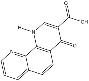
1,4-DPCA(1,4-二氢菲咯啉-4-酮-3-羧酸)是脯氨酰羟化酶 (PHD) 的小分子抑制剂[1]
缺氧诱导因子 1α (HIF-1α) 是 MRL 小鼠自发再生愈合过程中发现的中心调节因子。在常氧条件下,HIF-1α蛋白通过PHD介导的羟基化作用降解[1]。 研究表明,通过1,4-DPCA抑制PHD来稳定HIF-1α,可以模拟再生型MRL小鼠中发现的关键促再生机制,促进有氧糖酵解、细胞迁移、炎症和干细胞标志物表达等过程[1]。 将难溶性1,4-DPCA配制成生物相容性水凝胶中的微晶,可以实现持续的局部递送,并避免晶体与细胞直接接触可能产生的细胞毒性[1]。 该研究表明,使用1,4-DPCA/水凝胶对HIF-1α进行药理学稳定,可以在通常不具备再生能力的哺乳动物(瑞士韦伯斯特小鼠)受伤后诱导再生愈合反应[1]。 |
| 分子式 |
C13H8N2O3
|
|
|---|---|---|
| 分子量 |
240.22
|
|
| 精确质量 |
240.053
|
|
| CAS号 |
331830-20-7
|
|
| 相关CAS号 |
|
|
| PubChem CID |
459803
|
|
| 外观&性状 |
White to off-white solid powder
|
|
| 密度 |
1.5±0.1 g/cm3
|
|
| 沸点 |
474.5±45.0 °C at 760 mmHg
|
|
| 闪点 |
240.8±28.7 °C
|
|
| 蒸汽压 |
0.0±1.2 mmHg at 25°C
|
|
| 折射率 |
1.727
|
|
| LogP |
3.28
|
|
| tPSA |
83.05
|
|
| 氢键供体(HBD)数目 |
2
|
|
| 氢键受体(HBA)数目 |
5
|
|
| 可旋转键数目(RBC) |
1
|
|
| 重原子数目 |
18
|
|
| 分子复杂度/Complexity |
419
|
|
| 定义原子立体中心数目 |
0
|
|
| SMILES |
C1=CC2=C(C3=C(C=C2)C(=O)C(=CN3)C(=O)O)N=C1
|
|
| InChi Key |
XZZHOJONZJQARN-UHFFFAOYSA-N
|
|
| InChi Code |
InChI=1S/C13H8N2O3/c16-12-8-4-3-7-2-1-5-14-10(7)11(8)15-6-9(12)13(17)18/h1-6H,(H,15,16)(H,17,18)
|
|
| 化学名 |
|
|
| 别名 |
|
|
| HS Tariff Code |
2934.99.9001
|
|
| 存储方式 |
Powder -20°C 3 years 4°C 2 years In solvent -80°C 6 months -20°C 1 month |
|
| 运输条件 |
Room temperature (This product is stable at ambient temperature for a few days during ordinary shipping and time spent in Customs)
|
| 溶解度 (体外实验) |
|
|||
|---|---|---|---|---|
| 溶解度 (体内实验) |
配方 1 中的溶解度: ≥ 0.92 mg/mL (3.83 mM) (饱和度未知) in 10% DMSO + 90% Corn Oil (这些助溶剂从左到右依次添加,逐一添加), 澄清溶液。
例如,若需制备1 mL的工作液,可将100 μL 9.2 mg/mL 澄清 DMSO 储备液加入到 900 μL 玉米油中并混合均匀。 配方 2 中的溶解度: 10 mg/mL (41.63 mM) in 50% PEG300 50% Saline (这些助溶剂从左到右依次添加,逐一添加), 悬浊液; 超声助溶。 *生理盐水的制备:将 0.9 g 氯化钠溶解在 100 mL ddH₂O中,得到澄清溶液。 请根据您的实验动物和给药方式选择适当的溶解配方/方案: 1、请先配制澄清的储备液(如:用DMSO配置50 或 100 mg/mL母液(储备液)); 2、取适量母液,按从左到右的顺序依次添加助溶剂,澄清后再加入下一助溶剂。以 下列配方为例说明 (注意此配方只用于说明,并不一定代表此产品 的实际溶解配方): 10% DMSO → 40% PEG300 → 5% Tween-80 → 45% ddH2O (或 saline); 假设最终工作液的体积为 1 mL, 浓度为5 mg/mL: 取 100 μL 50 mg/mL 的澄清 DMSO 储备液加到 400 μL PEG300 中,混合均匀/澄清;向上述体系中加入50 μL Tween-80,混合均匀/澄清;然后继续加入450 μL ddH2O (或 saline)定容至 1 mL; 3、溶剂前显示的百分比是指该溶剂在最终溶液/工作液中的体积所占比例; 4、 如产品在配制过程中出现沉淀/析出,可通过加热(≤50℃)或超声的方式助溶; 5、为保证最佳实验结果,工作液请现配现用! 6、如不确定怎么将母液配置成体内动物实验的工作液,请查看说明书或联系我们; 7、 以上所有助溶剂都可在 Invivochem.cn网站购买。 |
| 制备储备液 | 1 mg | 5 mg | 10 mg | |
| 1 mM | 4.1629 mL | 20.8143 mL | 41.6285 mL | |
| 5 mM | 0.8326 mL | 4.1629 mL | 8.3257 mL | |
| 10 mM | 0.4163 mL | 2.0814 mL | 4.1629 mL |
1、根据实验需要选择合适的溶剂配制储备液 (母液):对于大多数产品,InvivoChem推荐用DMSO配置母液 (比如:5、10、20mM或者10、20、50 mg/mL浓度),个别水溶性高的产品可直接溶于水。产品在DMSO 、水或其他溶剂中的具体溶解度详见上”溶解度 (体外)”部分;
2、如果您找不到您想要的溶解度信息,或者很难将产品溶解在溶液中,请联系我们;
3、建议使用下列计算器进行相关计算(摩尔浓度计算器、稀释计算器、分子量计算器、重组计算器等);
4、母液配好之后,将其分装到常规用量,并储存在-20°C或-80°C,尽量减少反复冻融循环。
计算结果:
工作液浓度: mg/mL;
DMSO母液配制方法: mg 药物溶于 μL DMSO溶液(母液浓度 mg/mL)。如该浓度超过该批次药物DMSO溶解度,请首先与我们联系。
体内配方配制方法:取 μL DMSO母液,加入 μL PEG300,混匀澄清后加入μL Tween 80,混匀澄清后加入 μL ddH2O,混匀澄清。
(1) 请确保溶液澄清之后,再加入下一种溶剂 (助溶剂) 。可利用涡旋、超声或水浴加热等方法助溶;
(2) 一定要按顺序加入溶剂 (助溶剂) 。
Treatment with P4HA inhibitor attenuates breast cancer cell proliferation and invasiveness.BMC Cancer. 2014; 14: 1. |
|---|
Reducing P4HA2 expression or inhibiting its activity impairs collagen deposition.BMC Cancer. 2014; 14: 1. |
Knockdown of P4HA2 suppresses tumor growth and metastasisin vivo.BMC Cancer. 2014; 14: 1. |
InvivoChem的所有产品仅用于作科学研究,不面向患者销售
Copyright 2020 InvivoChem LLC | All Rights Reserved 粤ICP备20063088号-1


 COA
COA


 463611831
