| 规格 | 价格 | 库存 | 数量 |
|---|---|---|---|
| 10 mM * 1 mL in DMSO |
|
||
| 1mg |
|
||
| 2mg |
|
||
| 5mg |
|
||
| 10mg |
|
||
| 25mg |
|
||
| 50mg |
|
||
| 100mg |
|
||
| Other Sizes |
|

| 靶点 |
SHIP1/SH2 domain-containing inositol-5′-phosphatase 1 (IC50 = 2.5 μM)
3α-Aminocholestane (3AC) therapy substantially reduces OPM2 cell viability. When compared to OPM2 cells, RPMI8226 and U266 cells exhibit much lower sensitivity to 3α-Aminocholestane treatment; yet, viability is significantly reduced at doses of ≥12.5 μM. After being treated for 36 hours with 3α-Aminocholestane, the proportion of cells in the S phase is significantly decreased, and the number of cells in the G2/M phase increases. On the other hand, in the less proliferative RPMI8226 and U266 cells, treatment with 3α-Aminocholestane blocks cell cycle progression in the G0 /G1 phase and results in a lower percentage of cells progressing through the S phase[2]. |
|---|---|
| 体外研究 (In Vitro) |
3α-氨基胆甾烷 (3AC) 疗法显着降低 OPM2 细胞活力。与 OPM2 细胞相比,RPMI8226 和 U266 细胞对 3α-氨基胆甾烷治疗的敏感性要低得多;然而,剂量≥12.5 μM 时活力显着降低。 3α-氨基胆甾烷处理36小时后,S期细胞比例明显下降,G2/M期细胞数量增加。另一方面,在增殖能力较低的 RPMI8226 和 U266 细胞中,用 3α-氨基胆甾烷治疗可阻断 G0 /G1 期的细胞周期进程,并导致进入 S 期的细胞百分比较低[2]。
3α-氨基胆甾烷 (3AC) 选择性抑制 INPP5D (SHIP1) 的酶活性,IC50 约为 2.5 µmol/l,但对相关磷酸酶 INPP5L1 (SHIP2) 和 PTEN 的抑制活性不显著 (IC50 >20 µmol/l)。[1] 用 3α-氨基胆甾烷 (3AC) 处理患者来源的 Ph+ ALL 细胞可强烈诱导 SYK 的超活化(Y352位点磷酸化)。[1] 用 3α-氨基胆甾烷 (3AC) 处理患者来源的 TKI 耐药 Ph+ ALL 细胞可在四天内诱导细胞死亡。[1] 剂量反应分析显示,3α-氨基胆甾烷 (3AC) 对患者来源的 Ph+ ALL 细胞具有选择性毒性(IC50=2.8 µmol/l;n=5),而对成熟 B 细胞淋巴瘤细胞毒性较低(n=5)。[1] 用 SYK 抑制剂 PRT06207 预处理 Ph+ ALL 细胞,可在很大程度上保护细胞免受 3α-氨基胆甾烷 (3AC) 诱导的死亡,证明 Syk 的超活化是诱导细胞死亡所必需的。[1] 3α-氨基胆甾烷 (3AC) 在所有六例测试的、在 TKI 治疗下复发的 Ph+ ALL 病例(包括三例因 BCR-ABL1[T315I] 突变导致全局性 TKI 耐药的病例)中均诱导了大规模的细胞死亡(>95%)。相比之下,TKI 伊马替尼在 BCR-ABL1[T315I] 病例中无效。[1] |
| 体内研究 (In Vivo) |
在 OPM2 攻击后,发现 3α-氨基胆甾烷 (3AC) 会导致体内多发性骨髓瘤 (MM) 生长减少(通过血浆中游离人 Igλ 轻链的量来测量)。此外,通过人 HLA-ABC 标记检测,与载体对照相比,用 3-氨基胆甾烷处理的小鼠的外周血显示出较少的循环 OPM2 细胞。最值得注意的是,用 3α-氨基胆甾烷治疗的小鼠在肿瘤攻击后的存活率大大提高。当用3α-氨基胆甾烷治疗的小鼠对治疗没有反应时,发现MM肿瘤有SHIP2的过度表达,这与体外处理OPM2细胞时相似,这意味着SHIP2表达较高的肿瘤细胞可能会被SHIP1选择抑制[2]。
用 3α-氨基胆甾烷 (3AC) 治疗携带 TKI 耐药患者来源(BCR-ABL1[T315I])Ph+ ALL 细胞的 NOD/SCID 移植受体小鼠,能显著延长总生存期(P=0.0002,对数秩检验)并通过生物发光成像显示降低白血病负荷。[1] |
| 酶活实验 |
磷酸酶酶活性检测[2]
荧光偏振法如前所述。简而言之,重组SHIP1或SHIP2在潜在化学抑制剂存在的情况下与其底物PtdIns(3,4,5)P3混合。将反应产物与ptdins (3,4)P2检测蛋白和荧光PI(3,4)P2探针混合。新合成的ptdins (3,4)P2取代了检测蛋白,从而增强了混合物中未结合的荧光探针,并降低了平均极化单位。因此,确定的SHIP抑制剂(2-苯基苯并[h]喹啉-4-基)-[2]哌啶基-甲醇盐酸盐(1PIE), 1-[(氯苯基)甲基]-2-甲基-5-(甲基硫)- 1h -吲哚-3-乙胺盐酸盐(2PIQ)和(2-adamantan-1-基-6,8-二氯喹啉-4-基)-吡啶-2-甲醇盐酸盐(6PTQ)随后通过孔雀石绿法或荧光偏振法检测重组SHIP1或SHIP2对游离磷酸盐产生的抑制作用。为了证明SHIP1和SHIP2对其他磷酸酶的选择性,我们从OPM2细胞中免疫沉淀SHIP1和肌醇5-磷酸酶ocl。为此,OPM2细胞在ip裂解缓冲液(20 mmol/L Tris、150 mmol/L NaCl、1 mmol/L EDTA、1 mmol/L EGTA、1% Triton × 100、1 mmol/L苯基甲基磺酰氟和Halt蛋白酶抑制剂)中裂解,并使用小鼠IgG抗体免疫沉淀SHIP1或OCRL。用免疫沉淀(IP)裂解缓冲液洗涤4次,用tris缓冲盐水(TBS)/MgCl2 (10 mmol/L)洗涤1次,用TBS/MgCl2重悬。将SHIP抑制剂(200 μmol/L)加入微球5 min,免疫沉淀的SHIP1在100 μmol/L PtdIns(3,4,5)P3 存在下孵育,免疫沉淀的OCRL在100 μmol/L PtdIns(4,5)P2存在下孵育30 min。按照厂家说明加入孔雀石绿溶液,20 min后读板。3α-氨基胆甾(3AC)的鉴定见前文。 研究引用指出,3α-氨基胆甾烷 (3AC) 选择性抑制 INPP5D (SHIP1) 的酶活性,IC50 约为 2.5 µmol/l,但对相关磷酸酶 INPP5L1 (SHIP2) 和 PTEN 的抑制活性不显著 (IC50 >20 µmol/l)。所提供的文本中未详细描述酶活性测定(如激酶活性、SPR、ITC、HTRF)的具体实验方案。[1] |
| 细胞实验 |
细胞活力测定[2]
随着化合物浓度的增加,细胞被处理三次或更多次。根据制造商的说明,用细胞计数试剂盒测定细胞活力。用化合物处理细胞的OD除以对照细胞的OD,以未处理细胞的百分比表示细胞活力。结果用三个单独实验的平均值±标准误差表示。在PIP加回实验中,用10 μmol/L SHIP抑制剂处理MCF-7细胞2 h,洗净细胞,加入新鲜培养基。细胞在不含(0 μmol/L)或含有(10或20 μmol/L) PtdIns(3,4)P2-diC16 (P-3416)或PtdIns(3,5)P2-diC16 (P-3516)的条件下培养36 h,用Dojindo细胞计数试剂盒测定细胞活力。 细胞活力测定[1] 将10万个人ALL细胞以50 μl的培养基接种于96孔板的每孔中。将伊马替尼或其他抑制剂稀释后,在100 μl培养基中按指定浓度孵育。3 d后,用细胞计数试剂盒-8测定活细胞数。以载药处理细胞的基线值作为参考(设为100%)计算折叠变化。 流式细胞术[1] 流式细胞术中使用的抗体见补充表6。对于细胞周期分析,根据制造商的说明使用BrdU流式细胞术试剂盒或Click-iT EdU流式细胞术检测试剂盒。为了评估细胞内ROS水平,将ALL细胞与1 μM 5-(和6-)氯甲基-2 ',7 ' -二氯二氢荧光素(CM-H2DCFDA)在37℃下孵育7分钟,使染料被ROS氧化。用PBS洗涤后,将细胞在37°C的PBS中再孵育15分钟,使细胞内酯酶使氧化形式的CM-H2DCFDA完全去乙酰化。然后通过流式细胞术直接分析荧光水平,对活细胞进行门控。 Western blotting [1] 细胞缓冲液中添加蛋白酶抑制剂鸡尾酒和磷酸酶抑制剂鸡尾酒套装II,用于裂解细胞。每个样品10 μg的蛋白裂解物在微型预制凝胶上分离,并转移到硝化纤维素膜上。蛋白质检测采用一抗、碱性磷酸酶偶联二抗和化学发光底物。一抗的详细情况见补充表7。 小鼠细胞的集落形成试验[1] 本实验使用了10,000个bcr - abl1转化的ALL细胞或100,000个cml样细胞。将细胞重悬于小鼠MethoCult培养基中,并在直径3cm的培养皿上镀上一盘水以防止蒸发。7 ~ 14天后,计数菌落。 用于信号激活的 Western blot 分析:用 3α-氨基胆甾烷 (3AC)(10 µmol/l)处理患者来源的 Ph+ ALL 细胞指定时间点(0、4、8、16、60 分钟)。然后裂解细胞,蛋白质裂解物通过凝胶电泳分离,转膜,并使用针对 SYK (Y352)、SRC (Y416)、BTK (Y223) 和 PLCγ2 (Y1217) 磷酸化形式的抗体进行检测,以评估近端 pre-BCR 信号传导的超活化。[1] 用于细胞活力测定:将 100,000 个人类 ALL 细胞接种在 96 孔板中。将 3α-氨基胆甾烷 (3AC) 稀释并在指定浓度下孵育。3 天后,使用细胞计数试剂盒测定细胞活力。以载体处理细胞的基线值为参考计算倍数变化。[1] 为了测试 SYK 超活化在 3α-氨基胆甾烷 (3AC) 诱导死亡中的作用,在添加 3α-氨基胆甾烷 (3AC)(7.5 µmol/l)之前,用 SYK 抑制剂 PRT06207(2.5 µmol/l)预处理 ALL 细胞两天,并监测活力。[1] 用不同浓度的 3α-氨基胆甾烷 (3AC) 处理患者来源的 Ph+ ALL 细胞(n=5)和成熟 B 细胞淋巴瘤细胞(n=5),生成剂量反应曲线以确定 IC50 值。[1] |
| 动物实验 |
3α-氨基胆甾烷悬浮于 0.3% Klucel/H2O 溶液中,浓度为 11.46 mM,通过腹腔注射 100 μL 溶液给药。NOD/SCID/γcIL2R (NSG) 小鼠 OPM2 肿瘤攻毒研究 [2]
将 1 × 10⁷ 个 OPM2 细胞腹腔注射到 NOD/SCID/γcIL2R (NSG) 小鼠(美国缅因州巴港杰克逊实验室)中,6 小时后,小鼠接受 3α-氨基胆甾烷 (3AC) 或载体的初始注射。3α-氨基胆甾烷 (3AC) 悬浮于 0.3% Klucel/H2O 溶液中,浓度为 11.46 mmol/L,通过腹腔注射 100 μL 溶液给药。载体对照组小鼠注射100 μL 0.3% Klucel/H2O溶液。处理组小鼠体内3α-氨基胆甾烷(3AC)的最终浓度为60 μmol/L。随后,小鼠在接下来的6天内每天接受3α-氨基胆甾烷(3AC)或载体治疗,之后在剩余的15周生存研究中每周治疗两次。在某些情况下,当兽医认为小鼠濒死并建议实施安乐死后,切除载体组或3α-氨基胆甾烷(3AC)组小鼠的肿瘤,并制备单细胞悬液,用于SHIP2表达的Western blot分析。 小鼠外周血中人Igλ轻链的酶联免疫吸附试验[2] 在OPM2攻击4周后,将小鼠采血至血清收集管中,并在5000g离心5分钟后收集血细胞沉淀,获得血清。使用Biovendor公司的Ig轻链检测试剂盒,按照制造商的说明测定人Igλ轻链的含量。 小鼠血液中循环OPM2细胞的检测[2] 在OPM2攻击4周后,将小鼠采血至血液收集管中,裂解红细胞。将白细胞与抗CD16/32抗体孵育以阻断Fc受体结合,然后用抗人HLA-ABC抗体(克隆W6/32)进行染色。样本采集使用LSRII流式细胞仪(Becton Dickinson公司),采集完成后,通过排除DAPI(二氨基苯基吲哚)染色阳性的细胞,将死细胞排除在分析之外。 患者来源的Ph+ ALL细胞(例如,携带BCR-ABL1[T315I]突变的BLQ5细胞系)经编码萤火虫荧光素酶的慢病毒载体转导。然后将这些细胞通过尾静脉注射注入亚致死剂量照射的NOD/SCID小鼠体内。[1] 携带白血病异种移植瘤的小鼠每日腹腔注射3α-氨基胆甾烷(3AC)(50 mg/kg)或载体对照。[1] 使用IVIS系统进行生物发光成像,在体内监测白血病进展。在成像前 15 分钟腹腔注射 D-荧光素。[1] 采用 Kaplan-Meier 分析比较治疗组和对照组小鼠的总生存期。[1] |
| 参考文献 | |
| 其他信息 |
B细胞的选择取决于B细胞抗原受体(BCR)信号强度的中间水平:信号强度低于最低阈值(例如,无功能性BCR)或高于最高阈值(例如,自身反应性BCR)均会导致阴性选择。约25%的急性淋巴细胞白血病(ALL)病例中,细胞携带致癌的BCR-ABL1酪氨酸激酶(费城染色体阳性),该激酶模拟组成型激活的pre-BCR信号。目前的治疗方法主要集中于开发更有效的酪氨酸激酶抑制剂,以将致癌信号抑制到低于生存最低阈值的水平。我们检验了以下假设:靶向过度激活(高于最高阈值)将激活清除自身反应性B细胞的缺失检查点,并选择性地杀死ALL细胞。我们通过检测小鼠 BCR-ABL1 细胞中近端前 B 细胞受体 (pre-BCR) 信号传导的各个组成部分发现,Syk 酪氨酸激酶活性的逐步增加是诱导细胞死亡的必要且充分条件。Syk 过度激活在功能上等同于 ALL 细胞上自身反应性 BCR 的急性激活。尽管发生了致癌转化,这种基本的阴性选择机制在 ALL 细胞中仍然有效。与正常前 B 细胞不同,患者来源的 ALL 细胞高表达抑制性受体 PECAM1、CD300A 和 LAIR1。遗传学研究表明,Pecam1、Cd300a 和 Lair1 通过募集抑制性磷酸酶 Ptpn6(参考文献 7)和 Inpp5d(参考文献 8)来调节致癌信号强度,从而发挥关键作用。我们利用一种新型小分子INPP5D(也称为SHIP1)抑制剂,证实了SYK的药理学过度激活和B细胞阴性选择的参与代表了一种克服人类ALL耐药性的潜在新策略。
针对INPP5D的小分子抑制剂3-α-氨基胆甾烷(3AC)(扩展数据图10f)选择性地抑制了INPP5D(SHIP1;IC50 ~2.5 μmol/l)的酶活性,但对相关的磷酸酶INPP5L1(SHIP2)和PTEN(IC50 >20 μmol/l)没有影响。用3AC处理患者来源的Ph+ ALL细胞可诱导SYK的强烈过度激活(图4a)。在患者来源的髓系CML样本中,Syk活性的基线水平非常低,且对3AC治疗无反应(扩展数据图10g)。对患者来源的Ph+ ALL细胞中3AC介导的INPP5D抑制的生化表征显示,近端pre-BCR信号分子被强效且短暂地过度激活(图4a)。用3AC处理患者来源的TKI耐药Ph+ ALL细胞,可在4天内诱导细胞死亡。重要的是,用SYK抑制剂(PRT06207)预处理Ph+ ALL细胞可显著保护Ph+ ALL细胞免受3AC诱导的细胞死亡(图4b),表明Syk的过度激活是诱导细胞死亡所必需的。剂量反应分析显示,与成熟B细胞淋巴瘤(n=5;扩展数据图10h)相比,3AC对患者来源的Ph+ ALL细胞具有选择性毒性(IC50=2.8 μmol/l;n=5)。接下来,我们研究了6例Ph+ ALL患者的药物反应,这些患者均在TKI治疗后复发,其中包括3例因BCR-ABL1T315I突变而出现全身性TKI耐药的病例。正如预期的那样,TKI伊马替尼治疗对BCR-ABL1T315I病例无效(扩展数据图10i)。相反,无论BCR-ABL1突变状态如何,3AC均能在所有6例Ph+ ALL病例中诱导大量细胞死亡(>95%)(扩展数据图10i)。同样,用3AC治疗携带TKI耐药患者来源(BCR-ABL1T315I)Ph+ ALL细胞的NOD/SCID移植受体小鼠,显著延长了总生存期(P=0.0002,log-rank检验;图4c),并降低了白血病负荷(图4d)。虽然还需要进一步研究来优化针对该通路的药物靶向治疗,但这些实验表明,SYK 的瞬时过度激活和阴性 B 细胞选择的参与是克服 Ph+ ALL 耐药性的一种强有力的新策略。[1] 许多肿瘤表现出磷脂酰肌醇 3-激酶 (PI3K)-PtdIns(3,4,5)P(3)-蛋白激酶 B (PKB/Akt) 信号通路的激活增强。长期以来,人们认为脂质磷酸酶 SH2 结构域肌醇-5'-磷酸酶 1 (SHIP1) 和 SHIP2 通过水解 PtdIns(3,4,5)P(3) 生成 PtdIns(3,4)P(2) 来拮抗该通路诱导的生存信号,从而发挥肿瘤抑制作用。然而,越来越多的证据表明,PtdInd(3,4)P(2)能够激活Akt,并且是Akt激活所必需的,这提示SHIP1/2酶可能作为原癌基因发挥作用。我们近期报道了一种新型的SHIP1选择性化学抑制剂(3α-氨基胆甾烷[3AC]),该抑制剂能够杀伤恶性血液细胞。在本研究中,我们进一步探讨了3AC治疗对多发性骨髓瘤(MM)的生化影响,并证实SHIP1抑制可使MM细胞系停滞于细胞周期的G0/G1期或G2/M期,从而导致caspase激活和细胞凋亡。此外,我们还发现,用SHIP1抑制剂3AC治疗小鼠可阻断MM细胞的体内生长。此外,我们还鉴定了三种新型的泛SHIP1/2抑制剂,它们能够通过G2/M期阻滞、caspase激活和细胞凋亡诱导有效杀伤MM细胞。有趣的是,在缺乏SHIP1表达的SHIP2表达乳腺癌细胞中,泛SHIP1/2抑制也会降低活细胞数量,而添加外源性PtdIns(3,4)P(2)可以挽救这一现象。总之,本研究表明,抑制SHIP1和SHIP2可能在多种肿瘤类型的治疗中具有广泛的临床应用价值。 除了作为磷酸酶外,SHIP1还具有掩蔽受体尾部以防止其他信号蛋白募集的功能,或者作为Shc、DOK1和Grb2等蛋白的衔接蛋白,因此被认为可以降低Ras信号传导。理论上,用3AC阻断磷酸酶活性可能不会影响SHIP1的这些其他功能。然而,我们观察到,在MM细胞中,经3AC长期处理后,SHIP1蛋白表达降低,这表明这些支架蛋白功能可能不再发挥作用。最近的研究表明,SHIP-1磷酸化后会被泛素化并靶向蛋白酶体降解。然而,我们并未观察到在用3AC预处理后,IGF-1刺激的MM细胞中SHIP-1磷酸化水平的差异(未发表的观察结果,GM Fuhler)。因此,3AC处理后SHIP-1蛋白酶体降解的原因尚不清楚。[2] 3α-氨基胆甾烷 (3AC)是一种新型的小分子磷酸酶INPP5D (SHIP1)抑制剂。[1] 它代表了一种通过抑制负调控因子INPP5D来过度激活SYK激酶活性的药理学策略。这会激活B细胞内在的负选择检查点,防止酪氨酸激酶信号过度激活,从而导致前B细胞急性淋巴细胞白血病(ALL)细胞的选择性死亡。 [1] 这种机制有望克服耐药性,尤其是在TKI耐药的Ph+ ALL中,包括携带BCR-ABL1[T315I]突变的病例。[1] 对INPP5D抑制和随后的SYK过度激活的敏感性似乎是B系白血病细胞(如Ph+ ALL)特有的,在所测试的条件下,髓系白血病(如CML)或正常前B细胞中未观察到。[1] |
| 分子式 |
C27H49N
|
|
|---|---|---|
| 分子量 |
387.69
|
|
| 精确质量 |
387.386
|
|
| 元素分析 |
C, 83.65; H, 12.74; N, 3.61
|
|
| CAS号 |
2206-20-4
|
|
| 相关CAS号 |
|
|
| PubChem CID |
5351709
|
|
| 外观&性状 |
White to off-white solid powder
|
|
| 熔点 |
104.5-105.5℃ (methanol )
|
|
| LogP |
9.1
|
|
| tPSA |
26
|
|
| 氢键供体(HBD)数目 |
1
|
|
| 氢键受体(HBA)数目 |
1
|
|
| 可旋转键数目(RBC) |
5
|
|
| 重原子数目 |
28
|
|
| 分子复杂度/Complexity |
540
|
|
| 定义原子立体中心数目 |
9
|
|
| SMILES |
C[C@H](CCCC(C)C)[C@H]1CC[C@@H]2[C@@]1(CC[C@H]3[C@H]2CC[C@@H]4[C@@]3(CC[C@H](C4)N)C)C
|
|
| InChi Key |
RJNGJYWAIUJHOJ-FBVYSKEZSA-N
|
|
| InChi Code |
InChI=1S/C27H49N/c1-18(2)7-6-8-19(3)23-11-12-24-22-10-9-20-17-21(28)13-15-26(20,4)25(22)14-16-27(23,24)5/h18-25H,6-17,28H2,1-5H3/t19-,20+,21-,22+,23-,24+,25+,26+,27-/m1/s1
|
|
| 化学名 |
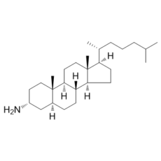
(3R,8R,9S,10S,13R,14S,17R)-10,13-dimethyl-17-((R)-6-methylheptan-2-yl)hexadecahydro-1H-cyclopenta[a]phenanthren-3-amine
|
|
| 别名 |
3α-Aminocholestane; 3AC; 3-AC; 2206-20-4; 3alpha-Aminocholestane; 3; A-Aminocholestane; (3alpha,5alpha)-Cholestan-3-amine; (3R,5S,8R,9S,10S,13R,14S,17R)-10,13-dimethyl-17-[(2R)-6-methylheptan-2-yl]-2,3,4,5,6,7,8,9,11,12,14,15,16,17-tetradecahydro-1H-cyclopenta[a]phenanthren-3-amine; 3??-Aminocholestane; 3+/--Aminocholestane; 3 AC
|
|
| HS Tariff Code |
2934.99.9001
|
|
| 存储方式 |
Powder -20°C 3 years 4°C 2 years In solvent -80°C 6 months -20°C 1 month |
|
| 运输条件 |
Room temperature (This product is stable at ambient temperature for a few days during ordinary shipping and time spent in Customs)
|
| 溶解度 (体外实验) |
|
|||
|---|---|---|---|---|
| 溶解度 (体内实验) |
配方 1 中的溶解度: ≥ 3.25 mg/mL (8.38 mM) (饱和度未知) in 10% EtOH + 40% PEG300 + 5% Tween80 + 45% Saline (这些助溶剂从左到右依次添加,逐一添加), 澄清溶液。
例如,若需制备1 mL的工作液,可将100 μL 32.5 mg/mL的澄清EtOH储备液加入到400 μL PEG300中并混合均匀;然后向上述溶液中加入50 μL Tween-80,混匀;加入450 μL生理盐水定容至1 mL。 *生理盐水的制备:将 0.9 g 氯化钠溶解在 100 mL ddH₂O中,得到澄清溶液。 配方 2 中的溶解度: ≥ 3.25 mg/mL (8.38 mM) (饱和度未知) in 10% EtOH + 90% Corn Oil (这些助溶剂从左到右依次添加,逐一添加), 澄清溶液。 例如,若需制备1 mL的工作液,可将 100 μL 32.5 mg/mL 澄清 EtOH 储备液添加到 900 μL 玉米油中并充分混合。 请根据您的实验动物和给药方式选择适当的溶解配方/方案: 1、请先配制澄清的储备液(如:用DMSO配置50 或 100 mg/mL母液(储备液)); 2、取适量母液,按从左到右的顺序依次添加助溶剂,澄清后再加入下一助溶剂。以 下列配方为例说明 (注意此配方只用于说明,并不一定代表此产品 的实际溶解配方): 10% DMSO → 40% PEG300 → 5% Tween-80 → 45% ddH2O (或 saline); 假设最终工作液的体积为 1 mL, 浓度为5 mg/mL: 取 100 μL 50 mg/mL 的澄清 DMSO 储备液加到 400 μL PEG300 中,混合均匀/澄清;向上述体系中加入50 μL Tween-80,混合均匀/澄清;然后继续加入450 μL ddH2O (或 saline)定容至 1 mL; 3、溶剂前显示的百分比是指该溶剂在最终溶液/工作液中的体积所占比例; 4、 如产品在配制过程中出现沉淀/析出,可通过加热(≤50℃)或超声的方式助溶; 5、为保证最佳实验结果,工作液请现配现用! 6、如不确定怎么将母液配置成体内动物实验的工作液,请查看说明书或联系我们; 7、 以上所有助溶剂都可在 Invivochem.cn网站购买。 |
| 制备储备液 | 1 mg | 5 mg | 10 mg | |
| 1 mM | 2.5794 mL | 12.8969 mL | 25.7938 mL | |
| 5 mM | 0.5159 mL | 2.5794 mL | 5.1588 mL | |
| 10 mM | 0.2579 mL | 1.2897 mL | 2.5794 mL |
1、根据实验需要选择合适的溶剂配制储备液 (母液):对于大多数产品,InvivoChem推荐用DMSO配置母液 (比如:5、10、20mM或者10、20、50 mg/mL浓度),个别水溶性高的产品可直接溶于水。产品在DMSO 、水或其他溶剂中的具体溶解度详见上”溶解度 (体外)”部分;
2、如果您找不到您想要的溶解度信息,或者很难将产品溶解在溶液中,请联系我们;
3、建议使用下列计算器进行相关计算(摩尔浓度计算器、稀释计算器、分子量计算器、重组计算器等);
4、母液配好之后,将其分装到常规用量,并储存在-20°C或-80°C,尽量减少反复冻融循环。
计算结果:
工作液浓度: mg/mL;
DMSO母液配制方法: mg 药物溶于 μL DMSO溶液(母液浓度 mg/mL)。如该浓度超过该批次药物DMSO溶解度,请首先与我们联系。
体内配方配制方法:取 μL DMSO母液,加入 μL PEG300,混匀澄清后加入μL Tween 80,混匀澄清后加入 μL ddH2O,混匀澄清。
(1) 请确保溶液澄清之后,再加入下一种溶剂 (助溶剂) 。可利用涡旋、超声或水浴加热等方法助溶;
(2) 一定要按顺序加入溶剂 (助溶剂) 。
Small molecule inhibition of Inpp5d induces hyperactivation of Syk and triggers a deletional checkpoint in pre-B ALL cells.Nature.2015May 21;521(7552):357-61. |
|---|
SHIP1 inhibition reduces viable cell numbers and either G2/M or G0/G1 cell cycle arrest.Mol Med.2012 Feb 10;18:65-75. |
SHIP1 inhibition affects apoptosis induction differently in MM cell lines.Mol Med.2012 Feb 10;18:65-75. |
InvivoChem的所有产品仅用于作科学研究,不面向患者销售
Copyright 2020 InvivoChem LLC | All Rights Reserved 粤ICP备20063088号-1


 COA
COA
")
")
")
 463611831
