| 规格 | 价格 | 库存 | 数量 |
|---|---|---|---|
| 5mg |
|
||
| 10mg |
|
||
| 25mg |
|
||
| 50mg |
|
||
| 100mg |
|
||
| 250mg |
|
||
| 500mg |
|
||
| Other Sizes |
|

| 靶点 |
Mps1/monopolar spindle 1 (IC50 = 0.63 nM)
|
|---|---|
| 体外研究 (In Vitro) |
BAY1217389 (BAY-1217389) 抑制 Mps1 激酶活性,IC50 值低于 10 nM,同时表现出优异的选择性。在细胞机制测定中,BAY 1217389 消除诺考达唑诱导的 SAC 活性并诱导有丝分裂过早退出,导致多核和肿瘤细胞死亡。 BAY 1217389 在体外有效抑制肿瘤细胞增殖[1]。
BAY 1161909和BAY1217389抑制Mps1激酶活性,IC50值低于10 nmol/L,同时显示出优异的选择性。在细胞机制分析中,两种Mps1抑制剂都消除了诺考达唑诱导的SAC活性,并诱导了有丝分裂的过早退出(“有丝分裂突破”),导致多核性和肿瘤细胞死亡。这两种化合物在体外均能有效抑制肿瘤细胞增殖(IC50 nmol/L范围)。[1] |
| 体内研究 (In Vivo) |
BAY1217389 (BAY-1217389) 在肿瘤异种移植研究中的单一疗法中取得了中等疗效。然而,根据其独特的作用模式,当与紫杉醇联合使用时,低剂量的 Mps1 抑制剂可减少紫杉醇诱导的有丝分裂停滞,同时减弱 SAC 活性。因此,在广泛的异种移植模型(包括那些表现出获得性或内在紫杉醇耐药性的模型)中,联合治疗在各自的 MTD 上比紫杉醇或 Mps1 抑制剂单一治疗大大提高了疗效。 BAY 1217389 显示出良好的耐受性,且不会增加紫杉醇单一疗法的毒性[1]。
在体内,Empesertib(BAY1161909)和BAY1217389在肿瘤异种移植物研究中的单一疗法中取得了中等疗效。然而,根据其独特的作用方式,当与紫杉醇联合使用时,低剂量的Mps1抑制剂通过削弱SAC活性来减少紫杉醇诱导的有丝分裂阻滞。因此,在广泛的异种移植物模型中,包括那些显示获得性或内在紫杉醇耐药性的模型中,联合治疗在各自的MTD上比紫杉醇或Mps1抑制剂单药治疗有显著提高的疗效。两种Mps1抑制剂均表现出良好的耐受性,且不会增加紫杉醇单一疗法的毒性。这些临床前研究结果验证了癌症治疗中废除SAC的创新概念,并证明了临床概念验证研究的合理性,这些研究评估了Mps1抑制剂BAY 1161909和BAY 1217389与抗有丝分裂癌症药物联合使用,以提高其疗效并潜在地克服耐药性[1]。 |
| 酶活实验 |
通过生物素化肽 (Biotin-Ahx-PWDPDDADITEILG-NH2) 的磷酸化,基于 TRFRET 的体外激酶测定可评估 BAY 1161909 或 BAY 1217389 对重组人 Mps1 的抑制作用。标准测定条件要求在两次孵育之间有 15 分钟的预孵育期。激酶和测试化合物,然后添加底物和 10 μM ATP 以启动酶反应。
激酶测定[1] 通过生物素化肽(生物素-Ahx-PWDPDDADITITEG-NH2)的磷酸化,在基于TR-FRET的体外激酶测定中评估了Empesertib(BAY1161909)或BAY1217389对重组人Mps1的抑制作用。激酶和测试化合物预孵育15分钟,然后通过添加底物和10μmol/L的ATP开始酶反应。有关更多详细信息,请参阅补充材料和方法。 激酶选择性分析[1] 使用Millipore激酶或DiscoveRx分析器筛选,对Empesertib(BAY1161909)和BAY1217389进行了针对一组激酶的反筛选。Empesertib(BAY1161909)最初在DiscoveRx激酶组中以1μmol/L的浓度进行测试,然后测定11种激酶的KD(补充表S1)。在Millipore激酶面板中以10μmol/L的浓度测试BAY 1161909,然后以1和0.1μmol/L的剂量重新测试,并测定JNK1alpha、JNK2alpha和JNK3的IC50(补充表S1)。BAY 1217389最初在DiscoveRx激酶面板中以1、0.1和0.01μmol/L的浓度进行测试(补充表S2)。 |
| 细胞实验 |
将细胞以每孔 1,000 至 5,000 个细胞的密度接种到 96 孔板中,并在补充有 10% FCS 的适当培养基中进行。 24 小时后,用 BAY1217389 (BAY-1217389) 或 BAY 1161909 的系列稀释液一式四份处理细胞。再过 96 小时后,用戊二醛固定贴壁细胞并用结晶紫染色。 IC50 值通过 4 参数拟合计算[1]。
多核试验。[2] 将U-2 OS(骨肉瘤ATCC:HTB-96)细胞以每孔2500个细胞的密度接种在20μL细胞培养基中的384孔微量滴定板中,并在37°C下孵育过夜。以100nmol/L的终浓度分三次加入恩帕替布(BAY1161909)或BAY1217389。在试验化合物存在下,细胞在37°C下处理0、24、48和72小时。此后,用4%(v/v)PFA固定细胞,用0.5%(v/v”)Triton X-100渗透,并用0.5%(v/v”)BSA在PBS中封闭。通过抗体标记检测α-微管蛋白结构。用山羊IgG阻断后,在阻断液中加入二抗。用PBS洗涤细胞,用Hoechst 33342标记细胞核。最后,洗涤细胞,用PBS覆盖,并在4°C下储存,直至图像采集。图像是用PerkinElmer Opera高内容分析阅读器获取的。 |
| 动物实验 |
小鼠:采用50日龄、平均体重20-22克的雌性无胸腺NMRI nu/nu小鼠进行肿瘤异种移植研究。当肿瘤大小达到20-40 mm²时(具体大小取决于肿瘤模型的生长情况),将动物随机分为治疗组和对照组(每组8-10只小鼠)。随后,分别给予小鼠灌胃给予赋形剂(70%聚乙二醇400、5%乙醇、25%溶剂)、BAY 1161909、BAY1217389和/或紫杉醇。将携带 A2780cis 肿瘤的雌性 NMRI 裸鼠分别接受紫杉醇(静脉注射,单次剂量 24 mg/kg)、BAY 1161909(口服,每日两次,连续两天,每次剂量 2.5 mg/kg)以及紫杉醇(静脉注射,单次剂量 24 mg/kg)联合 BAY 1161909(口服,每日两次,连续两天,每次剂量 1 mg/kg)治疗。这样可以分析体内多倍体和多核的诱导情况。
药代动力学研究[2] 在雄性 Wistar 大鼠和雌性 CD1 或 NMRI nu/nu 小鼠中进行了药代动力学研究。对于大鼠和小鼠的静脉注射研究,Empesertib (BAY1161909) 溶解于 1% DMSO、99% 血浆中;在大鼠口服给药研究中,BAY1217389 溶于 50% 聚乙二醇 (PEG) 400、10% 乙醇和 40% 水中;在小鼠口服给药研究中,BAY1217389 溶于 75% PEG 400、5% 乙醇和 25% 溶剂中。BAY1217389 溶于 50% PEG 400、10% 乙醇和 40% 水中,用于大鼠和小鼠的静脉注射和口服给药。在药代动力学研究中,分别于静脉给药后 2、5、15、30、45 分钟、1、2、4、6、8 和 24 小时,以及口服给药后 8、15、30、45 分钟、1、2、4、6、8 和 24 小时采集血浆样本,并用冰冷的乙腈 (1:5) 沉淀。采用液相色谱-串联质谱法(LC/MS-MS)分析上清液。药代动力学参数根据血浆浓度数据估算,例如,使用对数线性梯形法则估算AUC。最大血浆浓度(Cmax)及其达峰时间(Tmax)直接从浓度-时间曲线中获取。 动物疗效研究[2] 肿瘤异种移植研究中,使用50日龄、平均体重20至22克的雌性无胸腺NMRI nu/nu小鼠(Taconic),小鼠在适应环境14天后进行实验。小鼠全天24小时自由摄食饮水。取自指数生长期的细胞培养物的人肿瘤细胞,分别用100% Matrigel重悬,使A2780cis、NCI-H1299和SUM-149模型的最终浓度分别为2 × 10⁷、3 × 10⁷和5 × 10⁷个细胞/mL。将0.1 mL浓度分别为2 × 10⁶个A2780cis、3 × 10⁶个NCI-H1299或5 × 10⁶个SUM-149细胞的细胞悬液皮下植入无胸腺小鼠的腹股沟区域。从裸鼠体内连续传代培养获得的患者肿瘤外植体MAXF 1384或LU384的肿瘤组织块被切成直径4至5 mm的小块,并皮下移植到无胸腺小鼠的侧腹部。肿瘤面积(最长径与其垂直径的乘积)用游标卡尺测量,每周测定2-3次,并测定体重。肿瘤生长抑制率以治疗/对照比值(T/C)表示,该比值根据研究结束时的肿瘤面积计算得出。动物体重被用作治疗相关毒性的指标。体重下降超过20%被视为毒性反应。当肿瘤大小达到约20-40 mm²时(具体大小取决于肿瘤模型的生长情况),将动物随机分为治疗组和对照组(每组8-10只小鼠),并分别经口给予赋形剂(70% PEG 400、5%乙醇和25% Solutol)、Empesertib(BAY1161909)、BAY1217389和/或紫杉醇,具体给药方案见表格和图例。在联合治疗组中,Mps1抑制剂和紫杉醇在同一天给药,给药时间间隔为1小时。每只动物的治疗方案均根据其体重而定。 体内作用机制研究[2] 为了分析体内多倍体和多核诱导情况,将携带A2780cis肿瘤的雌性NMRI裸鼠(见上文)分别接受以下治疗:紫杉醇(静脉注射一次,24 mg/kg)、Empesertib(BAY1161909)(口服,每日两次,连续2天,每次2.5 mg/kg),以及紫杉醇(静脉注射一次,24 mg/kg)联合Empesertib(BAY1161909)(口服,每日两次,连续2天,每次1 mg/kg)。所有组的治疗均在肿瘤细胞接种后第14天,肿瘤大小达到60 mm²时开始。在治疗第1天首次施用Empesertib(BAY1161909)后4小时和8小时,以及在治疗第2天首次施用BAY1161909后4小时、8小时和24小时,分别制备肿瘤样本。每个时间点,每个治疗组分析3只动物。肿瘤样本经石蜡包埋和苏木精-伊红染色后,用于组织学检查。 |
| 药代性质 (ADME/PK) |
体内药代动力学参数[1]
在小鼠和大鼠中测定了药代动力学参数。向雄性CD1小鼠和雄性Wistar大鼠静脉注射0.5 mg/kg的BAY 1161909后,该化合物的血药清除率较低。两种动物的稳态分布容积(Vss)均较高,且终末半衰期较长。向雌性NMRI小鼠和雄性Wistar大鼠口服1 mg/kg和0.5 mg/kg后,4小时达到血浆峰浓度。小鼠和大鼠的口服生物利用度中等(表1)。分别向雌性CD1小鼠和雄性Wistar大鼠静脉注射1.0 mg/kg和0.5 mg/kg的BAY1217389。在所测试的动物中,血药清除率均较低。稳态分布容积(Vss)较高,且终末半衰期较长。将 BAY1217389 分别以口服方式给予雌性 NMRI 小鼠(1 mg/kg)和雄性 Wistar 大鼠(0.5 mg/kg)。血浆峰浓度出现在 1.5 至 7 小时之间。大鼠的口服生物利用度较高,小鼠的口服生物利用度中等(表 1)。我们的数据表明,我们已鉴定出两种具有良好药代动力学特征的新型 Mps1 激酶抑制剂,支持其进一步开发用于临床应用。 |
| 毒性/毒理 (Toxicokinetics/TK) |
目的:单极纺锤体1 (MPS1) 激酶抑制剂 BAY 1217389 (BAY) 与紫杉醇具有协同作用。本 I 期研究采用一种新型随机连续再评估方法 (rCRM) 评估 BAY 与紫杉醇联合用药的疗效,以期改进剂量确定。[2]
患者和方法:实体瘤患者被随机分配接受口服 BAY(每日两次,用药 2 天/停药 5 天)联合每周一次的紫杉醇 (90 mg/m2) 治疗,或接受紫杉醇单药治疗(第 1 个疗程)。剂量递增以 CRM 模型为指导。主要目标是评估安全性、确定 BAY 的最大耐受剂量 (MTD) 以及评估两种化合物的药代动力学特征。进行模拟以确定 rCRM 对剂量确定的贡献。[2] 结果:共纳入 75 例患者。主要的剂量限制性毒性是血液学毒性(55.6%)。BAY与紫杉醇联合用药的最大耐受剂量(MTD)确定为每日两次,每次64 mg。纳入对照组后,我们明确证实了≥3级中性粒细胞减少症是由较高的BAY暴露量引起的[AUC0-12 (P< 0.001)]。确定MTD后,我们纳入了19例接受该剂量治疗的乳腺癌患者进行剂量扩展研究。其他常见毒性包括恶心(45.3%)、疲乏(41.3%)和腹泻(40.0%)。在可评估的患者中,31.6%观察到总体确认的缓解。模拟结果表明,rCRM在确定真实MTD方面优于传统设计。[2] 结论:BAY与紫杉醇联合用药具有相当大的毒性,且无治疗窗。然而,rCRM设计使我们能够确定BAY的暴露-毒性关系。因此,我们认为 rCRM 可以改善 I 期试验中联合使用具有重叠毒性的药物时的剂量确定。 |
| 参考文献 |
|
| 其他信息 |
单极纺锤体1 (Mps1) 已被证实是激活纺锤体组装检查点 (SAC) 的关键激酶,以确保染色体正确分配至子细胞。本文报道了两种新型选择性Mps1抑制剂BAY 1161909和BAY 1217389的结构和功能表征,这两种抑制剂分别属于不同的化学类别。BAY 1161909和BAY 1217389对Mps1激酶活性的抑制IC50值均低于10 nmol/L,且具有优异的选择性。在细胞机制实验中,两种Mps1抑制剂均能抑制诺考达唑诱导的SAC活性,并诱导细胞过早退出有丝分裂(“有丝分裂突破”),最终导致多核细胞形成和肿瘤细胞死亡。两种化合物在体外均能有效抑制肿瘤细胞增殖(IC50值在nmol/L范围内)。在体内,BAY 1161909 和 BAY 1217389 在肿瘤异种移植研究中单药治疗取得了中等疗效。然而,与其独特的作用机制相符,当与紫杉醇联合使用时,低剂量的 Mps1 抑制剂可通过减弱 SAC 活性来降低紫杉醇诱导的有丝分裂阻滞。因此,在多种异种移植模型中,包括那些表现出获得性或固有性紫杉醇耐药性的模型,联合治疗在各自的最大耐受剂量 (MTD) 下显著提高了疗效,优于紫杉醇或 Mps1 抑制剂单药治疗。两种 Mps1 抑制剂均表现出良好的耐受性,且不会增加紫杉醇单药治疗的毒性。这些临床前研究结果验证了 SAC 抑制这一癌症治疗创新理念,并为评估 Mps1 抑制剂 BAY 1161909 和 BAY 1217389 与抗有丝分裂抗癌药物联合用药以提高疗效并可能克服耐药性提供了临床概念验证研究的依据。Mol Cancer Ther; 15(4); 583-92。
|
| 分子式 |
C27H24F5N5O3
|
|---|---|
| 分子量 |
561.5032
|
| 精确质量 |
561.179
|
| 元素分析 |
C, 57.75; H, 4.31; F, 16.92; N, 12.47; O, 8.55
|
| CAS号 |
1554458-53-5
|
| 相关CAS号 |
1554458-53-5
|
| PubChem CID |
78320750
|
| 外观&性状 |
White to off-white solid powder
|
| 密度 |
1.5±0.1 g/cm3
|
| 折射率 |
1.617
|
| LogP |
4.64
|
| tPSA |
89.78
|
| 氢键供体(HBD)数目 |
2
|
| 氢键受体(HBA)数目 |
11
|
| 可旋转键数目(RBC) |
9
|
| 重原子数目 |
40
|
| 分子复杂度/Complexity |
867
|
| 定义原子立体中心数目 |
0
|
| SMILES |
FC(C([H])([H])C([H])([H])N([H])C1=C([H])C(=NN2C1=NC([H])=C2C1C([H])=C([H])C(=C(C([H])([H])[H])C=1[H])C(N([H])C1([H])C([H])([H])C1([H])[H])=O)OC1C([H])=C([H])C(=C(C=1F)F)OC([H])([H])[H])(F)F
|
| InChi Key |
WNEILUNVMHVMPH-UHFFFAOYSA-N
|
| InChi Code |
InChI=1S/C27H24F5N5O3/c1-14-11-15(3-6-17(14)26(38)35-16-4-5-16)19-13-34-25-18(33-10-9-27(30,31)32)12-22(36-37(19)25)40-21-8-7-20(39-2)23(28)24(21)29/h3,6-8,11-13,16,33H,4-5,9-10H2,1-2H3,(H,35,38)
|
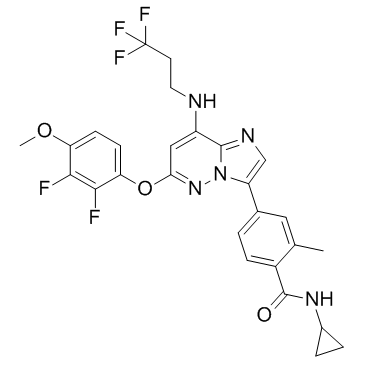
| 化学名 |
N-cyclopropyl-4-[6-(2,3-difluoro-4-methoxyphenoxy)-8-(3,3,3-trifluoropropylamino)imidazo[1,2-b]pyridazin-3-yl]-2-methylbenzamide
|
| 别名 |
BAY-1217389; BAY 1217389; 1554458-53-5; BAY1217,389; BAY-1217,389; Bay 1217,389; Benzamide, N-cyclopropyl-4-[6-(2,3-difluoro-4-methoxyphenoxy)-8-[(3,3,3-trifluoropropyl)amino]imidazo[1,2-b]pyridazin-3-yl]-2-methyl-; M964LB1114; N-cyclopropyl-4-[6-(2,3-difluoro-4-methoxyphenoxy)-8-(3,3,3-trifluoropropylamino)imidazo[1,2-b]pyridazin-3-yl]-2-methylbenzamide; UNII-M964LB1114; BAY1217389
|
| HS Tariff Code |
2934.99.9001
|
| 存储方式 |
Powder -20°C 3 years 4°C 2 years In solvent -80°C 6 months -20°C 1 month |
| 运输条件 |
Room temperature (This product is stable at ambient temperature for a few days during ordinary shipping and time spent in Customs)
|
| 溶解度 (体外实验) |
DMSO: ~100 mg/mL (~178.1 mM)
Ethanol: ~8 mg/mL (~14.3 mM) |
|---|---|
| 溶解度 (体内实验) |
配方 1 中的溶解度: 2.5 mg/mL (4.45 mM) in 10% DMSO + 40% PEG300 + 5% Tween80 + 45% Saline (这些助溶剂从左到右依次添加,逐一添加), 悬浮液;超声助溶。
例如,若需制备1 mL的工作液,可将100 μL 25.0 mg/mL澄清DMSO储备液加入到400 μL PEG300中,混匀;然后向上述溶液中加入50 μL Tween-80,混匀;加入450 μL生理盐水定容至1 mL。 *生理盐水的制备:将 0.9 g 氯化钠溶解在 100 mL ddH₂O中,得到澄清溶液。 配方 2 中的溶解度: ≥ 2.5 mg/mL (4.45 mM) (饱和度未知) in 10% DMSO + 90% Corn Oil (这些助溶剂从左到右依次添加,逐一添加), 澄清溶液。 例如,若需制备1 mL的工作液,可将 100 μL 25.0 mg/mL 澄清 DMSO 储备液加入到 900 μL 玉米油中并混合均匀。 请根据您的实验动物和给药方式选择适当的溶解配方/方案: 1、请先配制澄清的储备液(如:用DMSO配置50 或 100 mg/mL母液(储备液)); 2、取适量母液,按从左到右的顺序依次添加助溶剂,澄清后再加入下一助溶剂。以 下列配方为例说明 (注意此配方只用于说明,并不一定代表此产品 的实际溶解配方): 10% DMSO → 40% PEG300 → 5% Tween-80 → 45% ddH2O (或 saline); 假设最终工作液的体积为 1 mL, 浓度为5 mg/mL: 取 100 μL 50 mg/mL 的澄清 DMSO 储备液加到 400 μL PEG300 中,混合均匀/澄清;向上述体系中加入50 μL Tween-80,混合均匀/澄清;然后继续加入450 μL ddH2O (或 saline)定容至 1 mL; 3、溶剂前显示的百分比是指该溶剂在最终溶液/工作液中的体积所占比例; 4、 如产品在配制过程中出现沉淀/析出,可通过加热(≤50℃)或超声的方式助溶; 5、为保证最佳实验结果,工作液请现配现用! 6、如不确定怎么将母液配置成体内动物实验的工作液,请查看说明书或联系我们; 7、 以上所有助溶剂都可在 Invivochem.cn网站购买。 |
| 制备储备液 | 1 mg | 5 mg | 10 mg | |
| 1 mM | 1.7809 mL | 8.9047 mL | 17.8094 mL | |
| 5 mM | 0.3562 mL | 1.7809 mL | 3.5619 mL | |
| 10 mM | 0.1781 mL | 0.8905 mL | 1.7809 mL |
1、根据实验需要选择合适的溶剂配制储备液 (母液):对于大多数产品,InvivoChem推荐用DMSO配置母液 (比如:5、10、20mM或者10、20、50 mg/mL浓度),个别水溶性高的产品可直接溶于水。产品在DMSO 、水或其他溶剂中的具体溶解度详见上”溶解度 (体外)”部分;
2、如果您找不到您想要的溶解度信息,或者很难将产品溶解在溶液中,请联系我们;
3、建议使用下列计算器进行相关计算(摩尔浓度计算器、稀释计算器、分子量计算器、重组计算器等);
4、母液配好之后,将其分装到常规用量,并储存在-20°C或-80°C,尽量减少反复冻融循环。
计算结果:
工作液浓度: mg/mL;
DMSO母液配制方法: mg 药物溶于 μL DMSO溶液(母液浓度 mg/mL)。如该浓度超过该批次药物DMSO溶解度,请首先与我们联系。
体内配方配制方法:取 μL DMSO母液,加入 μL PEG300,混匀澄清后加入μL Tween 80,混匀澄清后加入 μL ddH2O,混匀澄清。
(1) 请确保溶液澄清之后,再加入下一种溶剂 (助溶剂) 。可利用涡旋、超声或水浴加热等方法助溶;
(2) 一定要按顺序加入溶剂 (助溶剂) 。
| NCT Number | Recruitment | interventions | Conditions | Sponsor/Collaborators | Start Date | Phases |
| NCT02366949 | Completed | Drug: BAY1217389 Drug: Paclitaxel |
Medical Oncology | Bayer | February 27, 2015 | Phase 1 |
InvivoChem的所有产品仅用于作科学研究,不面向患者销售
Copyright 2020 InvivoChem LLC | All Rights Reserved 粤ICP备20063088号-1


 COA
COA
 463611831
