| 规格 | 价格 | 库存 | 数量 |
|---|---|---|---|
| 10 mM * 1 mL in DMSO |
|
||
| 1mg |
|
||
| 5mg |
|
||
| 10mg |
|
||
| 25mg |
|
||
| 50mg |
|
||
| 100mg |
|
||
| 250mg |
|
||
| 500mg |
|
||
| Other Sizes |
| 靶点 |
STING/stimulator of interferon genes
|
|---|---|
| 体外研究 (In Vitro) |
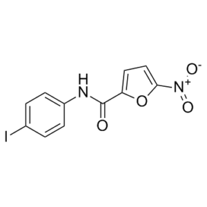
C-176 显着抑制 STING 介导的 IFNβ 报告基因活性,但不抑制 RIG-I 或 TBK1 介导的 IFNβ 报告基因活性。 C-176 预处理可显着抑制 CMA 介导的 I 型 IFN 和 IL-6 血清水平诱导[1]。
|
| 体内研究 (In Vivo) |
C-176(每只小鼠 750/375 nmol C-176,溶于 200 μL 玉米油)不会引起相当大的毒性,可大大降低 CMA 介导的 I 型 IFN 和 IL-6 血液水平的激活[1]。 Trex1−/− 小鼠中没有明显的明显毒性症状,C-176 显着降低 I 型 IFN 的血清水平,并强烈抑制心脏中的炎症标志物[1]。在 Trex1−/− 小鼠中,C-176 显着降低了许多全身炎症指标 [
|
| 酶活实验 |
Competition assay/竞争分析[1]
将表达Flag-STING的HEK293T细胞与指定化合物一起孵育,1小时后加入C-176-AL 1小时。将细胞收集在PBS中,通过C-176-AL-介导的STING标记的凝胶内分析进行分析(见“基于凝胶的化合物与STING结合的分析”)。 基于凝胶的化合物与STING结合分析[1] 表达Flag-STING的HEK293T细胞在无血清培养基中与C-176-AL、C-175-AZ、叠氮碘乙酰胺或H-151-AL一起孵育,收集在PBS中,通过反复冷冻和解冻裂解。用新制备的“点击试剂”混合物处理43微升裂解细胞,该混合物含有三(苄基三唑甲基)胺(TBTA)(每个样品3μl,3 mM在1:4 DMSO:t-ButOH中)、四甲基罗丹明(TAMRA)叠氮化物、SiR叠氮化物或SiR炔(每个样品2μl,1.25 mM在DMSO中),以及新制得的CuSO4(每个样品1μl)和三-(2-羧乙基)盐酸膦(TCEP)(每个样本1μl,在室温下孵育30分钟。通过加入还原性样品缓冲液淬灭反应。使用Fusion-FX可视化凝胶内荧光,并使用Fusion-capt高级采集软件进行分析。 与亚琥珀酸二琥珀酰亚胺酯交联[1] 表达Flag-mmSTING的HEK293T细胞在有或没有C-176(1μM)的情况下孵育1小时,并用DMSO或CMA(250μg ml-1)处理2小时。在室温下,在PBS中用DMSO中新鲜制备的1 mM二琥珀酰亚胺基琥珀酸盐(DSS)进行交联1小时。 [3H]-棕榈酸酯代谢标记[1] 指示标志-STING构建体在HEK293T细胞中表达。对于代谢标记,细胞在用10mM Hepes缓冲的格拉斯哥最低必需培养基中饥饿1小时,pH 7.4,用C-178或C-176(1μM)缓冲。然后在C-178或C-176存在下,在200μCi ml−1[3H]-棕榈酸(9,10-3H(N)) 的IM中孵育细胞2小时,并用CMA(250μg ml−1)刺激或不刺激。对于免疫沉淀,细胞在PBS中洗涤三次,在4°C下在以下缓冲液(0.5%Nonidet P-40、500 mM Tris pH 7.4、20 mM EDTA、10 mM NaF、2 mM苯甲脒和蛋白酶抑制剂混合物)中裂解30分钟,并在5000 rpm下离心3分钟。上清液在4°℃下与适当的抗体(抗Flag、抗转铁蛋白受体和抗钙蛋白酶)和G琼脂糖珠一起孵育过夜。对于放射性标记实验,在免疫沉淀后,在4-20%梯度SDS-PAGE之前,将洗涤过的珠子在90°C的还原样品缓冲液中孵育5分钟。SDS-PAGE后,将凝胶在固定剂溶液(25%异丙醇、65%H2O、10%乙酸)中孵育,并与信号增强剂Amplify NAMP100一起孵育30分钟。使用放射自显影显示放射性标记产物,并使用台风成像仪进行定量。 试试AI翻译 笔记 |
| 细胞实验 |
免疫沉淀[1]
多西环素在HEK293T细胞中诱导Flag-STING表达过夜。细胞与或不与C-178或C-176(1μM)一起孵育1小时,并用DMSO或CMA(250μg ml−1)处理2小时。细胞在PBS中洗涤,在裂解缓冲液(50 mM HEPES、150 mM NaCl、10%甘油、1 mM MgCl、1 mM CaCl、1%Brij-58和蛋白酶抑制剂混合物)中裂解30分钟。在4°C下使用抗Flag M2亲和凝胶琼脂糖凝胶对Flag-STING进行免疫沉淀2小时。在裂解缓冲液和PBS中严格洗涤后,完全去除上清液,在进行SDS-PAGE之前,将树脂在样品缓冲液中煮沸。为了免疫沉淀内源性STING,将脾细胞在上述裂解缓冲液中裂解,并与抗STING(RD System AF6516)和G琼脂糖珠一起孵育过夜。在PBS中洗涤珠子,并进行基于凝胶的C-176-AL与STING结合分析。 mmSTING的完整质量测量[1] 多西环素在HEK293T细胞中诱导Flag-mmSTING或Flag-mmS廷G(C91S)表达过夜。细胞用或不用C-178或C-176(1μM)处理30分钟,并在裂解缓冲液(20 mM Hepes、150 mM NaCl、10%甘油和1%DDM)中裂解。Flag-STING在4°C下用抗Flag M2亲和凝胶琼脂糖凝胶免疫沉淀2小时。根据制造商的说明,使用Flag肽洗脱沉淀的蛋白质。在岛津MS2020上进行蛋白质质谱分析,该MS2020连接到Nexerra UHPLC系统,配备Waters ACQUITY UPLC BEH C4 1.7-μm,2.1×50mm柱。缓冲液A为0.05%甲酸水溶液,缓冲液B为0.05%甲酸乙腈溶液。在6.0分钟内,缓冲液B的分析梯度为10%至90%,流速为0.75 ml min−1。从300-2000 Da收集质谱,并使用MagTran软件对光谱进行解卷积。 |
| 动物实验 |
Animal/Disease Models: WT type mice.
Doses: 750/375 nmol C-176 per mouse in 200 μL corn oil (~1.34/0.67 mg/mL). Route of Administration: Intraperitoneally, once. Experimental Results: Dramatically decreased Serum levels of type I IFNs and IL-6. Mice and in vivo studies[1] C57BL/6J mice (stock number 000664) were purchased from Jackson Laboratories. TREX1-deficient mice were a gift from T. Lindahl31 and were backcrossed for >10 generations to C57BL/6NJ. Mice were maintained under specific-pathogen-free (SPF) conditions at EPFL. For the pharmacokinetic studies, wild-type mice were injected intraperitoneally with 750 nmol C-176 per mouse in 200 μl corn oil. Blood was collected at 30 min, 2 h and 4 h and serum C-176 levels were measured by mass spectrometry (liquid chromatography–high-resolution mass spectrometry). To assess the in vivo inhibitory effect of C-176, wild-type mice (8–12 weeks of age) were injected either with vehicle or C-176. After 1 h or 4 h, CMA was administered at a concentration of 224 mg kg−1. Four hours later, mice were euthanized and the serum was collected to measure CMA-induced cytokine levels. To assess the in vivo inhibitory effect of H-151, wild-type mice were injected intraperitoneally with 750 nmol H-151 per mouse in 200 μl 10% Tween-80 in PBS. After 1 h CMA (112 mg kg−1) was administered, and after 4 h mice were euthanized and the serum was collected. The efficacy study in Trex1−/− mice was conducted as follows: mice (2–5 weeks of age) were injected with 7.5 μl of C-176 or DMSO dissolved in 85 μl corn oil twice per day for 11 consecutive days. Mice were euthanized by anaesthetization in a CO2 chamber followed by cervical dislocation. For toxicology studies, 8-week-old mice were injected daily with 562.5 nmol of C-176 for 2 weeks. This study was conducted in two separate stages. A total of 198 rats (300–350 g, 9–11 weeks, male) (Stage 1, 90 rats; Stage 2, 108 rats) were used for the experiments (as shown in Figure 1). In total, 7 rats were found dead over the course of experimentation (within 24 h after modeling), and 6 were excluded based on behavioral exclusion criteria (at 30 day after modeling). Animals were monitored for health and killed via cervical dislocation with secondary decapitation.[2] Stage 1[2] To investigate the role of STING, the selective STING antagonist C-176 or STING agonist ADU-S100 was administered via intraperitoneal injection to investigate STING signaling following TBI in the first stage. The rats were randomly divided into four groups (n = 18 rats/group): sham + vehicle1 group, TBI + vehicle1 group, TBI + ADU-S100 group, TBI + C-176 group. Rats were randomized using a web-based random group generator (www.pubmed.de/tools/zufallsgenerator). Data acquisition and analyses were blinded to the experimenter. Corn oil (cat. No. ST1177; Beyotime) as vehicle1 was used to dissolve ADU-S100 and C-176. Vehicle1 (corn oil) were administered via intraperitoneal injection at the same time points after TBI or sham modeling. Cerebral tissues for western blot assay were collected (n = 6 rats/group) 24 h after the injection. Neurobehavioral tests, including open field test, force swimming test, and novel object recognition test, were performed 30 days after the TBI induced by a weight-drop model (n = 12 rats/group). In addition, cerebral tissues for Nissl and immunofluorescence staining (n = 6 rats/group) and for ELISA (n = 6 rats/group) were also collected (Figure 1a). Stage 2[2] In the second stage, a selective activator of NLRP3 (nigericin) was administered via intracerebroventricular injection to elucidate NLRP3 as a downstream factor in TBI. The rats were divided into five groups (n = 18 rats/group): TBI + ADU-S100 + MCC950 group, TBI + ADU-S100 + VX765 group, TBI + C-176 + nigericin group, TBI +C-176+ vehicle2 group, and TBI + ADU-S100 + vehicle2 group. 10% ethyl alcohol and 90% corn oil as vehicle2 was used to dissolve nigericin, MCC950 and VX-765, and administered via intracerebroventricular injection. Tissues for western blot assay were collected 24 h after the TBI (n = 6 rats/group). After that, behavioral tests (n = 12 rats/group), Nissl, and immunofluorescence staining (n = 6 rats/group) and ELISA (n = 6 rats/group) were performed 30 days post-modeling (Figure 1b). The remaining rats were killed via cervical dislocation. Drug administration Nigericin, MCC950 or VX765 was administered through intracerebroventricular injection 30 mins before modeling using a needle attached to a microsyringe (5 μl). The injection was made using the following coordinates from bregma: 0.3 mm posterior, 1.0 mm lateral, and 2.5 mm ventral. Nigericin (2 μg/2 μl), MCC950 (2 μg/2 μl) or VX765 (2 μg/2 μl) was infused at a rate of 0.667 μl/min using a microinfusion pump (TJ-4A/SL0107-1A, LongerPump). Nigericin, MCC950 or VX765 was dissolved in 10% ethyl alcohol and 90% corn oil. The needle was held in the brain for an additional 10 min before being slowly removed. Finally, the burr hole was sealed using bone wax. The STING agonist ADU-S100 (0.5 mg/kg) and antagonist C-176(10 mg/kg) were administered via intraperitoneal injection immediately after the TBI. Drug doses used in this study were determined in a preliminary experiment based on previously published studies (Lee et al., 2021; Peng et al., 2020; Tsuchiya et al., 2019). |
| 参考文献 | |
| 其他信息 |
Aberrant activation of innate immune pathways is associated with a variety of diseases. Progress in understanding the molecular mechanisms of innate immune pathways has led to the promise of targeted therapeutic approaches, but the development of drugs that act specifically on molecules of interest remains challenging. Here we report the discovery and characterization of highly potent and selective small-molecule antagonists of the stimulator of interferon genes (STING) protein, which is a central signalling component of the intracellular DNA sensing pathway1,2. Mechanistically, the identified compounds covalently target the predicted transmembrane cysteine residue 91 and thereby block the activation-induced palmitoylation of STING. Using these inhibitors, we show that the palmitoylation of STING is essential for its assembly into multimeric complexes at the Golgi apparatus and, in turn, for the recruitment of downstream signalling factors. The identified compounds and their derivatives reduce STING-mediated inflammatory cytokine production in both human and mouse cells. Furthermore, we show that these small-molecule antagonists attenuate pathological features of autoinflammatory disease in mice. In summary, our work uncovers a mechanism by which STING can be inhibited pharmacologically and demonstrates the potential of therapies that target STING for the treatment of autoinflammatory disease.[1]
Long-term neurological deficits after severe traumatic brain injury (TBI), including cognitive dysfunction and emotional impairments, can significantly impair rehabilitation. Glial activation induced by inflammatory response is involved in the neurological deficits post-TBI. This study aimed to investigate the role of the stimulator of interferon genes (STING)-nucleotide-binding oligomerization domain-like receptor pyrin domain-containing-3 (NLRP3) signaling in a rodent model of severe TBI. Severe TBI models were established using weight-drop plus blood loss reinfusion model. Selective STING agonist ADU-S100 or antagonist C-176 was given as a single dose after modeling. Further, NLRP3 inhibitor MCC950 or activator nigericin, or caspase-1 inhibitor VX765, was given as an intracerebroventricular injection 30 min before modeling. After that, a novel object recognition test, open field test, force swimming test, western blot, and immunofluorescence assays were used to assess behavioral and pathological changes in severe TBI. Administration of C-176 alleviated TBI-induced cognitive dysfunction and emotional impairments, neuronal loss, and inflammatory activation of glia cells. However, the administration of STING agonist ADU-S100 exacerbated TBI-induced behavioral and pathological changes. In addition, STING activation exacerbated pyroptosis-associated neuroinflammation via promoting glial activation, as evidenced by increased cleaved caspase-1 and GSDMD N-terminal expression. In contrast, the administration of C-176 showed anti-pyroptotic effects. The neuroprotective effects of C-176 were partially reversed by the NLRP3 activator, nigericin. Collectively, glial STING is responsible for neuroinflammation post-TBI. However, pharmacologic inhibition of STING led to a remarkable improvement of neuroinflammation partly through suppressing NLRP3 signaling. The STING-NLRP3 signaling is a potential therapeutic target in TBI-induced neurological dysfunction.[2] |
| 分子式 |
C11H7IN2O4
|
|
|---|---|---|
| 分子量 |
358.09
|
|
| 精确质量 |
357.945
|
|
| 元素分析 |
C, 36.90; H, 1.97; I, 35.44; N, 7.82; O, 17.87
|
|
| CAS号 |
314054-00-7
|
|
| 相关CAS号 |
|
|
| PubChem CID |
1103958
|
|
| 外观&性状 |
Light yellow to yellow solid powder
|
|
| 密度 |
1.9±0.1 g/cm3
|
|
| 沸点 |
361.2±37.0 °C at 760 mmHg
|
|
| 闪点 |
172.3±26.5 °C
|
|
| 蒸汽压 |
0.0±0.8 mmHg at 25°C
|
|
| 折射率 |
1.714
|
|
| LogP |
3.38
|
|
| tPSA |
88.1
|
|
| 氢键供体(HBD)数目 |
1
|
|
| 氢键受体(HBA)数目 |
4
|
|
| 可旋转键数目(RBC) |
2
|
|
| 重原子数目 |
18
|
|
| 分子复杂度/Complexity |
326
|
|
| 定义原子立体中心数目 |
0
|
|
| SMILES |
IC1C=CC(=CC=1)N([H])C(C1=CC=C([N+](=O)[O-])O1)=O
|
|
| InChi Key |
JBIKQXOZLBLMKI-UHFFFAOYSA-N
|
|
| InChi Code |
InChI=1S/C11H7IN2O4/c12-7-1-3-8(4-2-7)13-11(15)9-5-6-10(18-9)14(16)17/h1-6H,(H,13,15)
|
|
| 化学名 |
N-(4-iodophenyl)-5-nitrofuran-2-carboxamide
|
|
| 别名 |
|
|
| HS Tariff Code |
2934.99.03.00
|
|
| 存储方式 |
Powder -20°C 3 years 4°C 2 years In solvent -80°C 6 months -20°C 1 month |
|
| 运输条件 |
Room temperature (This product is stable at ambient temperature for a few days during ordinary shipping and time spent in Customs)
|
| 溶解度 (体外实验) |
|
|||
|---|---|---|---|---|
| 溶解度 (体内实验) |
配方 1 中的溶解度: 1.67 mg/mL (4.66 mM) in 10% DMSO + 40% PEG300 + 5% Tween80 + 45% Saline (这些助溶剂从左到右依次添加,逐一添加), 悬浮液; 超声和加热处理
例如,若需制备1 mL的工作液,可将100 μL 16.7 mg/mL澄清的DMSO储备液加入到400 μL PEG300中,混匀;再向上述溶液中加入50 μL Tween-80,混匀;然后加入450 μL生理盐水定容至1 mL。 *生理盐水的制备:将 0.9 g 氯化钠溶解在 100 mL ddH₂O中,得到澄清溶液。 配方 2 中的溶解度: ≥ 0.25 mg/mL (0.70 mM) (饱和度未知) in 10% EtOH + 40% PEG300 + 5% Tween80 + 45% Saline (这些助溶剂从左到右依次添加,逐一添加), 澄清溶液。 例如,若需制备1 mL的工作液,可将 100 μL 2.5 mg/mL 澄清 EtOH 储备液加入到400 μL PEG300 中,混匀;再向上述溶液中加入50 μL Tween-80,混匀;然后加入450 μL 生理盐水定容至1 mL。 *生理盐水的制备:将 0.9 g 氯化钠溶解在 100 mL ddH₂O中,得到澄清溶液。 View More
配方 3 中的溶解度: ≥ 0.25 mg/mL (0.70 mM) (饱和度未知) in 10% EtOH + 90% (20% SBE-β-CD in Saline) (这些助溶剂从左到右依次添加,逐一添加), 澄清溶液。 配方 4 中的溶解度: 0.25 mg/mL (0.70 mM) in 10% EtOH + 90% Corn Oil (这些助溶剂从左到右依次添加,逐一添加), 悬浊液; 超声助溶。 例如,若需制备1 mL的工作液,您可以将 100 μL 2.5 mg/mL 澄清 EtOH 储备液添加到 900 μL 玉米油中并充分混合。 配方 5 中的溶解度: 10 mg/mL (27.93 mM) in Cremophor EL (这些助溶剂从左到右依次添加,逐一添加), 澄清溶液; 超声助溶. 1、请先配制澄清的储备液(如:用DMSO配置50 或 100 mg/mL母液(储备液)); 2、取适量母液,按从左到右的顺序依次添加助溶剂,澄清后再加入下一助溶剂。以 下列配方为例说明 (注意此配方只用于说明,并不一定代表此产品 的实际溶解配方): 10% DMSO → 40% PEG300 → 5% Tween-80 → 45% ddH2O (或 saline); 假设最终工作液的体积为 1 mL, 浓度为5 mg/mL: 取 100 μL 50 mg/mL 的澄清 DMSO 储备液加到 400 μL PEG300 中,混合均匀/澄清;向上述体系中加入50 μL Tween-80,混合均匀/澄清;然后继续加入450 μL ddH2O (或 saline)定容至 1 mL; 3、溶剂前显示的百分比是指该溶剂在最终溶液/工作液中的体积所占比例; 4、 如产品在配制过程中出现沉淀/析出,可通过加热(≤50℃)或超声的方式助溶; 5、为保证最佳实验结果,工作液请现配现用! 6、如不确定怎么将母液配置成体内动物实验的工作液,请查看说明书或联系我们; 7、 以上所有助溶剂都可在 Invivochem.cn网站购买。 |
| 制备储备液 | 1 mg | 5 mg | 10 mg | |
| 1 mM | 2.7926 mL | 13.9630 mL | 27.9259 mL | |
| 5 mM | 0.5585 mL | 2.7926 mL | 5.5852 mL | |
| 10 mM | 0.2793 mL | 1.3963 mL | 2.7926 mL |
1、根据实验需要选择合适的溶剂配制储备液 (母液):对于大多数产品,InvivoChem推荐用DMSO配置母液 (比如:5、10、20mM或者10、20、50 mg/mL浓度),个别水溶性高的产品可直接溶于水。产品在DMSO 、水或其他溶剂中的具体溶解度详见上”溶解度 (体外)”部分;
2、如果您找不到您想要的溶解度信息,或者很难将产品溶解在溶液中,请联系我们;
3、建议使用下列计算器进行相关计算(摩尔浓度计算器、稀释计算器、分子量计算器、重组计算器等);
4、母液配好之后,将其分装到常规用量,并储存在-20°C或-80°C,尽量减少反复冻融循环。
计算结果:
工作液浓度: mg/mL;
DMSO母液配制方法: mg 药物溶于 μL DMSO溶液(母液浓度 mg/mL)。如该浓度超过该批次药物DMSO溶解度,请首先与我们联系。
体内配方配制方法:取 μL DMSO母液,加入 μL PEG300,混匀澄清后加入μL Tween 80,混匀澄清后加入 μL ddH2O,混匀澄清。
(1) 请确保溶液澄清之后,再加入下一种溶剂 (助溶剂) 。可利用涡旋、超声或水浴加热等方法助溶;
(2) 一定要按顺序加入溶剂 (助溶剂) 。