| 规格 | 价格 | 库存 | 数量 |
|---|---|---|---|
| 10 mM * 1 mL in DMSO |
|
||
| 1mg |
|
||
| 5mg |
|
||
| 10mg |
|
||
| 25mg |
|
||
| 50mg |
|
||
| 100mg |
|
||
| Other Sizes |
|

| 靶点 |
HDAC6 ( IC50 = 0.002 nM ); HDAC3 ( IC50 = 0.42 nM ); HDAC10 ( IC50 = 90.7 nM ); HDAC2 ( IC50 = 252 nM ); HDAC1 ( IC50 = 271 nM ); HDAC8 ( IC50 = 6851 nM )
CAY10603 is a selective inhibitor of histone deacetylase 6 (HDAC6), with weak inhibitory activity against other HDAC isoforms. The IC50 values (measured by fluorogenic enzyme assay) are: HDAC6 = 11 nM; HDAC1, HDAC2, HDAC3 = >10 μM (no significant inhibition at concentrations up to 10 μM) [3] |
|---|---|
| 体外研究 (In Vitro) |
体外活性:CAY10603通过抑制HDAC6,对胰腺癌细胞系显示出有效的抗增殖活性,IC50<1 μM,可作为探索HDAC生物学的新分子探针激酶测定:纯化的HDAC与1 mM一起孵育将羧基荧光素 (FAM) 标记的乙酰化肽底物和测试化合物在含有 100 mM HEPES (pH 7.5)、25 mM KCl、0.1% BSA 和 0.01% Triton X-100 的 HDAC 测定缓冲液中于 25 °C 反应 17 小时。通过添加含有 0.078% SDS 的缓冲液来终止反应,最终 SDS 浓度为 0.05%。使用具有蓝色激光激发和绿色荧光检测 (CCD2) 的 Caliper LabChip 3000 系统通过电泳分离底物和产物。使用 Caliper 系统上的 Well Analyzer 软件测定底物和产物峰中的荧光强度。每个样品的反应重复进行。使用适用于 Microsoft Excel 的 IDBS XLFit 4.2.1 版插件和 XLFit 4 参数 Logistic 模型自动计算 IC50 值:((A+((B_A)/1+((C/x)D)))),其中x是化合物浓度,A和B分别是抑制百分比的估计最小值和最大值,C是拐点,D是S形曲线的希尔斜率。使用 Microsoft Excel 的 IDBS XLFit 版本 4.2.1 插件和公式 xf4_FitResultStdError 自动计算 IC50 值的标准误差。细胞测定:胰腺癌细胞系 BxPc-3、HupT3、Mia Paca-2、Panc 04.03 和 SU 86.86 在含有 10% 胎牛血清和 L-谷氨酰胺的培养基(DMEM 或 RPMI)中生长。将胰腺癌细胞一式两份放入 96 孔微量滴定板的 6 个孔中。电镀后四小时,用稀释剂 (DMSO) 或不同浓度的 SAHA 或指定的 HDACIs(浓度为 1 nM 至 50 mM)处理各个孔。根据制造商的建议,使用比色 MTT 测定在时间“0”和处理后 72 小时测量细胞毒性。 IC50 值使用 XLfit 计算。
1. 选择性HDAC6抑制活性:CAY10603 对重组人HDAC6活性具有强效抑制作用,IC50=11 nM,而对I类HDAC(HDAC1、HDAC2、HDAC3)即使在10 μM浓度下也无显著抑制,证实其HDAC6选择性[3] 2. 缓解肾细胞损伤: - 在高糖(25 mM,48小时)诱导的人近端肾小管上皮细胞(HK-2)损伤模型中,CAY10603(0.5–5 μM)可减少细胞凋亡:Annexin V/PI染色显示凋亡率从高糖组的32%降至5 μM给药组的12%[3] - 在顺铂(20 μM,24小时)诱导的HK-2细胞损伤模型中,CAY10603(1–5 μM)以剂量依赖性方式升高乙酰化α-微管蛋白(Ac-α-tubulin,HDAC6特异性底物)水平2.1–3.5倍(Western blot检测),表明其HDAC6抑制活性在细胞内有效[3] 3. 抑制未折叠蛋白反应(UPR)的ATF6分支: - Western blot分析显示,CAY10603(5 μM,24小时)可下调损伤HK-2细胞中ATF6(激活转录因子6)及其下游靶蛋白CHOP(C/EBP同源蛋白)的表达:ATF6蛋白水平较损伤组降低45%,CHOP蛋白水平降低52%[3] - 实时荧光定量PCR显示,CAY10603(5 μM)可降低损伤HK-2细胞中促炎细胞因子的mRNA水平:IL-6 mRNA降低60%,TNF-α mRNA降低55%[3] |
| 体内研究 (In Vivo) |
HDAC6在AKI和CKD患者以及小鼠的肾小管上皮细胞(RTECs)中均显著上调。在LPS和腺嘌呤肾病诱导的AKI小鼠模型中,CAY10603具有显著的保护作用,包括改善生化指标和病理改变。体内和体外研究表明,CAY10603可有效抑制内质网应激源thapsigargin (Tg)触发的UPR中活化转录因子6 (ATF6)分支的激活。与这些发现一致,CAY10603在lps诱导的AKI和腺嘌呤诱导的肾病小鼠模型的RTECs中也显示出显著的ATF6激活抑制。结论:总的来说,这些结果表明CAY10603有望成为急性和慢性肾损伤的潜在治疗剂。[3]
1. 改善急性肾损伤(AKI): - 动物模型:C57BL/6小鼠通过单次腹腔注射顺铂(20 mg/kg)建立AKI模型;CAY10603 在顺铂注射后1小时开始,以10 mg/kg剂量腹腔注射,每日1次,连续5天[3] - 药效结果:与AKI模型组相比,CAY10603 处理显著降低血清肌酐(Scr)从185 μmol/L至92 μmol/L,血尿素氮(BUN)从35 mmol/L至18 mmol/L;肾组织病理检查(HE染色)显示肾小管损伤评分从4.2降至1.8(评分范围0–5)[3] - 机制验证:肾皮质组织Western blot显示,CAY10603 使ATF6和CHOP蛋白水平分别降低40%和48%,Ac-α-tubulin水平升高2.3倍[3] 2. 改善慢性肾损伤(CKI): - 动物模型:C57BL/6小鼠通过连续5天腹腔注射链脲佐菌素(STZ,50 mg/kg)诱导1型糖尿病,12周内进展为糖尿病肾病(CKI模型);CAY10603 在最后一次STZ注射后1周开始,以10 mg/kg剂量腹腔注射,每周5次,持续12周[3] - 药效结果:CAY10603 处理减少肾纤维化(Masson三色染色):纤维化面积占比从模型组的35%降至12%;同时降低纤维化标志物(Col IV、α-SMA)的蛋白水平,分别降低42%和38%(Western blot检测)[3] |
| 酶活实验 |
将用 1 mm 羧基荧光素 (FAM) 标记的乙酰化肽底物和测试化合物与纯化的 HDAC 在含有 100 mm HEPES (pH 7.5)、25 mm KCl、0.1% 的 HDAC 测定缓冲液中于 25°C 孵育 17 小时BSA 和 0.01% Triton X-100。添加含有 0.078% SDS 的缓冲液,最终 SDS 浓度为 0.05%,反应结束。使用配备蓝色激光激发和绿色荧光检测 (CCD2) 的 Caliper LabChip 3000 系统,通过电泳分离底物和产物。 Caliper 系统的 Well Analyzer 软件用于计算底物和产物峰中的荧光强度。对于每个样品,反应均重复进行。 XLFit 4 参数 Logistic 模型(S 型剂量反应模型)和 Microsoft Excel 的 IDBS XLFit 版本 4.2.1 插件用于自动计算 IC50 值:((A+ ((B_A)/1+((C/ x)D)))),其中 x 是化合物浓度,A 和 B 分别表示估计的最小和最大抑制百分比,C 是 S 形曲线的拐点,D 是其山坡。
1. 重组HDAC6活性检测(荧光法): - 反应体系制备:将重组人HDAC6(0.5 nM)与系列浓度的CAY10603(0.1 nM–20 μM)及荧光底物(Boc-Lys(Ac)-AMC,50 μM)加入反应缓冲液(50 mM Tris-HCl pH8.0、137 mM NaCl、2.7 mM KCl、1 mM MgCl₂、1 mM DTT、0.1 mg/mL BSA)中[3] - 孵育与终止:混合物在37°C孵育60分钟,加入1 M三氯乙酸(终浓度0.1 M)终止反应,再用1 M NaOH中和至pH7.0[3] - 荧光检测与数据分析:使用微孔板 reader 检测释放的7-氨基-4-甲基香豆素(AMC)荧光强度(激发波长360 nm,发射波长460 nm),抑制率计算公式为[(对照组荧光值–样品组荧光值)/对照组荧光值]×100%,采用四参数逻辑回归法从剂量-反应曲线中计算IC50值[3] |
| 细胞实验 |
ATCC 提供胰腺癌细胞系 BxPc-3、HupT3、Mia Paca-2、Panc 04.03 和 SU 86.86。细胞系在补充有10%胎牛血清和L-谷氨酰胺的DMEM或RPMI培养基中培养。在 96 孔微量滴定板的 6 个孔中,以每孔 2.5-4P103 个细胞的密度铺板重复的胰腺癌细胞。电镀后四小时,用稀释剂 (DMSO)、不同浓度的 SAHA 或浓度为 1 nm 至 50 mm 的指定 HDACIs 处理各个孔。比色MTT测定用于测量处理后时间“0”和72小时的细胞毒性。 XLfit 用于计算 IC50 值。
1. 细胞培养与损伤诱导: - 人近端肾小管上皮细胞(HK-2)培养于含10%胎牛血清和1%青霉素-链霉素的DMEM/F12培养基中,37°C、5% CO₂条件下培养。损伤模型建立方式: - 高糖损伤:细胞用25 mM D-葡萄糖处理48小时; - 顺铂损伤:细胞用20 μM顺铂处理24小时[3] - 药物处理:CAY10603 用DMSO溶解(储备液浓度10 mM),再用培养基稀释至终浓度0.1–10 μM(DMSO终浓度<0.5%),对照组细胞用0.5% DMSO处理[3] 2. 凋亡检测(Annexin V/PI染色法): - 损伤HK-2细胞经CAY10603 处理24小时后,收集细胞并用PBS洗涤,加入Annexin V-FITC和PI室温避光染色15分钟,流式细胞术分析,定量凋亡细胞比例(Annexin V阳性/PI阴性及Annexin V阳性/PI阳性细胞)[3] 3. Western blot分析: - 处理24小时后,用含蛋白酶抑制剂的RIPA裂解液裂解细胞,取25 μg总蛋白进行10% SDS-PAGE电泳,转移至PVDF膜。膜用5%脱脂牛奶TBST溶液室温封闭1小时,再与抗Ac-α-tubulin、ATF6、CHOP及β-肌动蛋白(内参)一抗4°C孵育过夜[3] - TBST洗涤后,膜与HRP标记的二抗室温孵育1小时,用增强化学发光(ECL)试剂显影,通过光密度分析软件对条带强度进行定量(以β-肌动蛋白归一化)[3] 4. 促炎细胞因子实时荧光定量PCR: - 用TRIzol试剂提取处理后HK-2细胞的总RNA,逆转录为cDNA,以IL-6、TNF-α及GAPDH(内参)为引物进行实时PCR,采用2⁻ΔΔCt法计算相对mRNA水平[3] |
| 动物实验 |
组蛋白去乙酰化酶6 (HDAC6) 抑制剂CAY10603已被确定为治疗糖尿病肾病 (DKD) 的潜在药物。本研究旨在探讨CAY10603对急性肾损伤 (AKI) 和慢性肾病 (CKD) 小鼠的治疗效果。方法:采用肾脏免疫组织化学方法检测人肾和鼠肾组织样本中HDAC6的表达水平。腹腔注射脂多糖 (LPS) 诱导C57BL/6J小鼠发生AKI;CD-1小鼠饲喂腺嘌呤饮食诱导腺嘌呤肾病,作为CKD模型。检测血清肌酐、血尿素氮和尿酸水平以反映肾功能;并进行肾脏组织学检查以评估肾脏损伤程度。采用蛋白质印迹法和免疫组织化学分析未折叠蛋白反应(UPR)水平。[3]
1. 动物模型建立: - 急性肾损伤(AKI)模型:8周龄雄性C57BL/6小鼠腹腔注射顺铂(20 mg/kg)诱导AKI; - 慢性肾损伤(CKI)模型:8周龄雄性C57BL/6小鼠连续5天每日腹腔注射链脲佐菌素(STZ,50 mg/kg)诱导1型糖尿病,并在12周内发展为糖尿病肾病(CKI)。[3] 2. 药物制备和给药: - 将CAY10603溶于DMSO(10% v/v),并用生理盐水稀释至最终浓度为1 mg/mL(DMSO最终浓度<5%)。载体对照组为5% DMSO生理盐水[3] - AKI模型小鼠:在顺铂注射后1小时开始,腹腔注射CAY10603,剂量为10 mg/kg,每日一次,连续5天; - CKI模型小鼠:在最后一次STZ注射后1周开始,腹腔注射CAY10603,剂量为10 mg/kg,每周5次,连续12周[3] 3. 样本采集和分析: - 治疗结束后处死小鼠。通过眼眶静脉丛采集血液,使用商业生化试剂盒测定血清肌酐(Scr)和血尿素氮(BUN); - 肾脏被切除:一个肾脏用4%多聚甲醛固定,用于组织病理学染色(HE染色用于AKI,Masson三色染色用于CKI),另一个肾脏被解剖成肾皮质,用于Western blot分析[3] |
| 毒性/毒理 (Toxicokinetics/TK) |
1. 对正常细胞的体外细胞毒性:CAY10603(0.1–10 μM,48 小时)对正常 HK-2 细胞的活力(未诱导损伤)无显著影响,细胞活力 >90%(MTT 检测)[3]
2. 体内安全性:- 在 AKI 的 5 天治疗和 CKI 的 12 周治疗期间,CAY10603 治疗的小鼠体重未出现显著变化(例如,CKI 模型:最终体重 24.5 ± 1.3 g,而载体对照组为 23.8 ± 1.5 g); - 血清肝功能指标(ALT、AST)水平均在正常范围内,与载体对照组无显著差异(ALT:26 ± 4 U/L vs. 24 ± 3 U/L;AST:38 ± 5 U/L vs. 36 ± 4 U/L)[3] |
| 参考文献 |
|
| 其他信息 |
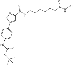
N-[4-[3-[[[7-(羟氨基)-7-氧代庚基]氨基]-氧代甲基]-5-异噁唑基]苯基]氨基甲酸叔丁酯是一种氨基甲酸酯。
利用腈氧化物环加成反应合成了一系列以苯基异噁唑为CAP基团的羟肟酸类HDAC抑制剂。其中鉴定出一种HDAC6选择性抑制剂,其效力约为2皮摩尔。部分化合物经检测抑制胰腺癌细胞生长的能力,发现其效力比SAHA高约10倍。这项研究为探索HDAC生物学提供了有价值的新型分子探针。[1] 组蛋白去乙酰化酶(HDAC)是癌症治疗的潜在靶点,第一代HDAC抑制剂目前正在进行癌症患者的临床试验。 HDAC6是许多与癌症相关的信号通路的关键调控因子,近年来已成为癌症治疗的一个极具吸引力的靶点。本研究发现,HDAC6在肺腺癌细胞系中过表达,且其表达水平与肺腺癌患者的预后呈负相关。HDAC6的过表达以去乙酰化酶活性依赖的方式促进肺腺癌细胞的增殖。HDAC6的过表达通过稳定表皮生长因子受体(EGFR)赋予细胞对吉非替尼的耐药性。CAY10603是一种高效且选择性的HDAC6抑制剂,其抑制HDAC6活性可抑制肺腺癌细胞的增殖并诱导细胞凋亡。CAY10603下调EGFR蛋白的表达水平,进而抑制EGFR信号通路的激活。此外,CAY10603 与吉非替尼协同作用,通过 EGFR 不稳定诱导肺腺癌细胞系凋亡。综上所述,我们的结果表明,抑制 HDAC6 可能是治疗肺腺癌的一种有前景的策略。[2] 基于化合物 12c(我们近期发现的一种 HDAC6/微管蛋白双重抑制剂)和 CAY10603(一种已知的 HDAC6 抑制剂),我们设计并合成了一系列 2-苯基噻唑类似物,作为潜在的组蛋白去乙酰化酶 6 (HDAC6) 抑制剂。其中,化合物 XP5 是最有效的 HDAC6 抑制剂,IC50 为 31 nM,且具有优异的 HDAC6 选择性(HDAC6 对 HDAC3 的选择性指数 SI = 338)。 XP5 对多种癌细胞系(包括 HDACi 耐药的 YCC3/7 胃癌细胞)均表现出较高的抗增殖活性(IC50 = 0.16-2.31 μM),优于 CAY10603。此外,XP5(50 mg/kg)在黑色素瘤肿瘤模型中显示出显著的抗肿瘤疗效,肿瘤生长抑制率 (TGI) 达到 63%,且未观察到明显的毒性。此外,XP5 与小分子 PD-L1 抑制剂联合使用时,可有效增强体内抗肿瘤免疫反应,表现为肿瘤浸润淋巴细胞数量增加和 PD-L1 表达水平降低。综上所述,以上结果表明 XP5 是一种有前景的 HDAC6 抑制剂,值得进一步研究。J Med Chem. 2022 Feb 10;65(3):2434-2457. 1.作用机制:CAY10603 通过选择性抑制 HDAC6 来缓解急性和慢性肾损伤。HDAC6 抑制可增加乙酰化 α-微管蛋白 (Ac-α-tubulin) 水平,从而减少 ATF6 的核转位,进而抑制未折叠蛋白反应 (UPR) 中 ATF6 分支的激活。这导致内质网应激降低、细胞凋亡减少,并抑制肾组织中的炎症和纤维化 [3] 2. 研究背景:HDAC6 在肾损伤模型(例如,顺铂诱导的 AKI、糖尿病性 CKI)中表达上调,而 ATF6-UPR 通路的激活会促进肾细胞死亡和纤维化。CAY10603 作为一种选择性 HDAC6 抑制剂,为肾损伤提供了一种潜在的治疗策略 [3] 3.临床状态:2024 年发表的文献表明,CAY10603 目前正处于肾损伤的临床前研究阶段;尚未报告任何临床试验、FDA 批准或适应症相关信息 [3] |
| 分子式 |
C22H30N4O6
|
|
|---|---|---|
| 分子量 |
446.5
|
|
| 精确质量 |
446.216
|
|
| 元素分析 |
C, 59.18; H, 6.77; N, 12.55; O, 21.50
|
|
| CAS号 |
1045792-66-2
|
|
| 相关CAS号 |
|
|
| PubChem CID |
24951314
|
|
| 外观&性状 |
White to light yellow solid powder
|
|
| 密度 |
1.2±0.1 g/cm3
|
|
| 折射率 |
1.563
|
|
| LogP |
1.94
|
|
| tPSA |
142.79
|
|
| 氢键供体(HBD)数目 |
4
|
|
| 氢键受体(HBA)数目 |
7
|
|
| 可旋转键数目(RBC) |
12
|
|
| 重原子数目 |
32
|
|
| 分子复杂度/Complexity |
616
|
|
| 定义原子立体中心数目 |
0
|
|
| SMILES |
O(C(N([H])C1C([H])=C([H])C(=C([H])C=1[H])C1=C([H])C(C(N([H])C([H])([H])C([H])([H])C([H])([H])C([H])([H])C([H])([H])C([H])([H])C(N([H])O[H])=O)=O)=NO1)=O)C(C([H])([H])[H])(C([H])([H])[H])C([H])([H])[H]
|
|
| InChi Key |
WWGBHDIHIVGYLZ-UHFFFAOYSA-N
|
|
| InChi Code |
InChI=1S/C22H30N4O6/c1-22(2,3)31-21(29)24-16-11-9-15(10-12-16)18-14-17(26-32-18)20(28)23-13-7-5-4-6-8-19(27)25-30/h9-12,14,30H,4-8,13H2,1-3H3,(H,23,28)(H,24,29)(H,25,27)
|
|
| 化学名 |
tert-butyl N-[4-[3-[[7-(hydroxyamino)-7-oxoheptyl]carbamoyl]-1,2-oxazol-5-yl]phenyl]carbamate
|
|
| 别名 |
CAY-10603; CAY10603; BML-281; tert-Butyl (4-(3-((7-(hydroxyamino)-7-oxoheptyl)carbamoyl)isoxazol-5-yl)phenyl)carbamate; CAY-10603; tert-butyl N-[4-[3-[[7-(hydroxyamino)-7-oxoheptyl]carbamoyl]-1,2-oxazol-5-yl]phenyl]carbamate; CHEMBL511749; compound 3 [PMID: 18642892]; CAY 10603
|
|
| HS Tariff Code |
2934.99.9001
|
|
| 存储方式 |
Powder -20°C 3 years 4°C 2 years In solvent -80°C 6 months -20°C 1 month |
|
| 运输条件 |
Room temperature (This product is stable at ambient temperature for a few days during ordinary shipping and time spent in Customs)
|
| 溶解度 (体外实验) |
|
|||
|---|---|---|---|---|
| 溶解度 (体内实验) |
配方 1 中的溶解度: ≥ 2.5 mg/mL (5.60 mM) (饱和度未知) in 10% DMSO + 40% PEG300 + 5% Tween80 + 45% Saline (这些助溶剂从左到右依次添加,逐一添加), 澄清溶液。
例如,若需制备1 mL的工作液,可将100 μL 25.0 mg/mL澄清DMSO储备液加入到400 μL PEG300中,混匀;然后向上述溶液中加入50 μL Tween-80,混匀;加入450 μL生理盐水定容至1 mL。 *生理盐水的制备:将 0.9 g 氯化钠溶解在 100 mL ddH₂O中,得到澄清溶液。 配方 2 中的溶解度: ≥ 2.5 mg/mL (5.60 mM) (饱和度未知) in 10% DMSO + 90% (20% SBE-β-CD in Saline) (这些助溶剂从左到右依次添加,逐一添加), 澄清溶液。 例如,若需制备1 mL的工作液,可将 100 μL 25.0 mg/mL澄清DMSO储备液加入900 μL 20% SBE-β-CD生理盐水溶液中,混匀。 *20% SBE-β-CD 生理盐水溶液的制备(4°C,1 周):将 2 g SBE-β-CD 溶解于 10 mL 生理盐水中,得到澄清溶液。 View More
配方 3 中的溶解度: ≥ 2.5 mg/mL (5.60 mM) (饱和度未知) in 10% DMSO + 90% Corn Oil (这些助溶剂从左到右依次添加,逐一添加), 澄清溶液。 配方 4 中的溶解度: 5% DMSO+50% PEG 300+ddH2O: 9mg/mL 配方 5 中的溶解度: 5 mg/mL (11.20 mM) in 50% PEG300 50% Saline (这些助溶剂从左到右依次添加,逐一添加), 悬浮液; 需要超声波加热并加热至 42°C。 *生理盐水的制备:将 0.9 g 氯化钠溶解在 100 mL ddH₂O中,得到澄清溶液。 1、请先配制澄清的储备液(如:用DMSO配置50 或 100 mg/mL母液(储备液)); 2、取适量母液,按从左到右的顺序依次添加助溶剂,澄清后再加入下一助溶剂。以 下列配方为例说明 (注意此配方只用于说明,并不一定代表此产品 的实际溶解配方): 10% DMSO → 40% PEG300 → 5% Tween-80 → 45% ddH2O (或 saline); 假设最终工作液的体积为 1 mL, 浓度为5 mg/mL: 取 100 μL 50 mg/mL 的澄清 DMSO 储备液加到 400 μL PEG300 中,混合均匀/澄清;向上述体系中加入50 μL Tween-80,混合均匀/澄清;然后继续加入450 μL ddH2O (或 saline)定容至 1 mL; 3、溶剂前显示的百分比是指该溶剂在最终溶液/工作液中的体积所占比例; 4、 如产品在配制过程中出现沉淀/析出,可通过加热(≤50℃)或超声的方式助溶; 5、为保证最佳实验结果,工作液请现配现用! 6、如不确定怎么将母液配置成体内动物实验的工作液,请查看说明书或联系我们; 7、 以上所有助溶剂都可在 Invivochem.cn网站购买。 |
| 制备储备液 | 1 mg | 5 mg | 10 mg | |
| 1 mM | 2.2396 mL | 11.1982 mL | 22.3964 mL | |
| 5 mM | 0.4479 mL | 2.2396 mL | 4.4793 mL | |
| 10 mM | 0.2240 mL | 1.1198 mL | 2.2396 mL |
1、根据实验需要选择合适的溶剂配制储备液 (母液):对于大多数产品,InvivoChem推荐用DMSO配置母液 (比如:5、10、20mM或者10、20、50 mg/mL浓度),个别水溶性高的产品可直接溶于水。产品在DMSO 、水或其他溶剂中的具体溶解度详见上”溶解度 (体外)”部分;
2、如果您找不到您想要的溶解度信息,或者很难将产品溶解在溶液中,请联系我们;
3、建议使用下列计算器进行相关计算(摩尔浓度计算器、稀释计算器、分子量计算器、重组计算器等);
4、母液配好之后,将其分装到常规用量,并储存在-20°C或-80°C,尽量减少反复冻融循环。
计算结果:
工作液浓度: mg/mL;
DMSO母液配制方法: mg 药物溶于 μL DMSO溶液(母液浓度 mg/mL)。如该浓度超过该批次药物DMSO溶解度,请首先与我们联系。
体内配方配制方法:取 μL DMSO母液,加入 μL PEG300,混匀澄清后加入μL Tween 80,混匀澄清后加入 μL ddH2O,混匀澄清。
(1) 请确保溶液澄清之后,再加入下一种溶剂 (助溶剂) 。可利用涡旋、超声或水浴加热等方法助溶;
(2) 一定要按顺序加入溶剂 (助溶剂) 。
|
|---|
|
|
InvivoChem的所有产品仅用于作科学研究,不面向患者销售
Copyright 2020 InvivoChem LLC | All Rights Reserved 粤ICP备20063088号-1


 COA
COA


 463611831
