| 规格 | 价格 | 库存 | 数量 |
|---|---|---|---|
| 1mg |
|
||
| 5mg |
|
||
| 10mg |
|
||
| 25mg |
|
||
| 50mg |
|
||
| 100mg |
|
||
| 250mg |
|
||
| Other Sizes |
|

| 靶点 |
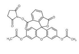
Fluorescent dye
Its mechanism is physicochemical: it relies on ubiquitous intracellular esterases (to generate fluorescence) and intracellular amines (for covalent binding). Consequently, any cell with active esterases (including most eukaryotic cells) can be labeled to track division or migration. |
|---|---|
| 体外研究 (In Vitro) |
CFDA-SE工作液的制备
1.1 制备储备液:将1毫克CFDA-SE溶解在0.1794毫升DMSO中,得到10毫摩尔的CFDA-SE储备液。 注意:储备液应在-20℃或-80℃下避光储存,避免反复冻融。 1.2 CFDA-SE工作溶液的制备 在无血清细胞培养中稀释储备溶液。 注意:请根据您的具体实验需求调整CFDA-SE工作液的浓度。 细胞染色 2.1 悬浮细胞:在4°C下以1000 g离心3-5分钟,丢弃上清液。用PBS洗两次,每次五分钟。 粘附细胞:丢弃细胞培养基,用胰蛋白酶分离细胞,产生单细胞悬浮液。在4°C下以1000 g离心3-5分钟,丢弃上清液。用PBS洗两次,每次五分钟。 2.2 加入1 mL CFDA-SE工作溶液并混合30分钟。 2.3 在4°C下以400 g离心3-4分钟。 2.4 用PBS洗涤细胞两次,每次5分钟。 2.5 将细胞重新悬浮在无血清培养基或PBS中,并使用荧光显微镜或流式细胞仪进行检测。 细胞增殖标记:CFSE可在体外用于标记细胞以进行增殖实验。它以非荧光形式自由扩散进入细胞,在细胞内被细胞内酯酶裂解,其琥珀酰亚胺基团不可逆地与细胞内氨基结合,形成荧光结合物并留存于细胞内。当细胞分裂时,荧光结合物平均分配到两个子代细胞中,子代细胞的荧光强度为亲代细胞的一半。通过流式细胞术可检测到CFSE标记的细胞增殖群体具有系列减半的荧光强度[1] CFSE(羧基荧光素琥珀酰亚胺酯)用作细胞内荧光染料,用于追踪细胞增殖。在细胞增殖实验中,即使在没有细胞分裂的情况下,细胞内的CFSE荧光强度(FI)也会因自然衰变和蛋白质周转而随时间降低。这种衰变表现出双相模式,并可以用数学模型进行描述。来自两名健康供体的未刺激PBMC培养物的实验数据显示,在160小时内,平均CFSE荧光强度随时间下降。[1] 该研究比较了多种数学模型来描述标记衰变动力学。与简单的指数衰减模型相比,包含多种损失率机制(例如,针对不同荧光偶联物的不同速率)或单一时间依赖性衰变速率(例如,Gompertz衰变模型,\( \frac{dx}{dt} = -c(x - x_a)e^{-kt} \) )的模型对观察到的CFSE衰变数据提供了更好的拟合。[1] CFSE 用于通过流式细胞术追踪增殖细胞,可区分多达八次或更多次连续细胞分裂。已应用于研究B细胞、T细胞和造血前体细胞分化过程中与分裂相关的表型和功能变化。可与PE、PerCP或PE/Cy5标记的抗体联合使用进行免疫表型分析。[2] CFSE 的染色强度与浓度呈线性关系,在未分裂细胞中,荧光强度在最初1–2天内下降约60%,之后在数周至数月内保持稳定。[2] 该染料已用于研究缓慢分裂的胶质母细胞瘤细胞、红系祖细胞增殖、抗原特异性T细胞克隆以及多发性硬化症中的神经抗原特异性T细胞。[2] 在体外,CFSE主要用于评估淋巴细胞的增殖动力学。研究表明,在IL-2等细胞因子刺激下,CFSE标记的人NK细胞或T细胞会发生增殖,表现为CFSE荧光强度的逐代衰减。该技术灵敏度高,能区分增殖的子代细胞与未增殖的祖细胞,并可用于评估细胞毒性T淋巴细胞(CTL)的特异性杀伤活性(如利用CFSE高低染色的靶细胞混合体进行体内外杀伤检测)。 |
| 体内研究 (In Vivo) |
CFDA-SE是一种细胞内绿色荧光染料,可用于检测体内菌株定殖。
方法:用于测定肠道中的菌株定植。 1.将菌株重新悬浮在PBS中,以获得108 CFU/mL的细胞密度。 2.将CFDA-SE(1 mM;10μL;20分钟;37°C;黑暗/避光)加入1 mL细菌悬浮液中。 3.离心(13000 g,10分钟,4°C),用无菌PBS洗涤混合物3次,以去除多余的CFDA-SE。 4.将菌株重新悬浮在无菌PBS(108 CFU/mL)中,并向SD大鼠施用1 mL细菌悬浮液。灌胃后第1天和第3天,对麻醉的大鼠进行荧光成像,然后处死大鼠,取不同的肠道切片进行成像。 5.使用IVIS Lumina III智能成像系统进行荧光成像。 细胞追踪:在体内,CFSE可在活细胞中以可测量的浓度维持数周,无论细胞类型或激活状态如何。它可用于追踪体内细胞的分裂和增殖过程,提供均匀的标记,且对细胞的细胞内机制几乎没有不利影响[2] CFSE 标记的细胞可在体内过继转移(如小鼠尾静脉注射),并追踪数月。已用于研究B细胞在无T细胞情况下的分裂、混合淋巴细胞反应中的同种异体反应以及移植物抗宿主反应的调控。[2] 在体内,CFSE被广泛用于追踪免疫细胞的归巢和增殖,以及评估疫苗或药物的免疫效果。例如,将CFSE标记的抗原特异性靶细胞过继转移至免疫小鼠体内,通过检测靶细胞CFSE信号的丢失(表明被杀伤),可评估CD8+ T细胞的体内杀伤效率。此外,通过脑室注射CFSE可标记胚胎脑内的巨噬细胞等特定细胞群,用于谱系追踪研究。 |
| 酶活实验 |
CFSE主要用于细胞标记,在非细胞体系中应用较少。若需确认标记效率或染料特性,常规流程为:将CFSE粉末溶解于无水DMSO中制备储存液(例如5mM)。在实验体系中检测荧光激发/发射光谱(约492nm/517nm)。若需与蛋白偶联,可将CFSE与含胺基的蛋白在pH 7.4-8.0的PBS缓冲液中室温避光共孵育30分钟,通过离心或透析去除未结合的染料。
|
| 细胞实验 |
本单元中描述的技术使用细胞内荧光标记羧基荧光素二乙酸酯琥珀酰亚胺酯(CFSE)来追踪增殖细胞。共价结合的CFSE在子细胞之间平均分配,从而可以区分连续几轮的细胞分裂。该技术适用于体外细胞分裂,也适用于过继转移细胞的体内分裂,可以分辨连续八代或更多代。CFSE寿命长,可以在细胞转移后几个月内进行分析,并且具有与荧光素相同的光谱特征,因此与藻红蛋白或其他相容的荧光染料结合的单克隆抗体可用于对分裂细胞进行免疫表型。此外,还提供了关于第二代染料Cell Trace Violet(CTV)的信息,该染料由405nm蓝色激光激发。CTV在化学上与CFSE有关,但允许氩激光的488nm线用于其他探针[2]。
细胞标记与增殖检测:制备细胞悬液,加入适量的CFSE溶液,在适宜温度下孵育一定时间,使CFSE进入细胞。然后洗涤细胞,去除未结合的CFSE。将标记后的细胞在适宜条件下培养,在不同时间点,利用流式细胞术测量细胞的荧光强度,根据荧光强度的变化分析细胞的增殖、分裂等情况[1] CFSE标记细胞的标准化流程涉及其前体羧基二乙酸荧光素琥珀酰亚胺酯(CFDA-SE)。CFDA-SE由于其亲脂性,可被动扩散穿过细胞膜并被细胞摄取。进入细胞后,细胞内酯酶(推测为乙酰酯酶)催化其乙酸酯水解,将其转化为高荧光且膜不透性的CFSE。CFSE的琥珀酰亚胺酯部分随后与细胞内蛋白质上的胺基反应,形成被保留的稳定荧光偶联物(如CF-R2),以及被丢失或降解的不稳定偶联物(如CF-R1)。在初始暴露和染色后,冲洗细胞培养物以去除多余的标记物。[1] 对于衰变研究,来自两名供体的外周血单个核细胞(PBMC)按照标准流程用CFSE染色,但未进行刺激以诱导分裂。这确保了任何观察到的荧光损失都是由于自然衰变过程。在160小时内,使用流式细胞术在24个不同的时间点对细胞进行三次重复测量,并记录每个样品的平均总荧光强度(FI)。[1] 将细胞重悬于PBS/0.1% BSA中,浓度为5 × 10⁷ 细胞/ml。加入CFSE至终浓度10 µM,37°C孵育10分钟。用冰预冷的RPMI 1640/10% FBS冰上孵育5分钟以终止染色。用培养基或注射液洗涤细胞三次。细胞在适当条件下培养或进行过继转移。收获的细胞可用抗体进行免疫表型染色,并通过流式细胞仪分析(激发光488 nm,发射光用525 nm带通滤光片收集)。[2] 为提高染色均匀性,可将CFSE在PBS/0.1% BSA中稀释至20 µM,再与等体积的2×浓缩细胞悬液混合。[2] 主要采用CFSE稀释法:1. 制备单细胞悬液(如PBMC),用预热的PBS重悬。2. 加入CFSE(终浓度通常为0.5-5μM),在37°C下避光孵育10-15分钟。3. 加入过量的冷完全培养基(含血清)终止反应,冰上孵育5分钟。4. 洗涤细胞两次以去除残余染料。5. 将标记的细胞置于培养板中,加入刺激因子(如抗CD3/CD28抗体、IL-2等)进行培养。6. 培养3-7天后,收集细胞进行流式细胞术检测,通过分析荧光强度的逐代减半来量化增殖指数。 |
| 动物实验 |
对于过继性细胞转移,将CFSE标记的细胞(例如,每只小鼠1 × 10⁷至5 × 10⁷个细胞)经尾静脉注射到小鼠或大鼠体内。在脾脏等淋巴器官中追踪细胞,并在转移后不同时间点通过流式细胞术进行分析。[2]
用于体内细胞毒性检测(In Vivo CTL Assay):1. 标记靶细胞:将来自供体小鼠的脾细胞分为两群,分别用低浓度(如0.5μM)和高浓度(如5μM)CFSE标记。2. 制备抗原肽脉冲:将其中一群(高浓度标记)用特异性抗原肽脉冲处理,另一群(低浓度标记)作为对照(未脉冲)。3. 混合细胞:将两群细胞按1:1比例混合。4. 过继转移:通过尾静脉或眼眶后注射将混合细胞输入已免疫的受体小鼠体内。5. 检测:通常在4-24小时后处死小鼠,取出脾脏检测细胞悬液中的CFSE信号。特异性杀伤导致对应肽脉冲的CFSE高标峰降低或消失。 |
| 药代性质 (ADME/PK) |
CFSE荧光强度在非分裂细胞中最初1-2天内会因不稳定结合成分的分解代谢而下降约60%,之后会保持稳定数周至数月。[2]
该染料在体内具有较长的半衰期,因此可以在移植后数月内进行追踪。[2] CFSE在体内的药代动力学显著依赖于其标记的细胞,而非游离染料。游离的CFSE遇水极易水解,在血浆中半衰期极短。一旦标记到细胞上,CFSE通过琥珀酰亚胺基与细胞内蛋白共价结合,变得非常稳定。在未分裂的静息细胞中,CFSE标记可持续数周而不衰减;细胞分裂时,荧光强度随分裂次数增加而呈对数衰减。其在体内的清除主要取决于被标记细胞的周转速率。 |
| 毒性/毒理 (Toxicokinetics/TK) |
高浓度的CFSE会影响细胞活力和分裂。最佳染色效果应使荧光强度相对于未染色细胞增加3-3.5个对数级。染色过程中加入蛋白质(0.1% BSA)可提高细胞活力并减少细胞聚集。在较低温度(冰上或室温)下染色也可能提高细胞活力。[2]
CFSE可改变某些细胞表面标志物的表达,或降低某些单克隆抗体的染色效果,这可能是由于空间位阻所致。[2] CFSE具有一定细胞毒性,这与其标记浓度和作用时间相关。高浓度的CFSE或长时间暴露会损害细胞功能,导致激活诱导的细胞死亡(AICD)或抑制细胞增殖。为了平衡标记效率与细胞活力,建议优化染料的浓度和孵育条件(如使用较低浓度标记,如0.5-2μM),并在标记后充分洗涤。对于原代淋巴细胞,使用含高浓度血清的培养基淬灭并洗涤,可有效降低毒性。 |
| 参考文献 | |
| 其他信息 |
我们针对细胞增殖实验中以细胞内染料羧基荧光素琥珀酰亚胺酯 (CFSE) 为染色剂的情况,开发了一系列标记衰减模型。我们使用从两名健康患者收集的数据来验证这些模型,并将其与 Akiake 信息准则 (AIC) 进行比较。数据中多种衰减速率的特征可以通过时间依赖性衰减模型(例如逻辑斯蒂模型和 Gompertz 模型)进行表征和解释。[1] 胰腺导管腺癌 (PDA) 仍然是一种致命的疾病,尽管近年来免疫疗法在其他恶性肿瘤的治疗中取得了许多成功,但治愈率仍然很低。由于人类疾病中效应 T 细胞大量浸润,我们假设,精确模拟 PDA 的免疫微环境将有助于我们研究可克服的免疫抑制机制,从而获得治疗益处。为此,我们利用新鲜 PDA 的活体精确切割切片,开发了一种类器官培养系统。我们证实,培养的胰腺切片在至少6天的培养后仍能保持其基线形态、表面积和微环境,并通过MTT法以及Ki-67和cleaved-Caspase-3抗体的免疫组化染色证实了切片的存活。在整个培养期间,免疫细胞(包括T细胞(CD3+、CD8+和FOXP3+)、巨噬细胞(CD68+、CD163+和HLA-DR+)以及基质肌成纤维细胞(αSMA+))均存在。对胰腺导管腺癌(PDA)切片培养前后蛋白质组的全面分析表明,大多数已鉴定的免疫蛋白在培养过程中保持稳定。药物(星形孢菌素,STS和环己酰亚胺,CHX)对PDA切片培养的细胞毒性作用证实,该系统可用于评估药物治疗后的功能反应和细胞存活率,且该评估结果具有治疗时间和剂量依赖性。我们利用多色免疫荧光技术对活体切片进行染色,以检测癌细胞(EpCAM+)和免疫细胞(CD11b+和CD8+)。最终,我们证实自体CFSE标记的脾细胞能够迅速迁移至共培养的肿瘤切片中。因此,本研究表明,肿瘤切片培养可用于研究胰腺导管腺癌(PDA)的免疫微环境。[4]
CFSE是一种无色、非荧光、非极性分子。它具有一个可特异性结合细胞的琥珀酰亚胺基团和一个可非酶促水解的羧基荧光素二乙酸酯基团。它是一种良好的活细胞标记物。在免疫学实验中,使用CFSE标记细胞可以替代传统的形态学观察和3H-TdR掺入法等方法,从而提高研究水平并拓展新型免疫学技术的应用[2]。 CFSE是由其前体CFDA-SE在细胞内生成的活性荧光形式。它是增殖实验中绘制细胞分裂历史的重要工具。CFSE与长寿命的细胞内蛋白共价结合(形成CF-R2)后,荧光标记可在活细胞中保留数周,从而实现长期追踪。荧光强度随时间推移而降低(标记衰减)是由于染料的自然衰减以及与其结合的蛋白质的周转所致[1]。 本文讨论的主要应用是基于流式细胞术的细胞增殖实验中CFSE的使用。每次细胞分裂都会对CFSE荧光进行系列稀释,从而建立测量的荧光强度与细胞分裂次数之间的相关性。准确模拟CFSE荧光自然衰减对于根据此类数据正确估算细胞增殖和死亡率至关重要。[1]文献中用于量化CFSE标记衰减的数学模型包括代表不同细胞内荧光物质(CFDA-SE、CFSE、CF-R1、CF-R2)转化和衰减的常微分方程组,以及使用指数、Logistic或Gompertz衰减动力学的简化单方程模型。Gompertz衰减模型具有时间依赖性衰减速率,与简单的指数模型相比,它能更准确地拟合观察到的双相衰减数据。[1]CFSE(羧基荧光素二乙酸琥珀酰亚胺酯)是一种细胞渗透性荧光素染料,它与细胞内胺基共价结合,从而在细胞分裂时均匀分配到子细胞中。它在 488 nm 处被激发,并在约 525 nm 处发射,与荧光素滤光片组兼容。[2] 第二代染料 Cell Trace Violet (CTV) 在 405 nm 处被激发,具有类似的细胞分裂追踪能力,并且可将 488 nm 通道用于其他探针。[2] CFSE 数据可用于建模软件(例如 ModFit)以计算前体细胞频率和增殖指数。[2] |
| 分子式 |
C29H19NO11
|
|---|---|
| 分子量 |
557.461268663406
|
| 精确质量 |
557.095
|
| 元素分析 |
C, 62.48; H, 3.44; N, 2.51; O, 31.57
|
| CAS号 |
150347-59-4
|
| 外观&性状 |
Off-white to yellow solid powder
|
| 密度 |
1.6±0.1 g/cm3
|
| 沸点 |
757.9±70.0 °C at 760 mmHg
|
| 熔点 |
152-154ºC(lit.)
|
| 闪点 |
412.2±35.7 °C
|
| 蒸汽压 |
0.0±2.6 mmHg at 25°C
|
| 折射率 |
1.701
|
| LogP |
0.5
|
| tPSA |
151.81
|
| SMILES |
C12(OC(=O)C3=CC(C(=O)ON4C(=O)CCC4=O)=CC=C13)C1C=CC(OC(=O)C)=CC=1OC1C=C(OC(=O)C)C=CC2=1
|
| InChi Key |
JGPOSNWWINVNFV-UHFFFAOYSA-N
|
| InChi Code |
InChI=1S/C29H19NO11/c1-14(31)37-17-4-7-20-23(12-17)39-24-13-18(38-15(2)32)5-8-21(24)29(20)22-11-16(3-6-19(22)28(36)40-29)27(35)41-30-25(33)9-10-26(30)34/h3-8,11-13H,9-10H2,1-2H3
|
| 化学名 |
(2,5-dioxopyrrolidin-1-yl) 3',6'-diacetyloxy-1-oxospiro[2-benzofuran-3,9'-xanthene]-5-carboxylate
|
| 别名 |
5(6-Carboxyfluorescein diacetate succinimidyl ester; CFDA-SE; 5(6-CFDA N-succinmidyl ester;
|
| HS Tariff Code |
2934.99.9001
|
| 存储方式 |
Powder -20°C 3 years 4°C 2 years In solvent -80°C 6 months -20°C 1 month 注意: 请将本产品存放在密封且受保护的环境中(例如氮气保护),避免吸湿/受潮和光照。 |
| 运输条件 |
Room temperature (This product is stable at ambient temperature for a few days during ordinary shipping and time spent in Customs)
|
| 溶解度 (体外实验) |
DMSO : ~50 mg/mL (~89.69 mM)
|
|---|---|
| 溶解度 (体内实验) |
配方 1 中的溶解度: ≥ 2.08 mg/mL (3.73 mM) (饱和度未知) in 10% DMSO + 40% PEG300 + 5% Tween80 + 45% Saline (这些助溶剂从左到右依次添加,逐一添加), 澄清溶液。
例如,若需制备1 mL的工作液,可将100 μL 20.8 mg/mL澄清DMSO储备液加入400 μL PEG300中,混匀;然后向上述溶液中加入50 μL Tween-80,混匀;加入450 μL生理盐水定容至1 mL。 *生理盐水的制备:将 0.9 g 氯化钠溶解在 100 mL ddH₂O中,得到澄清溶液。 配方 2 中的溶解度: ≥ 2.08 mg/mL (3.73 mM) (饱和度未知) in 10% DMSO + 90% Corn Oil (这些助溶剂从左到右依次添加,逐一添加), 澄清溶液。 例如,若需制备1 mL的工作液,可将 100 μL 20.8 mg/mL 澄清 DMSO 储备液加入到 900 μL 玉米油中并混合均匀。 请根据您的实验动物和给药方式选择适当的溶解配方/方案: 1、请先配制澄清的储备液(如:用DMSO配置50 或 100 mg/mL母液(储备液)); 2、取适量母液,按从左到右的顺序依次添加助溶剂,澄清后再加入下一助溶剂。以 下列配方为例说明 (注意此配方只用于说明,并不一定代表此产品 的实际溶解配方): 10% DMSO → 40% PEG300 → 5% Tween-80 → 45% ddH2O (或 saline); 假设最终工作液的体积为 1 mL, 浓度为5 mg/mL: 取 100 μL 50 mg/mL 的澄清 DMSO 储备液加到 400 μL PEG300 中,混合均匀/澄清;向上述体系中加入50 μL Tween-80,混合均匀/澄清;然后继续加入450 μL ddH2O (或 saline)定容至 1 mL; 3、溶剂前显示的百分比是指该溶剂在最终溶液/工作液中的体积所占比例; 4、 如产品在配制过程中出现沉淀/析出,可通过加热(≤50℃)或超声的方式助溶; 5、为保证最佳实验结果,工作液请现配现用! 6、如不确定怎么将母液配置成体内动物实验的工作液,请查看说明书或联系我们; 7、 以上所有助溶剂都可在 Invivochem.cn网站购买。 |
| 制备储备液 | 1 mg | 5 mg | 10 mg | |
| 1 mM | 1.7939 mL | 8.9693 mL | 17.9385 mL | |
| 5 mM | 0.3588 mL | 1.7939 mL | 3.5877 mL | |
| 10 mM | 0.1794 mL | 0.8969 mL | 1.7939 mL |
1、根据实验需要选择合适的溶剂配制储备液 (母液):对于大多数产品,InvivoChem推荐用DMSO配置母液 (比如:5、10、20mM或者10、20、50 mg/mL浓度),个别水溶性高的产品可直接溶于水。产品在DMSO 、水或其他溶剂中的具体溶解度详见上”溶解度 (体外)”部分;
2、如果您找不到您想要的溶解度信息,或者很难将产品溶解在溶液中,请联系我们;
3、建议使用下列计算器进行相关计算(摩尔浓度计算器、稀释计算器、分子量计算器、重组计算器等);
4、母液配好之后,将其分装到常规用量,并储存在-20°C或-80°C,尽量减少反复冻融循环。
计算结果:
工作液浓度: mg/mL;
DMSO母液配制方法: mg 药物溶于 μL DMSO溶液(母液浓度 mg/mL)。如该浓度超过该批次药物DMSO溶解度,请首先与我们联系。
体内配方配制方法:取 μL DMSO母液,加入 μL PEG300,混匀澄清后加入μL Tween 80,混匀澄清后加入 μL ddH2O,混匀澄清。
(1) 请确保溶液澄清之后,再加入下一种溶剂 (助溶剂) 。可利用涡旋、超声或水浴加热等方法助溶;
(2) 一定要按顺序加入溶剂 (助溶剂) 。
 Apotracker Red
 18:1-18:1-C11 BODIPY 505/515 TG
 18:1-6:0 DNP-C11 BODIPY 505/515 TG
 Anisoin
InvivoChem的所有产品仅用于作科学研究,不面向患者销售
Copyright 2020 InvivoChem LLC | All Rights Reserved 粤ICP备20063088号-1


 COA
COA
 463611831
