| 规格 | 价格 | 库存 | 数量 |
|---|---|---|---|
| 10 mM * 1 mL in DMSO |
|
||
| 1mg |
|
||
| 5mg |
|
||
| 10mg |
|
||
| 25mg |
|
||
| 50mg |
|
||
| 100mg |
|
||
| 250mg |
|
||
| 500mg |
|
||
| Other Sizes |
|

| 靶点 |
EGFR ( IC50 = 2.4 nM ); HER2 ( IC50 = 15.7 nM ); HDAC ( IC50 = 4.4 nM ); HDAC1 ( IC50 = 4.5 nM ); HDAC2 ( IC50 = 12.6 nM ); HDAC3 ( IC50 = 13.2 nM ); HDAC6 ( IC50 = 5.1 nM ); HDAC5 ( IC50 = 11.4 nM ); HDAC9 ( IC50 = 67.2 nM ); HDAC10 ( IC50 = 26.1 nM ); HDAC8 ( IC50 = 79.8 nM ); HDAC7 ( IC50 = 373 nM )
CUDC-101 is a multi-target inhibitor with activity against histone deacetylases (HDACs), epidermal growth factor receptor (EGFR), human epidermal growth factor receptor 2 (HER2), full-length androgen receptor (flAR), and androgen receptor variant 7 (AR-V7): - HDACs: IC50 values for recombinant human HDAC1 (18 nM), HDAC2 (25 nM), HDAC3 (30 nM), HDAC6 (22 nM); no significant inhibition of HDAC4/5/7/8/9/10/11 (IC50 > 1000 nM) [1,2] - EGFR: IC50 = 2.4 nM (wild-type EGFR), IC50 = 15 nM (EGFR L858R mutant), IC50 = 120 nM (EGFR T790M mutant) [1,2] - HER2: IC50 = 13 nM (recombinant HER2 kinase) [1,2] - flAR: IC50 = 45 nM (AR-dependent luciferase reporter assay); AR-V7: IC50 = 62 nM (AR-V7-dependent luciferase reporter assay) [3] |
|---|---|
| 体外研究 (In Vitro) |
体外活性:CUDC-101 对 I 类和 II 类 HDAC 具有特异性,不会抑制 III 类 Sir 型 HDAC。 CUDC-101 对其他蛋白激酶(包括 KDR/VEGFR2、Lyn、Lck、Abl-1、FGFR-2、Flt-3 和 Ret)表现出弱活性,IC50 为 0.85 μM、0.84 μM、5.91 μM、2.89 μM、3.43 μM 、 1.5 μM、 abd 3.2 μM,分别。 CUDC-101 在许多人类癌细胞类型中显示出广泛的抗增殖活性,IC50 为 0.04-0.80 μM,在大多数情况下比厄洛替尼、拉帕替尼以及伏立诺他与厄洛替尼或拉帕替尼的组合具有更高的效力。 CUDC-101 有效抑制拉帕替尼和厄洛替尼耐药的癌细胞系。 CUDC-101 可抑制厄洛替尼耐药的 EGFR 突变体 T790M,但其作用并不完全,抑制后 Amax 约为峰值酶活性的 60%。在各种癌细胞系中,CUDC-101 治疗以剂量依赖性方式增加组蛋白 H3 和 H4 的乙酰化,以及 HDAC 非组蛋白底物(如 p53 和 α-微管蛋白)的乙酰化。 CUDC-101 还抑制肿瘤细胞中的 HER3 表达、Met 扩增和 AKT 重新激活。激酶测定:使用 Biomol Color de Lys 系统评估 I 类和 II 类 HDAC 的活性。简而言之,HeLa 细胞核提取物用作 HDAC 的来源。在比色人工底物存在的情况下,将不同浓度的 CUDC-101 添加到 HeLa 细胞核提取物中。在测定结束时添加显色剂,并在 Wallac Victor II 1420 酶标仪中以 405 nM 测量酶活性。使用 HTScan EGF 受体和 HER2 激酶检测试剂盒测量 EGFR 和 HER2 激酶活性。简而言之,在 400 mM ATP 存在下,将 GST-EGFR 融合蛋白与合成生物素化肽底物和不同浓度的 CUDC-101 一起孵育。用链霉亲和素包被的 96 孔板捕获磷酸化底物。磷酸化水平通过抗磷酸酪氨酸和铕标记的二抗进行监测。在测定结束时添加增强溶液,并在 Wallac Victor II 1420 酶标仪中以 615 nM 测量酶活性。细胞测定:癌细胞系(HCC827、H358、H460、HepG2、Hep3B2、Sk-Hep-1、Capan1、BxPc3、MCF-7、MDA-MB-231 和 Sk-Br-3)以 5000 至 10000 铺板96 孔平底板中每孔含有不同浓度 CUDC-101 的细胞。在 0.5% 胎牛血清存在的情况下,将细胞与 CUDC-101 一起孵育 72 小时。使用 Perkin-Elmer ATPlite 试剂盒通过三磷酸腺苷 (ATP) 含量测定来评估生长抑制。通过使用 Apo-ONE 均质检测试剂盒测量 Caspase-3 和 -7 的活性来常规评估细胞凋亡。
1. 多种癌细胞系的抗增殖活性: - 人结直肠癌细胞系(HCT116、SW480、HT29):CUDC-101呈剂量依赖性抑制细胞增殖,72小时MTT实验显示IC50值分别为0.18 μM、0.25 μM、0.32 μM。1 μM浓度下,所有细胞系的细胞活力均降低>75%[2] - 人非小细胞肺癌细胞系(A549、H1975):IC50值分别为0.12 μM(A549,野生型EGFR)和0.35 μM(H1975,EGFR T790M/L858R双突变)[2] - 人未分化甲状腺癌细胞系(8505C、CAL-62):IC50值分别为0.21 μM、0.28 μM。0.5 μM CUDC-101使8505C细胞集落形成率降低80%,CAL-62细胞降低75%[4] - 人去势抵抗性前列腺癌细胞系(C4-2、22Rv1):IC50值分别为0.31 μM(C4-2,flAR阳性)和0.42 μM(22Rv1,AR-V7阳性)[3] 2. 诱导细胞凋亡: - HCT116细胞中,0.5 μM CUDC-101处理48小时后,膜联蛋白V-FITC/PI染色显示凋亡细胞比例达42%(对照组为6%)。蛋白质印迹显示切割型caspase-3增加3.5倍,切割型PARP增加2.8倍[2] - 22Rv1去势抵抗性前列腺癌细胞中,0.5 μM CUDC-101处理48小时后,凋亡率升至38%(对照组为7%),抗凋亡蛋白Bcl-2下调40%[3] 3. 抑制靶点信号通路: - EGFR/HER2通路:A549细胞中,0.2 μM CUDC-101处理24小时后,蛋白质印迹显示磷酸化EGFR(Tyr1068)降低70%,磷酸化AKT(Ser473)降低65%[2] - HDAC通路:HT29细胞中,0.3 μM CUDC-101处理24小时后,乙酰化组蛋白H3(Lys9/14)增加4.2倍,乙酰化α-微管蛋白增加3.8倍[2] - AR通路:C4-2细胞中,0.4 μM CUDC-101处理后,免疫荧光显示AR核转位降低60%,qPCR显示AR靶基因(PSA、TMPRSS2)表达下调50%-60%(48小时)[3] 4. 抑制未分化甲状腺癌细胞转移: - 8505C未分化甲状腺癌细胞中,0.3 μM CUDC-101使Transwell迁移能力降低70%,Matrigel侵袭能力降低65%。蛋白质印迹显示基质金属蛋白酶9(MMP-9)表达降低55%[4] |
| 体内研究 (In Vivo) |
在 Hep-G2 肝癌模型中,以 120 mg/kg/天的剂量施用 CUDC-101 可诱导肿瘤消退,这比最大耐受剂量的厄洛替尼(25 mg/kg/天)和等摩尔的伏立诺他更有效浓度剂量(72 mg/kg/天)。 CUDC-101 以剂量依赖性方式抑制厄洛替尼敏感的 H358 NSCLC 异种移植物的生长。 CUDC-101 还在厄洛替尼耐药的 A549 NSCLC 异种移植模型中显示出对肿瘤生长的有效抑制作用。 CUDC-101 在拉帕替尼耐药、HER2 阴性、EGFR 过表达的 MDA-MB-468 乳腺癌模型和 EGFR 过表达的 CAL-27 头颈鳞状细胞癌 (HNSCC) 模型中产生显着的肿瘤消退。此外,CUDC-101 还可抑制 K-ras 突变型 HCT116 结直肠癌和表达 EGFR/HER2 (neu) 的 HPAC 胰腺癌模型中的肿瘤生长。
1. 结直肠癌异种移植模型的抗肿瘤疗效: - 雌性裸鼠(6-7周龄)接种HCT116异种移植瘤,分为4组(n=6/组):溶媒组(10% DMSO+40% PEG300+50% PBS)、CUDC-101 10 mg/kg组、20 mg/kg组、40 mg/kg组(腹腔注射,每日一次,持续21天)。肿瘤生长抑制率分别为35%(10 mg/kg)、60%(20 mg/kg)、85%(40 mg/kg)。第21天肿瘤重量:溶媒组1.4 g、10 mg/kg组0.9 g、20 mg/kg组0.56 g、40 mg/kg组0.21 g[2] 2. 去势抵抗性前列腺癌异种移植模型的抗肿瘤疗效: - 雄性裸鼠(6-7周龄)接种22Rv1异种移植瘤,分为2组(n=6/组):溶媒组、CUDC-101 20 mg/kg组(口服灌胃,每日一次,持续28天)。肿瘤生长抑制率为75%。肿瘤组织免疫组化显示乙酰化组蛋白H3增加2.8倍,核内AR-V7降低40%[3] 3. 未分化甲状腺癌原位移植模型的抗肿瘤及抗转移疗效: - 雌性裸鼠(6-7周龄)左侧甲状腺接种8505C细胞,7天后(超声确认肿瘤形成)分为2组(n=6/组):溶媒组、CUDC-101 30 mg/kg组(腹腔注射,每日一次,持续28天)。原发肿瘤重量降低70%,肺转移结节数从溶媒组的12±3个降至处理组的3±1个(H&E染色检测)[4] 4. 体内靶点调节: - 40 mg/kg CUDC-101处理21天的HCT116异种移植瘤组织中,蛋白质印迹显示乙酰化组蛋白H3增加3.2倍,磷酸化EGFR降低60%,磷酸化AKT降低55%[2] |
| 酶活实验 |
Biomol Color de Lys 方法用于评估 I 类和 II 类 HDAC 的作用。简而言之,HDAC 是从 HeLa 细胞的核提取物中获得的。在人工比色底物存在下,用不同浓度的 CUDC-101 处理 HeLa 细胞核提取物。测定结束时添加显色剂后,在 Wallac Victor II 1420 酶标仪中以 405 nM 测量酶活性。 HTScan EGF 受体和 HER2 激酶检测试剂盒用于测量 EGFR 和 HER2 激酶活性。简而言之,将 400 mM ATP 添加到含有不同浓度的 CUDC-101 和 GST-EGFR 融合蛋白的合成生物素化肽底物的孵育混合物中。链霉亲和素包被的 96 孔板用于捕获磷酸化底物。用抗磷酸酪氨酸和铕标记的二抗可测量磷酸化的量。实验结束时,添加增强溶液,并使用Wallac Victor II 1420酶标仪在615 nM处测量酶活性。
1. HDAC酶活性测定: - 将重组人HDAC亚型(HDAC1-3、6)与荧光底物Boc-Lys(Ac)-AMC在反应缓冲液(50 mM Tris-HCl pH 8.0、137 mM NaCl、2.7 mM KCl、1 mM MgCl2、1 mM DTT)中混合。加入不同浓度(1 nM-10 μM)的CUDC-101,混合物在37°C孵育60分钟。加入含胰蛋白酶的显影液切割去乙酰化底物,释放荧光AMC。在激发波长360 nm、发射波长460 nm处检测荧光强度,通过“相对活性百分比(vs溶媒组)-药物浓度对数”的非线性回归计算IC50值[1,2] 2. EGFR/HER2激酶活性测定: - 将重组人EGFR(野生型、L858R、T790M)或HER2激酶结构域与ATP(10 μM)、生物素化肽底物在激酶缓冲液(25 mM HEPES pH 7.5、10 mM MgCl2、1 mM DTT)中混合。加入CUDC-101(0.1 nM-100 nM),30°C孵育30分钟。采用链霉亲和素偶联铕与抗磷酸酪氨酸抗体的时间分辨荧光共振能量转移(TR-FRET)法检测磷酸化肽,计算激酶活性(磷酸化肽vs总肽的TR-FRET信号比),确定IC50值[1,2] 3. AR/AR-V7活性测定(荧光素酶报告基因法): - 向HEK293T细胞转染flAR或AR-V7表达质粒及AR响应性荧光素酶报告质粒(PSA-Luc)。24小时后,加入CUDC-101(10 nM-10 μM)与双氢睾酮(DHT,10 nM),继续孵育24小时。裂解细胞后,用 luminometer 检测荧光素酶活性,IC50定义为抑制50% DHT诱导荧光素酶活性的药物浓度[3] |
| 细胞实验 |
在 96 孔平底板中,癌细胞系以每孔 5000-10,000 个细胞和不同的 CUDC-101 浓度进行铺板。在 0.5% 胎牛血清存在下,将 CUDC-101 与细胞一起孵育 72 小时。使用 Perkin-Elmer ATPlite 试剂盒,通过三磷酸腺苷 (ATP) 含量测定来评估生长抑制。 Apo-ONE 均相检测试剂盒用于测量 Caspase-3 和 -7 的活性,以便常规评估细胞凋亡。
1. 细胞增殖实验(MTT法): - 将癌细胞(HCT116、SW480、A549、8505C、C4-2)以3×10³-5×10³个细胞/孔接种于96孔板,过夜孵育。加入CUDC-101(0.01 μM-10 μM),37°C(5% CO2)培养72小时。加入MTT试剂(5 mg/mL,10 μL/孔),继续孵育4小时。DMSO溶解甲臜晶体,570 nm处读取吸光度。细胞活力(%)=(处理组吸光度/对照组吸光度)×100,使用GraphPad Prism计算IC50[2,3,4] 2. 凋亡实验(膜联蛋白V-FITC/PI染色): - 将HCT116或22Rv1细胞以1×10⁶个细胞/孔接种于6孔板,用CUDC-101(0.1 μM-1 μM)处理48小时。收集细胞,冷PBS洗涤,重悬于结合缓冲液(10 mM HEPES pH 7.4、140 mM NaCl、2.5 mM CaCl2)。加入5 μL膜联蛋白V-FITC与10 μL PI,黑暗中室温孵育15分钟。通过流式细胞术(BD FACSCanto)分析凋亡细胞(膜联蛋白V+/PI-:早期凋亡;膜联蛋白V+/PI+:晚期凋亡)[2,3] 3. 信号通路标志物蛋白质印迹实验: - 用CUDC-101(0.1 μM-0.5 μM)处理细胞24小时,收集细胞并在含蛋白酶和磷酸酶抑制剂的RIPA缓冲液中裂解。BCA法测定蛋白浓度,20-30 μg蛋白经10-12% SDS-PAGE分离后转移至PVDF膜。膜用含5%脱脂牛奶的TBST封闭1小时,4°C下与一抗(乙酰化组蛋白H3、乙酰化α-微管蛋白、磷酸化EGFR、磷酸化AKT、切割型caspase-3、PARP、Bcl-2、MMP-9、β-肌动蛋白)孵育过夜。洗涤后,与HRP偶联二抗孵育1小时,ECL试剂显影条带[2,3,4] 4. 细胞迁移/侵袭实验(Transwell法): - 将8505C细胞(5×10⁴个细胞/孔)悬浮于无血清培养基,加入Transwell上室(迁移实验用未包被小室,侵袭实验用Matrigel包被小室)。CUDC-101(0.1 μM-0.5 μM)加入上下室(下室含10% FBS作为趋化因子)。培养24小时(迁移)或48小时(侵袭)后,甲醇固定下室表面细胞,结晶紫染色,显微镜下计数[4] 5. AR靶基因qPCR实验: - 用CUDC-101(0.2 μM-0.6 μM)处理C4-2细胞48小时,提取总RNA并逆转录为cDNA。以GAPDH为内参,采用qPCR检测AR靶基因(PSA、TMPRSS2)的相对表达量,计算方法为2^(-ΔΔCt)法[3] |
| 动物实验 |
将4至6周龄的雌性无胸腺裸鼠(nu/nu CD-1)右后侧皮下注射100-200 μL细胞悬液(含1至5×10⁶个细胞)。使用27G针头将100 μL细胞悬液直接注射到乳腺脂肪垫,以进行乳腺癌细胞的原位移植。根据实验方案,分别以不同剂量口服、腹腔注射或尾静脉注射CUDC-101、常规抗癌药物和载体。
1. HCT116 CRC异种移植模型:将6-7周龄的雌性裸鼠饲养于SPF级条件下。将5×10⁶个HCT116细胞(悬浮于0.1 mL PBS + 50% Matrigel中)皮下注射到右侧腹部。当肿瘤体积达到约 100 mm³ 时,将小鼠随机分为 4 组(每组 n=6):载体组(10% DMSO + 40% PEG300 + 50% PBS)、CUDC-101 10 mg/kg、20 mg/kg 和 40 mg/kg。药物通过腹腔注射给药,每日一次,持续 21 天。每周测量两次肿瘤体积(长 × 宽² / 2)和体重。研究结束时,收集肿瘤组织进行蛋白质印迹分析 [2] 2. 22Rv1 CRPC 异种移植模型: - 将 5×10⁶ 个 22Rv1 细胞(0.1 mL PBS/Matrigel)皮下注射到 6-7 周龄的雄性裸鼠体内。当肿瘤体积达到约 100 mm³ 时,将小鼠随机分为两组(每组 n=6):载体组(10% DMSO + 40% PEG300 + 50% PBS)和 CUDC-101 组(20 mg/kg)。药物通过灌胃法每日一次给药,持续 28 天。每周测量两次肿瘤体积和体重。研究结束时,收集肿瘤进行免疫组织化学分析 [3]。 3. 8505C ATC 原位异种移植模型:- 将 6-7 周龄的雌性裸鼠用异氟烷麻醉。将 1×10⁶ 个 8505C 细胞(0.05 mL PBS)注射到左侧甲状腺。 7天后(超声确认肿瘤形成),将小鼠随机分为两组(每组n=6):载体组和CUDC-101 30 mg/kg组。药物每日腹腔注射一次,持续28天。研究结束时,处死小鼠;称量原发性甲状腺肿瘤的重量,并取肺组织进行H&E染色以计数转移结节[4]。 4. 小鼠药代动力学研究:- 将雌性CD-1小鼠(20-25 g)分为两组(每个时间点n=3):腹腔注射(40 mg/kg CUDC-101)和口服(60 mg/kg CUDC-101)。药物溶于载体(10% DMSO + 40% PEG300 + 50% PBS)。分别于给药后 0.25、0.5、1、2、4、6、8 和 24 小时采集血样。采用离心法(3000×g,10 分钟,4°C)分离血浆,并用液相色谱-串联质谱法(LC-MS/MS)进行分析[2] |
| 药代性质 (ADME/PK) |
1. 血浆药代动力学参数(小鼠): - 腹腔注射(40 mg/kg):最大血浆浓度 (Cmax) = 9.2 μM (Tmax = 0.5 小时),末端半衰期 (t₁/₂) = 3.8 小时,AUC₀₋∞ = 32.5 μM·h [2]
- 口服(60 mg/kg):Cmax = 3.5 μM (Tmax = 1 小时),t₁/₂ = 4.1 小时,AUC₀₋∞ = 14.8 μM·h,口服生物利用度 = 23% [2] 2. 组织分布(小鼠,40 mg/kg 腹腔注射,给药后 1 小时): - 最高浓度:肝脏 (18.5 μM)、肾脏 (15.2 μM)、肿瘤(HCT116 异种移植瘤: 12.8 μM) - 中等浓度:肺 (8.6 μM)、脾 (7.3 μM) - 低浓度:脑 (0.9 μM)、血浆 (9.2 μM) - 肿瘤/血浆浓度比 = 1.4 [2] 3. 代谢: - 在人肝微粒体中,CUDC-101主要由CYP3A4(占总代谢的55%)和CYP2C9(25%)代谢。CYP1A2、CYP2C19或CYP2D6的代谢不明显。主要代谢产物通过LC-MS/MS鉴定为单羟基化和N-去烷基化产物[2] |
| 毒性/毒理 (Toxicokinetics/TK) |
1. 急性毒性(小鼠,单次腹腔注射):- 测试剂量:50 mg/kg、100 mg/kg、150 mg/kg、200 mg/kg(每组 n=6)。≤100 mg/kg 剂量组无死亡;150 mg/kg 剂量组 1/6 只小鼠死亡;200 mg/kg 剂量组 3/6 只小鼠死亡。100 mg/kg 剂量组在第 2 天观察到短暂性体重下降(初始体重的 6%),并在第 5 天完全恢复。≤75 mg/kg 剂量组未观察到临床症状(嗜睡、腹泻)[2]
2. 慢性毒性(大鼠,28 天腹腔注射给药):- 分组:0 mg/kg(赋形剂)、10 mg/kg、20 mg/kg、40 mg/kg(每组 n=8)。无死亡或显著的体重变化。血清生化:ALT、AST、肌酐和BUN均无变化。血液学:白细胞、红细胞、血小板和血红蛋白均无变化。组织病理学:肝脏、肾脏、脾脏、心脏和肺脏均未见异常病变[2] 3. 血浆蛋白结合:将CUDC-101(0.1 μM、1 μM、10 μM)加入人血浆中,并在37°C下孵育30分钟。采用超滤(30 kDa截留分子量)分离游离药物。使用LC-MS/MS测定超滤液和血浆中的药物浓度。在所有浓度下,血浆蛋白结合率均>98% [2] 4. 药物相互作用潜力: - 体外人肝微粒体:CUDC-101(1 μM、10 μM)不抑制CYP1A2、CYP2C9、CYP2C19、CYP2D6或CYP3A4(10 μM时抑制率<10%)。它不诱导人肝细胞中CYP3A4 mRNA的表达(诱导率<1.2 vs. 对照组)[2] |
| 参考文献 |
|
| 其他信息 |
CUDC-101 已用于多种癌症的治疗试验,包括肿瘤、肝癌、乳腺癌和胃癌等。
HDAC/EGFR/HER2 抑制剂 CUDC-101 是一种多靶点小分子抑制剂,可抑制组蛋白去乙酰化酶 (HDAC)、表皮生长因子受体酪氨酸激酶 (EGFR/ErbB1) 和人表皮生长因子受体 2 酪氨酸激酶 (HER2/neu 或 ErbB2),具有潜在的抗肿瘤活性。HDAC/EGFR/HER2 抑制剂 CUDC-101 可抑制这三种酶的活性,但其确切的作用机制目前尚不清楚。该药物可能通过同时协同抑制EGFR、Her2和HDAC,帮助克服EGFR和Her2抑制剂的耐药性。 通过将组蛋白去乙酰化酶(HDAC)抑制功能整合到表皮生长因子受体(EGFR)和人表皮生长因子受体2(HER2)抑制剂的药效团中,我们合成了一系列新型化合物,这些化合物具有强效的多靶点HDAC、EGFR和HER2抑制活性,并筛选出7-(4-(3-乙炔基苯基氨基)-7-甲氧基喹唑啉-6-氧基)-N-羟基庚酰胺8 (CUDC-101)作为候选药物,目前该药物正处于临床开发阶段。化合物8对HDAC、EGFR和HER2均表现出强效的体外抑制活性,IC50值分别为4.4 nM、2.4 nM和15.7 nM。在大多数测试的肿瘤细胞系中,化合物 8 表现出高效的抗增殖活性,其效力高于伏立诺他(SAHA)、厄洛替尼、拉帕替尼以及伏立诺他/厄洛替尼和伏立诺他/拉帕替尼的组合。体内实验表明,化合物 8 可促进多种癌症异种移植模型(包括非小细胞肺癌 (NSCLC)、肝癌、乳腺癌、头颈癌、结肠癌和胰腺癌)的肿瘤消退或抑制。这些结果提示,能够同时抑制 HDAC、EGFR 和 HER2 的单一化合物,可能通过干扰多个通路以及 HDAC 和 EGFR/HER2 抑制剂之间潜在的协同作用,在癌症治疗中比单药治疗更具优势。[1] 受体酪氨酸激酶抑制剂近年来已成为多种癌症的重要治疗药物。然而,由于肿瘤的异质性和动态性,这些药物的疗效常常受到低反应率和获得性耐药性的限制。为了克服这些局限性,我们开发了一种新型小分子化合物CUDC-101,它能同时抑制癌细胞中的组蛋白去乙酰化酶和受体激酶表皮生长因子受体(EGFR)及人表皮生长因子受体2(HER2)。由于其对组蛋白去乙酰化酶的综合抑制作用,CUDC-101能够协同阻断EGFR/HER2信号通路的关键调控因子,并减弱多种补偿通路,例如AKT、HER3和MET通路,这些通路使癌细胞能够逃避传统EGFR/HER2抑制剂的作用。CUDC-101对培养和移植的肿瘤细胞均表现出强大的抗增殖和促凋亡活性,这些肿瘤细胞对多种已获批准的单靶点药物敏感或耐药。我们的研究结果表明,CUDC-101有望显著改善单靶点药物无法有效控制的异质性和耐药性肿瘤的治疗。此外,它们还提供了一个框架,用于构建能够同时拮抗多个生化特性不同的致癌靶点的单个小分子,这提示了一种超越传统单靶点癌症疗法的通用范式。[2]去势抵抗性前列腺癌 (CRPC) 是一种雄激素受体 (AR) 依赖性疾病,预计 2015 年将导致超过 27,000 名美国人死亡。目前 CRPC 的治疗方法寥寥无几,因此迫切需要发现新药。我们发现 CUDC-101(一种 HER2/NEU、EGFR 和 HDAC 抑制剂)能够抑制全长 AR (flAR) 和 AR 变体 AR-V7。这一发现促使我们开展实验,以探究 CUDC-101 的哪些已知活性导致了对 flAR/AR-V7 信号通路的抑制。我们采用药理学和遗传学方法,发现 CUDC-101 对 flAR 和 AR-V7 的作用仅能被其他 HDAC 抑制剂或 HDAC 亚型 HDAC5 和 HDAC10 的沉默所复制。我们观察到,CUDC-101 处理或通过 RNAi 沉默 AR-V7 均能降低 AR-V7 靶基因 PSA 的转录,且不影响 22Rv1 细胞的活力。然而,当以细胞增殖为终点时,CUDC-101 的效果优于 AR-V7 沉默,这提示 CUDC-101 可能除了 AR-V7 外还具有其他靶点。为了验证这一假设,我们发现 CUDC-101 能增加细胞周期蛋白依赖性激酶抑制剂 p21 的表达,并降低癌基因 HER2/NEU 的表达。为了确定 CUDC-101 是否能抑制前列腺癌异种移植模型中的肿瘤生长,研究人员将该药物连续 14 天给予接种了 22Rv1 细胞的去势雄性 SCID 小鼠。与载体组相比,CUDC-101 显著抑制了异种移植瘤的生长,且未观察到明显的副作用。这些研究表明,CUDC-101 在体外和体内均能抑制 wtAR 和 AR-V7 的活性以及 22Rv1 细胞的生长。这些作用源于 CUDC-101 不仅能靶向与 flAR 和 AR-V7 活性降低相关的 HDAC 信号通路,还能靶向多种其他致癌通路。这些观察结果提示,采用类似的多靶点策略或许可以用于治疗去势抵抗性前列腺癌 (CRPC)。[3]作用机制:CUDC-101通过同时抑制三个关键通路发挥协同抗癌作用:(1) HDAC抑制(增加组蛋白乙酰化,上调抑癌基因);(2) EGFR/HER2抑制(阻断致癌酪氨酸激酶信号传导);(3) AR/AR-V7抑制(抑制雄激素依赖性和非依赖性前列腺癌的生长)。这种多靶点活性克服了多种癌症的单药耐药性[1,2,3,4]。 2. 临床前研究显示,CUDC-101优于单靶点抑制剂:与单药HDAC抑制剂(例如伏立诺他)或EGFR抑制剂(例如厄洛替尼)相比,CUDC-101在EGFR过表达/HDAC依赖性癌症(例如CRC、ATC)中显示出更强的抗肿瘤疗效。例如,在HCT116异种移植模型中,40 mg/kg的CUDC-101(肿瘤抑制率达85%)优于同等剂量的伏立诺他(抑制率40%)和厄洛替尼(抑制率35%)[2] 3. 潜在的临床适应症:基于临床前数据,CUDC-101正在评估用于治疗以下疾病:(1) EGFR/HER2过表达的实体瘤(CRC、ATC);(2) flAR/AR-V7表达的去势抵抗性前列腺癌(CRPC);(3) EGFR突变型非小细胞肺癌(NSCLC)(对T790M突变体的活性有限,但正在研究其与EGFR T790M抑制剂的协同作用)[2,3,4] 4.制剂注意事项:CUDC-101水溶性低,因此在体内研究中需要配制成DMSO/PEG300/PBS溶液。口服生物利用度(小鼠为23%)中等;未来的制剂优化(例如,纳米混悬液)可能提高口服吸收率[2] |
| 分子式 |
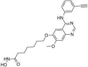
C24H26N4O4
|
|
|---|---|---|
| 分子量 |
434.49
|
|
| 精确质量 |
434.195
|
|
| 元素分析 |
C, 66.34; H, 6.03; N, 12.89; O, 14.73
|
|
| CAS号 |
1012054-59-9
|
|
| 相关CAS号 |
|
|
| PubChem CID |
24756910
|
|
| 外观&性状 |
White to yellow solid powder
|
|
| 密度 |
1.3±0.1 g/cm3
|
|
| 熔点 |
174-177ºC
|
|
| 折射率 |
1.638
|
|
| LogP |
2.84
|
|
| tPSA |
109.09
|
|
| 氢键供体(HBD)数目 |
3
|
|
| 氢键受体(HBA)数目 |
7
|
|
| 可旋转键数目(RBC) |
12
|
|
| 重原子数目 |
32
|
|
| 分子复杂度/Complexity |
624
|
|
| 定义原子立体中心数目 |
0
|
|
| SMILES |
O(C1=C(C([H])=C2C(C(=NC([H])=N2)N([H])C2=C([H])C([H])=C([H])C(C#C[H])=C2[H])=C1[H])OC([H])([H])[H])C([H])([H])C([H])([H])C([H])([H])C([H])([H])C([H])([H])C([H])([H])C(N([H])O[H])=O
|
|
| InChi Key |
PLIVFNIUGLLCEK-UHFFFAOYSA-N
|
|
| InChi Code |
InChI=1S/C24H26N4O4/c1-3-17-9-8-10-18(13-17)27-24-19-14-22(21(31-2)15-20(19)25-16-26-24)32-12-7-5-4-6-11-23(29)28-30/h1,8-10,13-16,30H,4-7,11-12H2,2H3,(H,28,29)(H,25,26,27)
|
|
| 化学名 |
7-[4-(3-ethynylanilino)-7-methoxyquinazolin-6-yl]oxy-N-hydroxyheptanamide
|
|
| 别名 |
CUDC-101; CUDC 101; 7-(4-(3-Ethynylphenylamino)-7-methoxyquinazolin-6-yloxy)-N-hydroxyheptanamide; CUDC101; 7-[[4-(3-Ethynylphenylamino)-7-methoxyquinazolin-6-yl]oxy]-N-hydroxyheptanamide; CHEMBL598797; 1A7Y9MP123; CUDC101
|
|
| HS Tariff Code |
2934.99.9001
|
|
| 存储方式 |
Powder -20°C 3 years 4°C 2 years In solvent -80°C 6 months -20°C 1 month |
|
| 运输条件 |
Room temperature (This product is stable at ambient temperature for a few days during ordinary shipping and time spent in Customs)
|
| 溶解度 (体外实验) |
|
|||
|---|---|---|---|---|
| 溶解度 (体内实验) |
配方 1 中的溶解度: ≥ 2.08 mg/mL (4.79 mM) (饱和度未知) in 10% DMSO + 40% PEG300 + 5% Tween80 + 45% Saline (这些助溶剂从左到右依次添加,逐一添加), 澄清溶液。
例如,若需制备1 mL的工作液,可将100 μL 20.8 mg/mL澄清DMSO储备液加入400 μL PEG300中,混匀;然后向上述溶液中加入50 μL Tween-80,混匀;加入450 μL生理盐水定容至1 mL。 *生理盐水的制备:将 0.9 g 氯化钠溶解在 100 mL ddH₂O中,得到澄清溶液。 配方 2 中的溶解度: ≥ 2.08 mg/mL (4.79 mM) (饱和度未知) in 10% DMSO + 90% (20% SBE-β-CD in Saline) (这些助溶剂从左到右依次添加,逐一添加), 澄清溶液。 例如,若需制备1 mL的工作液,可将 100 μL 20.8 mg/mL澄清DMSO储备液加入900 μL 20% SBE-β-CD生理盐水溶液中,混匀。 *20% SBE-β-CD 生理盐水溶液的制备(4°C,1 周):将 2 g SBE-β-CD 溶解于 10 mL 生理盐水中,得到澄清溶液。 View More
配方 3 中的溶解度: ≥ 2.08 mg/mL (4.79 mM) (饱和度未知) in 10% DMSO + 90% Corn Oil (这些助溶剂从左到右依次添加,逐一添加), 澄清溶液。 配方 4 中的溶解度: 15% Captisol: 30mg/mL 配方 5 中的溶解度: 16.67 mg/mL (38.37 mM) in 50% PEG300 50% Saline (这些助溶剂从左到右依次添加,逐一添加), 悬浊液; 超声助溶。 *生理盐水的制备:将 0.9 g 氯化钠溶解在 100 mL ddH₂O中,得到澄清溶液。 1、请先配制澄清的储备液(如:用DMSO配置50 或 100 mg/mL母液(储备液)); 2、取适量母液,按从左到右的顺序依次添加助溶剂,澄清后再加入下一助溶剂。以 下列配方为例说明 (注意此配方只用于说明,并不一定代表此产品 的实际溶解配方): 10% DMSO → 40% PEG300 → 5% Tween-80 → 45% ddH2O (或 saline); 假设最终工作液的体积为 1 mL, 浓度为5 mg/mL: 取 100 μL 50 mg/mL 的澄清 DMSO 储备液加到 400 μL PEG300 中,混合均匀/澄清;向上述体系中加入50 μL Tween-80,混合均匀/澄清;然后继续加入450 μL ddH2O (或 saline)定容至 1 mL; 3、溶剂前显示的百分比是指该溶剂在最终溶液/工作液中的体积所占比例; 4、 如产品在配制过程中出现沉淀/析出,可通过加热(≤50℃)或超声的方式助溶; 5、为保证最佳实验结果,工作液请现配现用! 6、如不确定怎么将母液配置成体内动物实验的工作液,请查看说明书或联系我们; 7、 以上所有助溶剂都可在 Invivochem.cn网站购买。 |
| 制备储备液 | 1 mg | 5 mg | 10 mg | |
| 1 mM | 2.3015 mL | 11.5077 mL | 23.0155 mL | |
| 5 mM | 0.4603 mL | 2.3015 mL | 4.6031 mL | |
| 10 mM | 0.2302 mL | 1.1508 mL | 2.3015 mL |
1、根据实验需要选择合适的溶剂配制储备液 (母液):对于大多数产品,InvivoChem推荐用DMSO配置母液 (比如:5、10、20mM或者10、20、50 mg/mL浓度),个别水溶性高的产品可直接溶于水。产品在DMSO 、水或其他溶剂中的具体溶解度详见上”溶解度 (体外)”部分;
2、如果您找不到您想要的溶解度信息,或者很难将产品溶解在溶液中,请联系我们;
3、建议使用下列计算器进行相关计算(摩尔浓度计算器、稀释计算器、分子量计算器、重组计算器等);
4、母液配好之后,将其分装到常规用量,并储存在-20°C或-80°C,尽量减少反复冻融循环。
计算结果:
工作液浓度: mg/mL;
DMSO母液配制方法: mg 药物溶于 μL DMSO溶液(母液浓度 mg/mL)。如该浓度超过该批次药物DMSO溶解度,请首先与我们联系。
体内配方配制方法:取 μL DMSO母液,加入 μL PEG300,混匀澄清后加入μL Tween 80,混匀澄清后加入 μL ddH2O,混匀澄清。
(1) 请确保溶液澄清之后,再加入下一种溶剂 (助溶剂) 。可利用涡旋、超声或水浴加热等方法助溶;
(2) 一定要按顺序加入溶剂 (助溶剂) 。
Link: https://clinicaltrials.gov/ct2/show/NCT00728793
Conditions:TumorsLink: https://clinicaltrials.gov/ct2/show/NCT01384799
Conditions:Head and Neck CancerLink: https://clinicaltrials.gov/ct2/show/NCT01702285
Conditions:Cancer
Title:A Phase Ib Expansion Study Investigating the Safety, Efficacy, and Pharmacokinetics of Intravenous CUDC-101 in Subjects With Advanced Head and Neck, Gastric, Breast, Liver and Non-small Cell Lung Cancer Tumors
Status:Completed
updateDate:2016-03-01
Ctid:NCT01171924
Link: https://clinicaltrials.gov/ct2/show/NCT01171924
Conditions:Head and Neck Cancer|Liver Cancer|Breast Cancer|Gastric Cancer|Non-Small Cell Lung Cancer |
|---|
 |
 |
 HDAC6-IN-18
 Mz325
Mz325
 WW437
WW437
 PI3Kα/HDAC6-IN-1
InvivoChem的所有产品仅用于作科学研究,不面向患者销售
Copyright 2020 InvivoChem LLC | All Rights Reserved 粤ICP备20063088号-1


 COA
COA
 463611831
