| 规格 | 价格 | 库存 | 数量 |
|---|---|---|---|
| 10 mM * 1 mL in DMSO |
|
||
| 1mg |
|
||
| 5mg |
|
||
| 10mg |
|
||
| 50mg |
|
||
| 100mg |
|
||
| 250mg |
|
||
| 500mg |
|
||
| Other Sizes |
|
| 靶点 |
hSGLT2 ( EC50 = 1.1 nM )
|
|---|---|
| 体外研究 (In Vitro) |
体外活性:达格列净对 hSGLT1 不敏感,IC50 为 1200 倍。达格列净对抗 hSGLT2 的效力比根皮苷强 32 倍,但对抗 hSGLT1 的效力比根皮苷低 4 倍。达格列净对 GLUT 转运蛋白具有高度选择性,在 20 μM 的无蛋白缓冲液中显示出 8-9% 的抑制作用,而在 4% 牛血清白蛋白存在的情况下几乎没有抑制作用。 Dapagliflozin 对 Caco-2 细胞膜具有良好的渗透性,是 P-糖蛋白 (P-gp) 的底物,但不是重要的 P-gp 抑制剂。 10 μM 的达格列净在大鼠、狗、猴和人血清中稳定。达格列净对人 P450 酶没有抑制反应或诱导作用。达格列净的体外代谢途径为葡萄糖醛酸化、羟基化和 O-脱乙基化 激酶测定:达格列净的 hSGLT2 的 EC50 值为 1.1 nM,hSGLT1 的 EC50 值为 1.4 μM,对应于 SGLT2 的 1200 倍选择性,而根皮苷的选择性为 10 倍。达格列净对大鼠 SGLT (rSGLT)2 和 hSGLT2 的抑制效力相当,但与 rSGLT1 相比,达格列净对 rSGLT2 的选择性下降至 200 倍 细胞测定:为了进行细胞存活测定,在与载体或达格列净一起孵育 24 小时后收集细胞预处理30分钟缺血,并用台盼蓝染色对存活细胞进行计数。存活百分比通过将处理的细胞的相对存活数除以未处理的细胞的存活数进行量化来确定。
体外活性。[1] 达格列净对hSGLT2的平均EC50为1.12 nmol/l,而根皮苷的EC50为35.6 nmol/l(表1)。与hSGLT1相比,达格列净和根皮苷的平均EC50值分别为1391和330 nmol/l,表明达格列净对hSGLT2的选择性很高(约1200倍)。达格列净对hSGLT2的效力是根皮苷的32倍,对hSGL41的效力比根皮苷低4倍。Dapagliflozin也是rSGLT2的强效选择性抑制剂,显示平均EC50值为3.0 nmol/l,与rSGLT1相比选择性约为200倍。在人脂肪细胞中检测到,达格列净对GLUT转运蛋白具有高度选择性,在20μmol/l的无蛋白缓冲液中显示出8-9%的抑制作用,在4%牛血清白蛋白存在的情况下几乎没有抑制作用(表2)。将蛋白质添加到该测定中以模拟血浆蛋白结合的体内条件。Phlorizin最低限度地抑制脂肪细胞GLUT活性;然而,根皮苷的苷元根皮素抑制GLUT活性约77%,无论检测中是否存在牛血清白蛋白。 细胞内镁浓度[5] 孵育24小时后,测量并计算细胞内镁浓度。如表3和图3所示,与第一次测量时(0分钟)的对照组相比,用达格列净治疗与细胞内镁浓度增加60%有关。单独使用AG1478和间充质治疗显著降低了细胞内镁浓度。达格列净与AG1478或间充质联合使用可降低镁浓度(均p<0.05)。在120分钟内,除80分钟外,达格列嗪组的镁浓度高于对照组。单独使用AG1478治疗在20、60、100和120分钟时与显著降低的浓度相关。单独使用间充质治疗在整个120分钟期间降低了镁浓度,但80分钟除外。联合治疗(达格列嗪与AG1478和达格列嗪与间充质)在大多数时间点显著降低了镁含量。 |
| 体内研究 (In Vivo) |
在高血糖链脲佐菌素 (STZ) 大鼠中口服 0.1 mg/kg 剂量后,达格列净可将血糖水平降低 55%,部分原因是 C-葡萄糖苷键赋予的代谢稳定性。达格列净表现出良好的吸收、分布、代谢和排泄 (ADME) 特性,并且具有口服生物利用度。 Dapagliflozin (1 mg/kg) 在正常大鼠给药后 24 小时内引起显着的剂量依赖性糖尿和尿量增加。 Dapagliflozin 会在 Zucker 糖尿病脂肪 (ZDF) 大鼠给药后 6 小时诱导尿糖和尿量排泄增加。即使治疗 2 周,达格列净仍可降低 ZDF 大鼠的空腹和餐后血糖水平,且没有任何肾或肝毒性标志。达格列净显着减少高血糖的发生,降低血糖。达格列净可以提高胰岛素敏感性,减少β细胞质量和胰腺功能受损的发展。
急性体内疗效。[2] 在正常大鼠中,达格列净给药引起明显的剂量依赖性糖尿(图1)和尿量增加,给药后24小时内,与赋形剂相比,1 mg/kg的尿糖增加了400倍,尿量增加了三倍。在正常大鼠的口服葡萄糖耐量试验中,在1和10mg/kg剂量下,达格列嗪给药与给药后1小时内曲线下葡萄糖面积的减少有关(图2),表明这种葡萄糖尿酸剂能够减少正常大鼠急性葡萄糖挑战后的葡萄糖波动。在单剂量口服达格列嗪的ZDF大鼠中,给药后6小时,尿糖和尿量排泄明显呈剂量依赖性增加(图3A),同时同一只大鼠在0.01-1.0 mg/kg的剂量下血糖降低(图4)。在24小时内检查时,尿糖排泄效应的明显剂量依赖性下降(图3B),所有剂量组的治疗大鼠的尿糖水平均比赋形剂治疗大鼠提高了两倍。在这些大鼠以1mg/kg的剂量重新进食一段时间后,在给药后24小时仍观察到降低血糖的疗效。在这些研究过程中没有观察到低血糖的证据。在1 mg/kg的剂量下,给药后1小时达格列净的血浆暴露量为1.2μmol/l,在实验的前6小时估计为5.2μmol·l-1·h-1。 慢性体内疗效。[2] 在两项慢性研究中的第一项中,Dapagliflozin剂量依赖性地降低了治疗第8天时禁食18小时的ZDF大鼠的空腹血糖水平,这是在前一次给药后24小时测量的(图5)。在治疗的第15天,大鼠禁食24小时,这种效果也很明显,在研究的第14天,喂食动物也有这种效果。这些数据表明,在为期2周的每日一次治疗方案中,降低FPG的疗效得以保持。与赋形剂处理的大鼠相比,没有发现体重变化,也没有观察到异常行为;老鼠似乎很好。没有测量到肾或肝毒性的标志物。[2] 在第二项慢性研究中,当在第15天最后一次给药后24小时进行测量时,与赋形剂治疗的大鼠相比,用0.5mg/kg dDapagliflozin治疗的ZDF大鼠在18小时的FPG水平下降了53%(表3)。在最后一次给药后的第三天,启动了一项高胰岛素血症正常血糖钳夹研究,以评估达格列嗪与赋形剂治疗的代谢影响。在基础阶段,与达格列嗪治疗的大鼠相比,赋形剂治疗的大白鼠的尿糖损失率明显更高(表3),这可能反映了赋形剂治疗大白鼠血浆葡萄糖水平升高的趋势。在大鼠最后一剂半衰期为4至5小时的化合物给药后48小时开始夹紧程序的事实(W.Humphreys,W.N.W.,未发表的数据)表明,夹紧程序期间的血浆药物水平可以忽略不计,尽管没有测量。因此,我们预计达格列嗪急性诱导的尿糖排泄对钳夹期间观察到的代谢效应没有显著影响。在此过程中,达格列嗪治疗的大鼠的尿量也显著减少。尽管与赋形剂处理的大鼠相比,达格列嗪治疗的大鼠在夹子的胰岛素输注阶段尿葡萄糖损失似乎有所减少,但这种差异在统计学上并不显著。[2] 与赋形剂处理的大鼠相比,在夹具的胰岛素输注阶段,达格列净处理的大白鼠的葡萄糖输注速率显著增加,表明全身葡萄糖利用率有所改善(表3)。与赋形剂处理的大鼠相比,达格列嗪治疗的大鼠在胰岛素输注阶段的葡萄糖产量也显著降低(表3)。此外,在达格列嗪治疗的大鼠中,钳夹期间肝脏对放射性标记葡萄糖的摄取显著增加,而骨骼肌或白色脂肪组织对葡萄糖的摄取没有显著变化。这些数据表明,用达格列净治疗2周可以改善ZDF大鼠葡萄糖产量的升高,并增强肝脏胰岛素敏感性。 从高脂肪喂养开始服用达格列净/Dapagliflozin,可以减少高血糖的发展;24天后,血糖为8.6±0.5 vs.13.3±1.3 mmol/l(与溶媒相比p<0.005),糖化血红蛋白为3.6±0.1 vs.4.8±0.26%(与赋形剂相比p<0.003)。达格列净改善了肥胖对照组的胰岛素敏感性指数:0.08±0.01比0.02±0.01(p<0.03)。DI改善到瘦对照组大鼠的水平(达格列净0.29±0.04;肥胖对照组0.15±0.01;瘦对照组0.28±0.01)。在达格列嗪治疗的大鼠中,β细胞质量的变化较小,与赋形剂治疗的大白鼠相比,胰岛形态有显著改善,尽管达格列嗪的平均β细胞质量没有变化。当动物已经中度高血糖时,开始达格列嗪治疗,结果相似。 结论:在2型糖尿病模型中,达格列净持续降糖可防止胰腺β细胞功能适应性的持续下降。[4] 成年大鼠在前3个月喂食富含果糖的饮食以诱导代谢综合征,然后用<Dapagliflozin达格列净 或含硫酸镁的饮用水治疗另外3个月。喂食果糖的动物胰岛素抵抗增加,低镁血症,尿镁排泄减少。达格列净治疗通过降低葡萄糖和胰岛素水平、增加血清镁水平和减少尿镁排泄来改善胰岛素抵抗。果糖喂养的动物血清维生素D和甲状旁腺激素水平降低,尽管补充了达格列嗪和镁,但水平仍然很低。在肾脏中,果糖喂养的动物的claudin-16、TRPM6/7和FXDY表达增加。达格列净增加了细胞内镁浓度,这种作用被TRPM6阻断剂和EGFR拮抗剂抑制。我们得出结论,高果糖摄入结合低镁饮食会导致代谢综合征和低镁血症。达格列嗪和硫酸镁补充剂都改善了代谢综合征的特征,并提高了血清镁水平。在果糖喂养的动物和服用达格列嗪和硫酸镁的动物中,claudin-16、TRPM6/7和FXYD2等镁转运蛋白的表达水平升高。达格列净增强肾小管细胞中TRPM6介导的跨上皮镁转运[5]。 |
| 酶活实验 |
选择性 SGLT 底物 α-甲基-D-吡喃葡萄糖苷 (AMG) 用于使用稳定表达人 SGLT2 (hSGLT2) 和人 SGLT1 (\hSGLT1) 的中国仓鼠卵巢 (CHO) 细胞开发转运测定。达格列净抑制 [14C]AMG 摄取的能力是在无蛋白质缓冲液中孵育两个小时的过程中测量的。通过将响应曲线拟合到经验四参数模型来找到半最大响应时的抑制剂浓度(或 EC50)。使用无蛋白缓冲液复制肾小球滤液的低蛋白环境,覆盖肾脏近端小管腔表面上的 SGLT 靶标。
SGLT1和SGLT2测定。[2] 使用标准细胞培养技术维持表达hSGLT1、hSGLT2、rSGLT1或rSGLT2的细胞。通过添加100μl/孔的含钠(HEPES/Tris pH 7.4、137 mmol/l NaCl、5.4 mmol/l KCl、2.8 mmol/l CaCl2、1.2 mmol/l MgSO4)、10μmol/l 14C-α-甲基-d-吡喃葡萄糖苷和抑制剂或DMSO载体的无蛋白测定缓冲液,启动96孔板中钠依赖性葡萄糖转运的测定,并将板在37°C下孵育2小时。钠依赖性14C-α-甲基-d-吡喃葡萄糖苷摄取量是通过从含钠条件下观察到的计数中减去无钠摄取条件下每分钟的计数来计算的。在钠存在的情况下,对八种浓度的抑制剂进行了三次检测,并通过比较含抑制剂的孔中每分钟的计数与仅含DMSO载体的孔中的每分钟计数来计算抑制百分比。在每次测定中平行评估Phlorizin。使用XL Fit将剂量反应曲线拟合到经验四参数模型中,以确定半最大反应时的抑制剂浓度(EC50)。 脂肪细胞葡萄糖摄取测定。[2] 在试验之前,将预分化的人脂肪细胞在Dulbecco改良的Eagle培养基中洗涤一次,该培养基为低葡萄糖,不含胎牛血清,并在37°C下孵育2小时。然后在不含葡萄糖的Krebs-Ringer碳酸氢盐HEPES缓冲液中洗涤细胞两次。测定缓冲液(100μl/孔)由不含胰岛素或100 nmol/l胰岛素的Krebs-Ringer碳酸氢盐HEPES缓冲液、10μmol/l 2-14C-脱氧-d-葡萄糖、抑制剂或细胞松弛素B以及DMSO对照(每组n=6)组成。细胞在37°C下孵育20分钟,在PBS中洗涤三次,并在50μl/孔的0.1 N NaOH中裂解。加入MicroScint-40,在TopCount闪烁计数器中计数细胞。通过比较含抑制剂孔中每分钟的计数与不含抑制剂孔的每分钟计数来计算抑制百分比。 |
| 细胞实验 |
在 30 分钟的缺血期间,使用载体或达格列净预处理 24 小时孵育期后,收集细胞用于细胞存活测定。然后使用台盼蓝染色对任何剩余的细胞进行计数。将处理的细胞的相对存活数除以未处理的细胞的存活数进行量化以获得存活百分比。
Caco-2通透性和P-gp相互作用。[3] 在pH 7.4、初始浓度为50μM时,达格列净在顶端(A)至基底外侧(B)方向的渗透系数为60 nm/s。该值与在人体中表现出中等吸收的化合物相当(Marino等人,2005)。在相同条件下,达格列嗪的平均B-to-A渗透系数值为227nm/s(BA/AB比为3.8)。在P-gp抑制剂存在的情况下,达格列嗪的a-to-B通透性为159 nm/s,B-to-a通透性为150 nm/s。。。 细胞培养和细胞内镁浓度测量。[5] 将NRK52E细胞接种在96孔荧光板上,并在以下条件下处理细胞24小时:1。常规培养基;2.10μM间充质;3.10μM AG1478;4.0.2μM达格列净;5.10μM间充质和0.2μM达格列嗪;6.10μM AG1478(0.2μM)和达格列嗪。然后将细胞与5μM Mg-Fura-2 AM在37°C下孵育60分钟,然后用所需的最终培养基洗涤三次。然后将细胞再孵育60分钟,以便在荧光测量之前完全脱除细胞内AM酯。所有实验重复4-6次,然后取平均值。 |
| 动物实验 |
将化合物溶于 5% 甲基吡咯烷酮、20% PEG400 和 20 mM 磷酸二钠溶液中;以 0.01-10 mg/kg(1 mL/kg)的剂量口服给药,随后给予 50% 葡萄糖溶液(2 g/kg)。实验对象为正常 Sprague Dawley 大鼠或链脲佐菌素诱导的雄性 Sprague Dawley 大鼠。实验方案的示意图可在 http://dx.doi.org/10.2337/db07-1472 的在线附录中找到。除非另有说明,动物可自由摄取食物和水,饲养室温度保持在 22°C,湿度为 50%,光照/黑暗周期为 12 小时。雄性 Sprague-Dawley 大鼠饲喂 Harlan Teklad 2018 饲料,实验时体重为 250 g。雄性Zucker糖尿病肥胖(ZDF)大鼠饲喂Purina 5008饲料。急性研究中,大鼠19周龄,平均体重422 g。第一项慢性研究中,雄性ZDF大鼠17周龄,平均体重399 g;第二项慢性研究中,雄性ZDF大鼠15周龄,平均体重397 g。所有ZDF大鼠研究均采用随机分组,各组间体重和血浆葡萄糖水平无统计学差异。采集血样后,于4℃、2500 rpm下离心10分钟(Eppendorf离心机)。取5 μL血浆,与25 μL生理盐水混合,使用Cobas Mira分析仪进行葡萄糖分析。所有尿量均被测量并记录。尿糖分析中,取5 μl尿液与250 μl生理盐水混合,使用Cobas Mira分析仪进行分析。
急性正常和糖尿病大鼠研究。[2] 所有动物实验中,给药溶剂为5% 1-甲基-2-吡咯烷酮、20%聚乙二醇和20 mmol/l磷酸二钠。葡萄糖耐量试验中,15只Sprague-Dawley大鼠禁食过夜(18小时),称重,尾尖采血(30-40 μl),随机分为五组(n = 3)。大鼠分别口服单剂量溶剂或药物(1 ml/kg;药物剂量0.01-10 mg/kg),随后口服50%葡萄糖水溶液(2 g/kg)。分别于给药后15、30、60分钟和24小时采血。这些研究中未测量胰岛素水平。 为了评估尿糖,将禁食过夜的Sprague-Dawley大鼠置于代谢笼中,收集24小时的基线尿液。称量大鼠体重后,随机分为五组(n = 3),分别口服单剂量赋形剂或药物(1 ml/kg;药物剂量为0.01–10 mg/kg),随后口服50%葡萄糖水溶液(2 g/kg)。给药后立即将大鼠放回代谢笼中,收集24小时尿液,并在葡萄糖负荷后1小时重新喂食。使用GraphPad Prism软件计算血浆葡萄糖浓度曲线下面积(AUC)相对于基线值的差值。尿糖和尿量数据均按每200克体重进行标准化。 为了评估ZDF大鼠的急性糖尿和血浆葡萄糖效应,将动物称重,在喂食状态下通过尾尖采血(40–50 μl),并随机分为四组(n = 6)。大鼠分别给予赋形剂或药物(1 ml/kg;药物剂量为0.01–1.0 mg/kg),并放入代谢笼中(无需事先适应)。分别在给药前以及给药后2、4、6和24小时采集血样。分别在给药后2、4、6和24小时收集尿液。6小时后允许动物重新进食。分析各时间点的血浆样本中葡萄糖和达格列净的含量。尿糖和尿量数据均按每400克体重进行标准化。这些研究中未测量胰岛素。 慢性糖尿病大鼠研究。[2] 在ZDF大鼠中进行了两项研究,以评估多剂量达格列净治疗2周后对餐后和空腹血糖(FPG)以及大鼠代谢谱的影响。在研究1中,大鼠禁食过夜,称重,从尾尖采血(40–50 μl),并随机分为四组(每组n = 6)。大鼠每天口服一次赋形剂或药物(1 ml/kg;0.01–1.0 mg/kg药物),持续14天。分别于第8天(禁食18小时)和第15天(禁食24小时)采集空腹体重和血浆样本,并于第14天(上次给药后24小时)采集餐后血浆样本。在研究2中,ZDF大鼠被随机分为两组(每组n=6),并分别每日一次口服给予赋形剂或药物(1 ml/kg;0.5 mg/kg药物),持续15天。在研究的第1、8和15天,从所有组别禁食18小时的大鼠尾部采血(40 μl),以测定血浆葡萄糖水平。本研究未测定空腹或餐后胰岛素水平。在第17天以及最后一次给予赋形剂或达格列净后48小时,对赋形剂组和达格列净组大鼠进行高胰岛素-正常血糖钳夹试验。这些研究中均未监测食物和水的摄入量。 在一项初步研究中,四只肥胖的雌性ZDF大鼠被喂食C13004高脂饮食12-15天,同时四只匹配的肥胖大鼠和四只瘦大鼠继续喂食RM1标准饲料。随后进行了一项高血糖钳夹试验,具体方法将在后续正文中描述。为了评估达格列净,在两批不同的大鼠中平行进行了两项独立的研究。在第一项研究(“预防”)中,从开始喂食高脂饮食起就给予达格列净(n = 14)。在第二项研究(“干预”)中,在开始喂食高脂饮食10天后,当动物出现中度高血糖时,开始给予达格列净治疗(n = 14)。达格列净(1 mg/kg,溶于水中,口服)或赋形剂每日一次,于上午 8:00 给药,持续 33 天。两项研究中,末次给药后 48 小时,所有动物禁食过夜,然后根据第 24 天的血糖和糖化血红蛋白 (gHb) 水平,将所有组别分为两个匹配的亚组。其中一个亚组(5-6 只动物)用于通过高血糖钳夹试验评估胰腺功能。其余动物吸入 5:1 的 CO₂:O₂ 混合气体使其失去知觉,以最大程度地减少通过心脏穿刺采集到乙二胺四乙酸 (EDTA) 抗凝血注射器中的血样对血糖和胰岛素水平的影响。胰腺被切除后,固定于10%中性缓冲福尔马林溶液中24-48小时,随后进行常规处理并包埋于石蜡中。[4] 血液样本采集和血浆检测[4] 在研究开始前以及第14天和第24天,通过测量清醒动物尾静脉采集的小样本中的糖化血红蛋白(gHb)和血糖水平来监测疾病进展。样本采集于达格列净给药前8:00。血浆胰岛素采用酶联免疫吸附试验(ELISA)进行测定。血浆C肽采用放射免疫分析法进行测定。采用罗氏模块化系统,通过甘油-3-磷酸氧化酶-对氨基苯酚法测定血浆甘油三酯。 本实验采用体重200-250 g的成年雄性Sprague Dawley大鼠。动物饲养于12小时恒定光照周期、温度21℃-23℃的条件下。动物可自由摄取水和食物。动物被随机分为对照组和果糖饮食组。果糖饮食组饲喂高果糖饮食(60%果糖,0.05%镁,w/w),对照组(n = 10)饲喂标准大鼠饲料,持续6个月。高果糖饮食组在饲喂3个月后开始药物治疗。动物被分为以下三组:1. 继续饲喂果糖6个月(FR,n = 10); 2. 继续喂食果糖饮食并接受达格列净治疗(FR+Dapa,1 mg·kg⁻¹·day⁻¹,灌胃给药;n = 10),持续3个月;3. 继续喂食果糖饮食并补充硫酸镁(FR+Mg,饮用水中添加296 mg/L的镁,台湾桃园生物科技有限公司;n = 10),持续3个月。每周两次采用间接尾套法测量每只动物的血压,至少测量5次并取平均值。每周测量体重。研究结束时,在独立的代谢笼中收集24小时尿液样本。随后处死大鼠,并从下腔静脉抽取血液样本进行生化分析。[5] |
| 药代性质 (ADME/PK) |
吸收、分布和排泄
空腹患者口服达格列净后,1小时内即可达到最大血药浓度。空腹状态下,口服达格列净后,通常在2小时内达到最大血浆浓度(Cmax)。在治疗剂量范围内,Cmax和AUC值随达格列净剂量的增加而呈比例增加。口服10 mg达格列净的绝对生物利用度为78%。与空腹状态相比,与高脂餐同服可使达格列净的Cmax降低高达50%,Tmax延长约1小时,但AUC无明显变化。这些变化不具有临床意义,达格列净可与食物同服或空腹服用。 达格列净及其相关代谢物主要经肾脏途径排泄。单次服用 50 mg [14C]-达格列净后,总放射性的 75% 和 21% 分别经尿液和粪便排出。尿液中,不到 2% 的剂量以原药形式排出。粪便中,约 15% 的剂量以原药形式排出。 分布容积估计为 118 L。 口服血浆清除率为 4.9 mL/min/kg,肾清除率为 5.6 mL/min。 代谢/代谢物 达格列净主要经葡萄糖醛酸化代谢为无活性的 3-O-葡萄糖醛酸苷代谢物 (60.7%)。达格列净还会产生另一种次要的葡萄糖醛酸化代谢物(5.4%)、一种去乙基化代谢物(<5%)和一种羟基化代谢物(<5%)。达格列净的代谢由细胞色素P-450 (CYP) 1A1、CYP1A2、CYP2A6、CYP2C9、CYP2D6、CYP3A4、尿苷二磷酸葡萄糖醛酸转移酶 (UGT) 1A9、UGT2B4 和 UGT2B7 介导。UGT1A9 介导达格列净转化为主要代谢物。 生物半衰期 单次口服 10 mg 达格列净后,其平均血浆末端半衰期 (t1/2) 约为 12.9 小时。健康受试者单次口服 50 mg 达格列净后,平均终末半衰期为 13.8 小时。达格列净 (2S,3R,4R,5S,6R)-2-(3-(4-乙氧基苄基)-4-氯苯基)-6-羟甲基-四氢-2H-吡喃-3,4,5-三醇(达格列净;BMS-512148)是一种强效的钠-葡萄糖协同转运蛋白 II 型抑制剂,在动物和人体内均有效,目前正在开发用于治疗 2 型糖尿病。为了筛选合适的化合物并预测其在临床上的药理学和体内分布行为,达格列净的临床前特性研究包括Caco-2细胞渗透性研究、细胞色素P450 (P450)抑制和诱导研究、P450反应表型分析、肝细胞代谢物鉴定以及在大鼠、犬和猴体内的药代动力学研究。研究发现,达格列净具有良好的Caco-2细胞膜渗透性。它是P-糖蛋白(P-gp)的底物,但并非显著的P-gp抑制剂。达格列净既不是人P450酶的抑制剂,也不是其诱导剂。达格列净与小鼠、大鼠、犬、猴和人肝细胞孵育后的体外代谢谱在性质上相似。大鼠肝细胞孵育实验显示达格列净的周转率最高,而达格列净在人肝细胞中最为稳定。体外观察到的主要代谢途径包括葡萄糖醛酸化、羟基化和O-去乙基化。临床前动物模型中达格列净的药代动力学参数显示,该化合物具有足够的口服暴露量、清除率和消除半衰期,符合人体每日单次给药的潜力。人体单次服用50 mg [(14)C]达格列净后的药代动力学结果显示,药物暴露量良好,清除率低,半衰期适宜,且未检测到具有显著药理活性或毒理学风险的代谢产物。[3] |
| 毒性/毒理 (Toxicokinetics/TK) |
妊娠期和哺乳期影响
◉ 哺乳期用药概述 目前尚无关于达格列净在哺乳期临床应用的信息。达格列净是一种不带电荷的分子,其血浆蛋白结合率为91%,因此不太可能以具有临床意义的量进入母乳。由于理论上存在对婴儿发育中肾脏的风险,生产商不建议在哺乳期使用达格列净。尤其是在哺乳新生儿或早产儿时,应优先选择其他药物。 ◉ 对母乳喂养婴儿的影响 截至修订日期,未找到相关的已发表信息。 ◉ 对哺乳和母乳的影响 截至修订日期,未找到相关的已发表信息。 蛋白结合 达格列净的蛋白结合率约为91%。肾功能或肝功能受损患者的蛋白质结合能力并未改变。 |
| 参考文献 |
|
| 其他信息 |
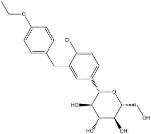
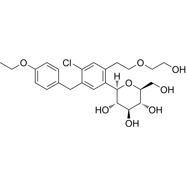
达格列净是一种C-糖苷类化合物,由β-D-葡萄糖组成,其端羟基被4-氯-3-(4-乙氧基苄基)苯基取代。它以丙二醇一水合物的形式用于改善2型糖尿病成人患者的血糖控制,需配合饮食和运动。它是一种降血糖药和钠-葡萄糖协同转运蛋白2 (SGLT2) 抑制剂。它是一种C-糖苷类化合物,属于芳香醚类,也是一氯苯类化合物。
达格列净是一种钠-葡萄糖协同转运蛋白2 (SGLT2) 抑制剂,也是首个获批的SGLT2抑制剂,用于治疗2型糖尿病。在成人患者中,达格列净与饮食和运动联合使用,可通过抑制肾单位近端小管对葡萄糖的重吸收并引起糖尿,从而帮助改善血糖控制。达格列净已被研究作为单药治疗或与胰岛素或其他口服降糖药联合使用。达格列净最初于2014年1月8日获得美国食品药品监督管理局(FDA)批准,用于改善2型糖尿病成人患者的血糖控制,需配合饮食和运动。2021年4月,达格列净获批用于降低慢性肾脏病成人患者的肾功能下降、肾衰竭、心血管死亡和心力衰竭住院的风险。 达格列净是一种钠-葡萄糖协同转运蛋白2抑制剂。达格列净的作用机制是作为钠-葡萄糖协同转运蛋白2抑制剂。 达格列净是一种选择性钠-葡萄糖协同转运蛋白2(SGLT2)抑制剂,具有降血糖活性。达格列净选择性且强效地抑制SGLT2,而非SGLT1(肠道中的葡萄糖协同转运蛋白)。 达格列净是一种小分子药物,其临床试验阶段最高为IV期(涵盖所有适应症),于2012年首次获批,目前有3项已获批适应症和37项在研适应症。 药效学 达格列净还能减少钠的重吸收,并增加钠向远端肾小管的输送。这可能影响多种生理功能,包括但不限于降低心脏的前负荷和后负荷、下调交感神经活性以及降低肾小球内压(据信这是由肾小管-肾小球反馈增强介导的)。在健康受试者和2型糖尿病患者服用达格列净后,均观察到尿糖排泄量增加。在2型糖尿病患者中,每日服用5或10毫克达格列净治疗12周后,第12周时每日尿糖排泄量约为70克。每日服用20毫克达格列净时,尿糖排泄量接近最大值。达格列净引起的尿糖排泄增加也会导致尿量增加。停用达格列净后,平均而言,10毫克剂量组的尿糖排泄量在约3天内恢复至基线水平。在一项针对健康受试者的研究中,每日服用高达150毫克(推荐最大剂量的15倍)的达格列净未引起具有临床意义的QTc间期延长。此外,在健康受试者中,单次服用高达 500 mg(推荐最大剂量的 50 倍)的达格列净后,未观察到对 QTc 间期有临床意义的影响。 C-芳基葡萄糖苷 6(达格列净)被鉴定为一种强效且选择性的 hSGLT2 抑制剂,在链脲佐菌素 (STZ) 诱导的高血糖大鼠模型中,其血糖水平可呈剂量依赖性地降低高达 55%。这些发现,结合其良好的 ADME 特性,促使人们对达格列净用于治疗 2 型糖尿病进行了临床评估。[1] 目的:抑制肠道和肾脏钠-葡萄糖协同转运蛋白 (SGLT) 被认为是一种治疗糖尿病的新方法。我们已在体外鉴定出达格列净是一种强效且选择性的肾脏钠-葡萄糖协同转运蛋白SGLT2抑制剂,并对其体外和体内药理学特性进行了表征。研究设计与方法:采用基于细胞的葡萄糖类似物摄取测定法评估达格列净抑制钠依赖性和易化葡萄糖转运活性的能力。在正常大鼠和糖尿病大鼠中进行急性及多剂量研究,以评估达格列净改善餐后和空腹血糖水平的能力。进行高胰岛素-正常血糖钳夹试验,以评估达格列净多剂量治疗后改善葡萄糖利用率的能力。结果:达格列净强效且选择性地抑制人SGLT2(肠道中主要的葡萄糖协同转运蛋白),而对人SGLT1抑制作用较弱,且对人脂肪细胞中的易化葡萄糖转运无显著抑制作用。在体内,达格列净可急性诱导正常大鼠和糖尿病大鼠肾脏葡萄糖排泄,改善正常大鼠的葡萄糖耐量,并降低Zucker糖尿病脂肪(ZDF)大鼠的血糖水平(单次口服剂量为0.1至1.0 mg/kg)。每日一次、持续两周的达格列净治疗(剂量范围为0.1至1.0 mg/kg)可显著降低空腹和餐后血糖水平,并显著提高葡萄糖利用率,同时显著降低葡萄糖生成。结论:这些数据表明,达格列净具有成为2型糖尿病有效治疗药物的潜力。 [2] 目的:探讨选择性钠-葡萄糖协同转运蛋白2 (SGLT2) 抑制剂达格列净降低血糖是否能预防或减轻胰腺功能下降和正常胰岛形态的破坏。方法:将7-8周龄的雌性Zucker糖尿病肥胖(ZDF)大鼠置于高脂饮食喂养。从高脂饮食开始或大鼠出现中度高血糖时,给予达格列净(1 mg/kg/天,口服),持续约33天。在麻醉状态下,采用高血糖钳夹技术评估动物(n = 5-6)的胰岛素敏感性和胰腺功能;使用处置指数(DI)量化β细胞功能,以校正胰岛素抵抗的代偿作用。我们从匹配的亚组(n = 7-8)中取出胰腺组织进行固定,并采用免疫组织化学方法研究了β细胞数量和胰岛形态。[4]代谢综合征(MetS)的患病率正在上升,MetS患者罹患心血管疾病和糖尿病的风险增加。低镁血症与MetS密切相关。据报道,钠-葡萄糖协同转运蛋白2(SGLT2)抑制剂可提高糖尿病患者的血清镁水平。我们利用MetS动物模型研究了肾脏镁处理的改变,并分析了SGLT2抑制剂的作用。成年大鼠在前3个月喂食富含果糖的饮食以诱导MetS,随后分别接受达格列净或含硫酸镁的饮用水治疗3个月。喂食果糖的动物表现出胰岛素抵抗增加、低镁血症和尿镁排泄减少。达格列净治疗通过降低血糖和胰岛素水平、提高血清镁水平和减少尿镁排泄,改善了胰岛素抵抗。果糖喂养动物的血清维生素D和甲状旁腺激素水平降低,即使补充达格列净和镁,这些水平仍然很低。在肾脏中,果糖喂养动物的claudin-16、TRPM6/7和FXDY表达增加。达格列净可增加细胞内镁浓度,而TRPM6阻断剂和EGFR拮抗剂可抑制这种作用。我们得出结论,高果糖摄入联合低镁饮食可诱发代谢综合征和低镁血症。达格列净和硫酸镁补充剂均可改善代谢综合征的特征并提高血清镁水平。在喂食果糖的动物以及服用达格列净和硫酸镁的动物中,claudin-16、TRPM6/7 和 FXYD2 等镁转运蛋白的表达水平升高。达格列净可增强肾小管细胞中 TRPM6 介导的跨上皮镁转运。[5] |
| 分子式 |
C21H25CLO6
|
|---|---|
| 分子量 |
408.87
|
| 精确质量 |
408.133
|
| 元素分析 |
C, 61.69; H, 6.16; Cl, 8.67; O, 23.48
|
| CAS号 |
461432-26-8
|
| 相关CAS号 |
Dapagliflozin ((2S)-1,2-propanediol, hydrate); 960404-48-2; Dapagliflozin-d5; 1204219-80-6
|
| PubChem CID |
9887712
|
| 外观&性状 |
White solid powder
|
| 密度 |
1.3±0.1 g/cm3
|
| 沸点 |
609.0±55.0 °C at 760 mmHg
|
| 闪点 |
322.1±31.5 °C
|
| 蒸汽压 |
0.0±1.8 mmHg at 25°C
|
| 折射率 |
1.614
|
| LogP |
4.42
|
| tPSA |
99.38
|
| 氢键供体(HBD)数目 |
4
|
| 氢键受体(HBA)数目 |
6
|
| 可旋转键数目(RBC) |
6
|
| 重原子数目 |
28
|
| 分子复杂度/Complexity |
472
|
| 定义原子立体中心数目 |
5
|
| SMILES |
ClC1C([H])=C([H])C(=C([H])C=1C([H])([H])C1C([H])=C([H])C(=C([H])C=1[H])OC([H])([H])C([H])([H])[H])[C@@]1([H])[C@@]([H])([C@]([H])([C@@]([H])([C@@]([H])(C([H])([H])O[H])O1)O[H])O[H])O[H]
|
| InChi Key |
JVHXJTBJCFBINQ-ADAARDCZSA-N
|
| InChi Code |
InChI=1S/C21H25ClO6/c1-2-27-15-6-3-12(4-7-15)9-14-10-13(5-8-16(14)22)21-20(26)19(25)18(24)17(11-23)28-21/h3-8,10,17-21,23-26H,2,9,11H2,1H3/t17-,18-,19+,20-,21+/m1/s1
|
| 化学名 |
(2S,3R,4R,5S,6R)-2-[4-chloro-3-[(4-ethoxyphenyl)methyl]phenyl]-6-(hydroxymethyl)oxane-3,4,5-triol
|
| 别名 |
Dapagliflozin; BMS 512148; BMS512148; Dapagliflozin; 461432-26-8; BMS-512,148; Forxiga; Farxiga; BMS 512,148; (2S,3R,4R,5S,6R)-2-(4-chloro-3-(4-ethoxybenzyl)phenyl)-6-(hydroxymethyl)tetrahydro-2H-pyran-3,4,5-triol; dapagliflozine; BMS-512148; trade name Farxiga in the US and Forxiga in the EU
|
| HS Tariff Code |
2934.99.9001
|
| 存储方式 |
Powder -20°C 3 years 4°C 2 years In solvent -80°C 6 months -20°C 1 month 注意: 请将本产品存放在密封且受保护的环境中,避免吸湿/受潮。 |
| 运输条件 |
Room temperature (This product is stable at ambient temperature for a few days during ordinary shipping and time spent in Customs)
|
| 溶解度 (体外实验) |
|
|||
|---|---|---|---|---|
| 溶解度 (体内实验) |
配方 1 中的溶解度: ≥ 2.5 mg/mL (6.11 mM) (饱和度未知) in 5% DMSO + 40% PEG300 + 5% Tween80 + 50% Saline (这些助溶剂从左到右依次添加,逐一添加), 澄清溶液。
*生理盐水的制备:将 0.9 g 氯化钠溶解在 100 mL ddH₂O中,得到澄清溶液。 配方 2 中的溶解度: ≥ 2.5 mg/mL (6.11 mM) (饱和度未知) in 5% DMSO + 95% (20% SBE-β-CD in Saline) (这些助溶剂从左到右依次添加,逐一添加), 澄清溶液。 *20% SBE-β-CD 生理盐水溶液的制备(4°C,1 周):将 2 g SBE-β-CD 溶解于 10 mL 生理盐水中,得到澄清溶液。 View More
配方 3 中的溶解度: ≥ 2.08 mg/mL (5.09 mM) (饱和度未知) in 10% DMSO + 40% PEG300 + 5% Tween80 + 45% Saline (这些助溶剂从左到右依次添加,逐一添加), 澄清溶液。 配方 4 中的溶解度: ≥ 2.08 mg/mL (5.09 mM) (饱和度未知) in 10% DMSO + 90% (20% SBE-β-CD in Saline) (这些助溶剂从左到右依次添加,逐一添加), 澄清溶液。 例如,若需制备1 mL的工作液,可将100μL 20.8mg/mL澄清的DMSO储备液加入到900μL 20%SBE-β-CD生理盐水中,混匀。 *20% SBE-β-CD 生理盐水溶液的制备(4°C,1 周):将 2 g SBE-β-CD 溶解于 10 mL 生理盐水中,得到澄清溶液。 配方 5 中的溶解度: ≥ 2.08 mg/mL (5.09 mM) (饱和度未知) in 10% DMSO + 90% Corn Oil (这些助溶剂从左到右依次添加,逐一添加), 澄清溶液。 例如,若需制备1 mL的工作液,可将100 μL 20.8 mg/mL 澄清 DMSO 储备液加入900 μL 玉米油中,混合均匀。 配方 6 中的溶解度: ≥ 0.5 mg/mL (1.22 mM) (饱和度未知) in 1% DMSO 99% Saline (这些助溶剂从左到右依次添加,逐一添加), 澄清溶液。 *生理盐水的制备:将 0.9 g 氯化钠溶解在 100 mL ddH₂O中,得到澄清溶液。 配方 7 中的溶解度: 30% PEG400+0.5% Tween80+5% Propylene glycol : 30 mg/mL 配方 8 中的溶解度: 33.33 mg/mL (81.52 mM) in PBS (这些助溶剂从左到右依次添加,逐一添加), 澄清溶液; 超声助溶 (<60°C). 1、请先配制澄清的储备液(如:用DMSO配置50 或 100 mg/mL母液(储备液)); 2、取适量母液,按从左到右的顺序依次添加助溶剂,澄清后再加入下一助溶剂。以 下列配方为例说明 (注意此配方只用于说明,并不一定代表此产品 的实际溶解配方): 10% DMSO → 40% PEG300 → 5% Tween-80 → 45% ddH2O (或 saline); 假设最终工作液的体积为 1 mL, 浓度为5 mg/mL: 取 100 μL 50 mg/mL 的澄清 DMSO 储备液加到 400 μL PEG300 中,混合均匀/澄清;向上述体系中加入50 μL Tween-80,混合均匀/澄清;然后继续加入450 μL ddH2O (或 saline)定容至 1 mL; 3、溶剂前显示的百分比是指该溶剂在最终溶液/工作液中的体积所占比例; 4、 如产品在配制过程中出现沉淀/析出,可通过加热(≤50℃)或超声的方式助溶; 5、为保证最佳实验结果,工作液请现配现用! 6、如不确定怎么将母液配置成体内动物实验的工作液,请查看说明书或联系我们; 7、 以上所有助溶剂都可在 Invivochem.cn网站购买。 |
| 制备储备液 | 1 mg | 5 mg | 10 mg | |
| 1 mM | 2.4458 mL | 12.2288 mL | 24.4577 mL | |
| 5 mM | 0.4892 mL | 2.4458 mL | 4.8915 mL | |
| 10 mM | 0.2446 mL | 1.2229 mL | 2.4458 mL |
1、根据实验需要选择合适的溶剂配制储备液 (母液):对于大多数产品,InvivoChem推荐用DMSO配置母液 (比如:5、10、20mM或者10、20、50 mg/mL浓度),个别水溶性高的产品可直接溶于水。产品在DMSO 、水或其他溶剂中的具体溶解度详见上”溶解度 (体外)”部分;
2、如果您找不到您想要的溶解度信息,或者很难将产品溶解在溶液中,请联系我们;
3、建议使用下列计算器进行相关计算(摩尔浓度计算器、稀释计算器、分子量计算器、重组计算器等);
4、母液配好之后,将其分装到常规用量,并储存在-20°C或-80°C,尽量减少反复冻融循环。
计算结果:
工作液浓度: mg/mL;
DMSO母液配制方法: mg 药物溶于 μL DMSO溶液(母液浓度 mg/mL)。如该浓度超过该批次药物DMSO溶解度,请首先与我们联系。
体内配方配制方法:取 μL DMSO母液,加入 μL PEG300,混匀澄清后加入μL Tween 80,混匀澄清后加入 μL ddH2O,混匀澄清。
(1) 请确保溶液澄清之后,再加入下一种溶剂 (助溶剂) 。可利用涡旋、超声或水浴加热等方法助溶;
(2) 一定要按顺序加入溶剂 (助溶剂) 。
Vitamin D and SGLT-2 Inhibitor in CPAP-naive Obstructive Sleep Apnea
CTID: NCT06690723
Phase: Phase 3 Status: Completed
Date: 2024-11-15
|
|
|
|
 SGLT2-IN-3
 SAR-7226
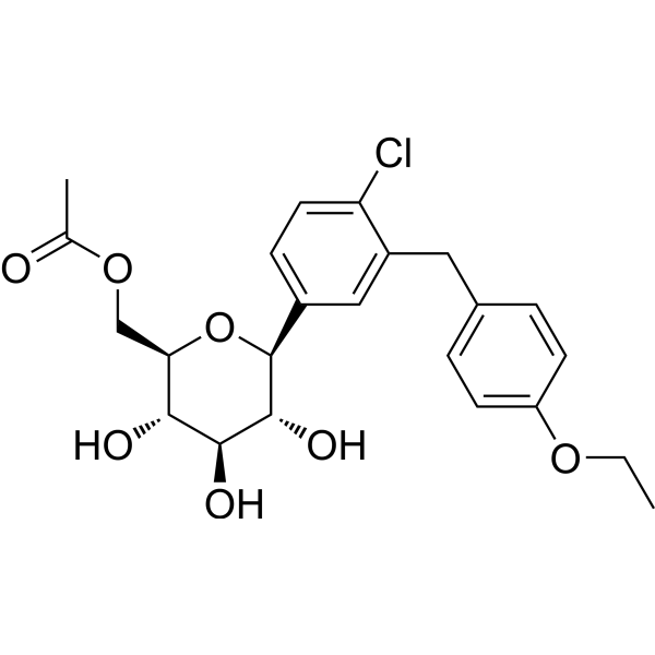 Dapagliflozin methyl acetate
 SGLT2-IN-4
InvivoChem的所有产品仅用于作科学研究,不面向患者销售
Copyright 2020 InvivoChem LLC | All Rights Reserved 粤ICP备20063088号-1


 COA
COA


 463611831
