| 规格 | 价格 | 库存 | 数量 |
|---|---|---|---|
| 5mg |
|
||
| 10mg |
|
||
| 25mg |
|
||
| 50mg |
|
||
| 100mg |
|
||
| 250mg |
|
||
| 500mg |
|
||
| Other Sizes |
|

| 靶点 |
Eg5 (IC50 = 200 nM)
The therapeutic target is mitotic Kinesin Eg5 (also known as KIF11)[1] The therapeutic target is mitotic Kinesin Eg5, which exerts effects by allosterically inhibiting the ATPase activity of the motor domain of Eg5[2] The therapeutic target is mitotic Kinesin Eg5[3] |
|---|---|
| 体外研究 (In Vitro) |
体外活性:Dimethylenastron 是一种喹唑啉硫酮类似物,是一种有效的、细胞渗透性的、特异性的、可逆的有丝分裂驱动蛋白 5 (Eg5) 抑制剂,Eg5 是微管刺激的 ATP 酶活性的有丝分裂马达。作为 Eg5 抑制剂 III,Dimethylenastron 具有潜在的抗癌活性,并且对驱动蛋白-1、-4、-7 和 -10 的 ATP 酶活性几乎没有影响。 Dimethylenastron 还抑制 HeLa 细胞和非洲爪蟾卵提取物中双极纺锤体的形成,并诱导细胞周期停滞 (~1 μM)。 Dimethylenastron(3 和 10 μM)处理 24 小时后浓度依赖性地抑制 PANC1 胰腺癌细胞中癌细胞的迁移能力,但在 24 小时至 72 小时不抑制癌细胞的增殖。 Dimethylenastron 还降低癌细胞的侵袭能力。激酶测定:Dimethylenastron 是一种有效的 Eg5 抑制剂,IC50 为 200 nM。 Dimethylenastron 对其他 5 个驱动蛋白亚家族(驱动蛋白 1/4/7/10 和一种未分组的 - 源自 4 种不同生物体)没有抑制作用。 Dimethylenastron (0.5, 1 μM) 会导致 HeLa 细胞中 G2/M 细胞的积累。细胞测定:Dimethylenastron 还可抑制 HeLa 细胞和非洲爪蟾卵提取物中双极纺锤体的形成,并诱导细胞周期停滞 (~1 μM)。 Dimethylenastron(3 和 10 μM)处理 24 小时后浓度依赖性地抑制 PANC1 胰腺癌细胞中癌细胞的迁移能力,但在 24 小时至 72 小时不抑制癌细胞的增殖。 Dimethylenastron 还降低癌细胞的侵袭能力。通过 Transwell 实验进行响应 Dimethylenastron 的细胞侵袭。 Transwell 过滤器的上表面涂有基质胶或纤连蛋白。将悬浮在 200 μL 无血清培养基中的细胞添加到室中,并将室放置在含有完全培养基的 24 孔板中。 37℃孵育24小时后,轻轻取出滤膜,用棉签除去滤膜上表面的基质胶。 Transwell滤膜下侧的细胞用4%多聚甲醛固定30分钟,用0.1%结晶紫染色10分钟,然后拍照。为了进行定量评估,入侵细胞的数量在每个过滤器的五个随机区域中进行计数。细胞侵袭的程度量化为药物治疗组中入侵细胞的数量除以对照组中入侵细胞的数量
胰腺癌细胞相关活性 1. 迁移抑制:Dimethylenastron以浓度依赖方式抑制人胰腺癌细胞系PANC1的迁移能力,3 μmol/L和10 μmol/L剂量处理24小时后,划痕愈合实验显示迁移细胞数量显著减少(5%血清或10%血清培养条件下均有此效应,P<0.05或P<0.01)[2] 2. 侵袭抑制:Dimethylenastron抑制PANC1细胞的侵袭能力,Transwell实验(基质胶或纤连蛋白包被滤膜)显示,3 μmol/L和10 μmol/L剂量处理24小时后,侵袭至滤膜下侧的细胞数量显著减少(P<0.05或P<0.01)[2] 3. 增殖抑制:Dimethylenastron处理PANC1细胞24小时对增殖无显著影响;处理72小时则以浓度依赖方式抑制细胞增殖(磺罗丹明B、MTT实验验证)[2] 4. 靶点抑制机制:分子模拟研究显示,Dimethylenastron可结合Eg5的马达结构域,通过降低ADP释放速率变构抑制Eg5的ATP酶活性;有无微管存在时,均能显著降低Eg5马达结构域的ADP释放速率(P<0.01)[2] |
| 体内研究 (In Vivo) |
Dimethylenastron (1.0 µmol) 会引起较轻微的疤痕,但与对照组相比,疱疹存活时间并未显着延长。 Dimethylenastron (1.0 µmol) 显示接受青光眼滤过手术治疗的兔子的眼内压比率显着降低,结膜下纤维化反应较轻微,但不明显减轻
青光眼滤过手术模型(新西兰白兔) 1. 给药方案与眼部反应:对37只雌性新西兰白兔行青光眼滤过手术,分别给予不同方案的Dimethylenastron结膜下注射:单剂量组(基线时单侧注射1.0 μmol或3.0 μmol)、多次给药组(基线+术后3天+7天注射1.0 μmol或3.0 μmol),对照组为仅手术、给予赋形剂(DMSO)[3] 2. 纤维化与滤过泡瘢痕:仅手术组、赋形剂组、3.0 μmol Dimethylenastron组出现滤过泡瘢痕;1.0 μmol组结膜下纤维化反应较轻,但滤过泡存活时长无显著延长(Kaplan-Meier log rank test,p=0.053)[3] 3. 眼压变化:所有组别眼压均与纤维化进程相关,术后14天内恢复至正常水平;1.0 μmol Dimethylenastron组眼压比值显著降低,但未充分改善手术结局[3] 4. 组织学验证:免疫组化(平滑肌肌动蛋白SMA、CD31)和组织学分析显示,1.0 μmol Dimethylenastron组结膜下纤维化反应较轻,但未达到显著改善手术效果的程度[3] |
| 酶活实验 |
Dimethylenastron 的 IC50 为 200 nM,使其成为强效 Eg5 抑制剂。其他五个驱动蛋白亚家族(驱动蛋白 1/4/7/10 和一个未分组的,源自四种不同的生物体)不受二甲那司酮抑制。在 HeLa 细胞中,二甲基黄酮 (0.5, 1 μM) 诱导细胞在 G2/M 期积累。
1. Eg5马达结构域ADP释放速率检测:采用MANT-ADP试剂检测Dimethylenastron对Eg5马达结构域ADP释放速率的影响,分别设置有/无微管的实验分组,定量测定不同浓度药物处理后ADP释放速率的变化,验证药物对Eg5 ATP酶活性的变构抑制作用;实验重复3次,结果以均值±标准差表示,采用统计学检验分析差异(P<0.01)[2] |
| 细胞实验 |
Transwell 测定测量细胞对二甲那琼反应的侵袭。将基质胶或纤连蛋白涂在 Transwell 过滤器的上表面。将悬浮在 200 μL 无血清培养基中的细胞添加到腔室中后,将腔室放入 24 孔板中,其中包含全部培养基。将滤膜从 37°C 的 24 小时孵育中轻轻取出后,用棉签除去滤膜上表面的基质胶。将 Transwell 滤膜下侧的细胞在 4% 多聚甲醛中固定 30 分钟并在 0.1% 结晶紫中染色 10 分钟后拍照。每个过滤器在五个随机区域中对入侵细胞的数量进行计数,以进行定量评估。药物治疗组的侵袭细胞数除以对照组的侵袭细胞数即可计算细胞侵袭的程度[2]。
胰腺癌细胞迁移实验(划痕愈合法) 1. 实验流程:将PANC1胰腺癌细胞接种于无血清培养基中培养至融合,用移液器枪头在细胞单层上制造划痕,分别加入含0、3、10 μmol/L Dimethylenastron且含10%或5%血清的培养基,培养24小时后拍摄划痕区域图像,计数迁移至划痕区域的细胞数量以量化迁移能力;实验重复3次,结果以均值±标准差表示,采用统计学检验分析与对照组的差异(P<0.05或P<0.01)[2] ### 胰腺癌细胞侵袭实验(Transwell法) 1. 实验流程:Transwell小室滤膜上侧分别包被基质胶或纤连蛋白,将经0、3、10 μmol/L Dimethylenastron处理的PANC1细胞接种至小室上腔,下腔加入含血清的培养基,培养24小时后固定并染色滤膜下侧的细胞,成像后计数侵袭细胞数量;实验重复3次,结果以均值±标准差表示,采用统计学检验分析与对照组的差异(P<0.05或P<0.01)[2] ### 胰腺癌细胞增殖实验 1. 磺罗丹明B(SRB)实验:将PANC1细胞接种于培养板,分别给予梯度浓度Dimethylenastron处理24小时或72小时,固定细胞后加入SRB染色液,洗脱未结合染料后用分光光度计检测吸光度值,计算细胞增殖百分比;实验重复3次,结果以均值±标准差表示[2] 2. MTT实验:操作流程与SRB实验一致,替换为MTT试剂检测细胞活力,计算增殖百分比;对比SRB与MTT实验数据,验证药物对增殖的抑制效应(P<0.05)[2] ### 胰腺癌细胞Eg5表达检测(免疫荧光法) 1. 实验流程:对PANC1、EPP85、BxPC3、CFPAC1、AsPAC1胰腺癌细胞系及正常胰腺上皮细胞进行免疫荧光染色,一抗靶向Eg5(红色荧光),DAPI染色细胞核(蓝色荧光),成像后定量分析Eg5的表达水平及核内分布;实验重复3次,结果显示胰腺癌细胞Eg5表达量为正常细胞的9-16倍(P<0.01)[2] |
| 动物实验 |
结膜缝合完成后,使用金属针(30 G)将以下药物之一注射到上直肌鼻侧缘的结膜下间隙:手术后,对照组的兔子不接受任何佐剂,其他组在基线时接受单侧结膜下注射二甲烯酮(1.0 µmol,3.0 µmol)或载体(DMSO,99.9%,10 mg/mL)。这意味着注射在手术后立即进行,另外两组分别在术后第3天和第7天再注射两次(1.0 µmol,3.0 µmol)[3]。
青光眼滤过手术动物实验(金吉拉兔) 1. 实验动物:37只雌性金吉拉兔(ChBBCH)[3] 2. 手术操作:进行模拟临床实践的青光眼滤过手术[3] 3. 给药方案: - 单剂量组:在手术基线时单侧结膜下注射1.0 μmol或3.0 μmol的二甲烯司琼,或仅注射赋形剂[3] - 多剂量组:在手术基线时、术后第3天和第7天单侧结膜下注射1.0 μmol或3.0 μmol的二甲烯司琼分别[3] 4. 观察期和终点:术后14天记录滤泡瘢痕和眼压变化等临床指标;于第14天处死动物,收集眼组织进行组织学和免疫组织化学分析(SMA、CD31染色)[3] |
| 毒性/毒理 (Toxicokinetics/TK) |
1. 眼毒性:结膜下注射3.0 μmol二甲烯司琼后,2例出现短暂性纤维蛋白反应;未观察到其他不良反应,如炎症或光学介质混浊;赋形剂(DMSO)耐受性良好[3]
|
| 参考文献 |
|
| 其他信息 |
1. 药物类别:二甲烯司琼是一种特异性抑制有丝分裂驱动蛋白Eg5的喹唑啉/硫酮类化合物[1][2][3]
2. 作用机制:二甲烯司琼通过变构作用与Eg5的运动结构域结合,降低ADP的释放速率,并抑制Eg5的ATPase活性;其抑制胰腺癌细胞迁移和侵袭的作用与抑制增殖无关[2] 3. 研发背景:Eg5在双极纺锤体组装中起着关键作用,其抑制剂在临床前研究中显示出显著的抗癌活性; Eg5在胰腺癌细胞中高表达(比正常细胞高9-16倍),提示其可作为胰腺癌的治疗靶点[2] 4. 应用潜力:二甲烯司琼可抑制胰腺癌细胞的迁移和侵袭,为胰腺癌化疗提供了一种新的机制;它可轻微减轻青光眼滤过手术中的结膜下纤维化,但现有浓度尚未充分改善手术效果[2][3] |
| 分子式 |
C16H18N2O2S
|
|
|---|---|---|
| 分子量 |
302.39
|
|
| 精确质量 |
302.108
|
|
| 元素分析 |
C, 63.55; H, 6.00; N, 9.26; O, 10.58; S, 10.60
|
|
| CAS号 |
863774-58-7
|
|
| 相关CAS号 |
|
|
| PubChem CID |
11609157
|
|
| 外观&性状 |
White to off-white solid powder
|
|
| 密度 |
1.3±0.1 g/cm3
|
|
| 沸点 |
475.6±55.0 °C at 760 mmHg
|
|
| 闪点 |
241.4±31.5 °C
|
|
| 蒸汽压 |
0.0±1.2 mmHg at 25°C
|
|
| 折射率 |
1.674
|
|
| LogP |
2.47
|
|
| tPSA |
93.45
|
|
| 氢键供体(HBD)数目 |
3
|
|
| 氢键受体(HBA)数目 |
3
|
|
| 可旋转键数目(RBC) |
1
|
|
| 重原子数目 |
21
|
|
| 分子复杂度/Complexity |
512
|
|
| 定义原子立体中心数目 |
0
|
|
| SMILES |
O=C1CC(C)(C)CC2NC(NC(C1=2)C1C=C(O)C=CC=1)=S
|
|
| InChi Key |
RUOOPLOUUAYNPY-UHFFFAOYSA-N
|
|
| InChi Code |
InChI=1S/C16H18N2O2S/c1-16(2)7-11-13(12(20)8-16)14(18-15(21)17-11)9-4-3-5-10(19)6-9/h3-6,14,19H,7-8H2,1-2H3,(H2,17,18,21)
|
|
| 化学名 |
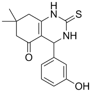
4-(3-hydroxyphenyl)-7,7-dimethyl-2-sulfanylidene-3,4,6,8-tetrahydro-1H-quinazolin-5-one
|
|
| 别名 |
Dimethylenastron
|
|
| HS Tariff Code |
2934.99.9001
|
|
| 存储方式 |
Powder -20°C 3 years 4°C 2 years In solvent -80°C 6 months -20°C 1 month |
|
| 运输条件 |
Room temperature (This product is stable at ambient temperature for a few days during ordinary shipping and time spent in Customs)
|
| 溶解度 (体外实验) |
|
|||
|---|---|---|---|---|
| 溶解度 (体内实验) |
配方 1 中的溶解度: ≥ 2.5 mg/mL (8.27 mM) (饱和度未知) in 10% DMSO + 40% PEG300 + 5% Tween80 + 45% Saline (这些助溶剂从左到右依次添加,逐一添加), 澄清溶液。
例如,若需制备1 mL的工作液,可将100 μL 25.0 mg/mL澄清DMSO储备液加入到400 μL PEG300中,混匀;然后向上述溶液中加入50 μL Tween-80,混匀;加入450 μL生理盐水定容至1 mL。 *生理盐水的制备:将 0.9 g 氯化钠溶解在 100 mL ddH₂O中,得到澄清溶液。 配方 2 中的溶解度: ≥ 2.5 mg/mL (8.27 mM) (饱和度未知) in 10% DMSO + 90% Corn Oil (这些助溶剂从左到右依次添加,逐一添加), 澄清溶液。 例如,若需制备1 mL的工作液,可将 100 μL 25.0 mg/mL 澄清 DMSO 储备液加入到 900 μL 玉米油中并混合均匀。 请根据您的实验动物和给药方式选择适当的溶解配方/方案: 1、请先配制澄清的储备液(如:用DMSO配置50 或 100 mg/mL母液(储备液)); 2、取适量母液,按从左到右的顺序依次添加助溶剂,澄清后再加入下一助溶剂。以 下列配方为例说明 (注意此配方只用于说明,并不一定代表此产品 的实际溶解配方): 10% DMSO → 40% PEG300 → 5% Tween-80 → 45% ddH2O (或 saline); 假设最终工作液的体积为 1 mL, 浓度为5 mg/mL: 取 100 μL 50 mg/mL 的澄清 DMSO 储备液加到 400 μL PEG300 中,混合均匀/澄清;向上述体系中加入50 μL Tween-80,混合均匀/澄清;然后继续加入450 μL ddH2O (或 saline)定容至 1 mL; 3、溶剂前显示的百分比是指该溶剂在最终溶液/工作液中的体积所占比例; 4、 如产品在配制过程中出现沉淀/析出,可通过加热(≤50℃)或超声的方式助溶; 5、为保证最佳实验结果,工作液请现配现用! 6、如不确定怎么将母液配置成体内动物实验的工作液,请查看说明书或联系我们; 7、 以上所有助溶剂都可在 Invivochem.cn网站购买。 |
| 制备储备液 | 1 mg | 5 mg | 10 mg | |
| 1 mM | 3.3070 mL | 16.5349 mL | 33.0699 mL | |
| 5 mM | 0.6614 mL | 3.3070 mL | 6.6140 mL | |
| 10 mM | 0.3307 mL | 1.6535 mL | 3.3070 mL |
1、根据实验需要选择合适的溶剂配制储备液 (母液):对于大多数产品,InvivoChem推荐用DMSO配置母液 (比如:5、10、20mM或者10、20、50 mg/mL浓度),个别水溶性高的产品可直接溶于水。产品在DMSO 、水或其他溶剂中的具体溶解度详见上”溶解度 (体外)”部分;
2、如果您找不到您想要的溶解度信息,或者很难将产品溶解在溶液中,请联系我们;
3、建议使用下列计算器进行相关计算(摩尔浓度计算器、稀释计算器、分子量计算器、重组计算器等);
4、母液配好之后,将其分装到常规用量,并储存在-20°C或-80°C,尽量减少反复冻融循环。
计算结果:
工作液浓度: mg/mL;
DMSO母液配制方法: mg 药物溶于 μL DMSO溶液(母液浓度 mg/mL)。如该浓度超过该批次药物DMSO溶解度,请首先与我们联系。
体内配方配制方法:取 μL DMSO母液,加入 μL PEG300,混匀澄清后加入μL Tween 80,混匀澄清后加入 μL ddH2O,混匀澄清。
(1) 请确保溶液澄清之后,再加入下一种溶剂 (助溶剂) 。可利用涡旋、超声或水浴加热等方法助溶;
(2) 一定要按顺序加入溶剂 (助溶剂) 。
|
|---|
|
|
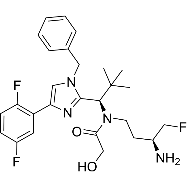 NVP-BQS481
 Mitotic kinesin-IN-3 hydrochloride
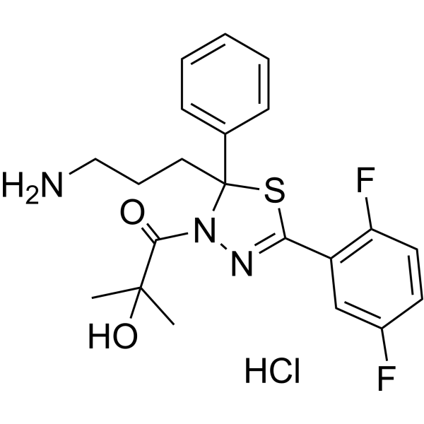
 Mitotic kinesin-IN-1 hydrochloride
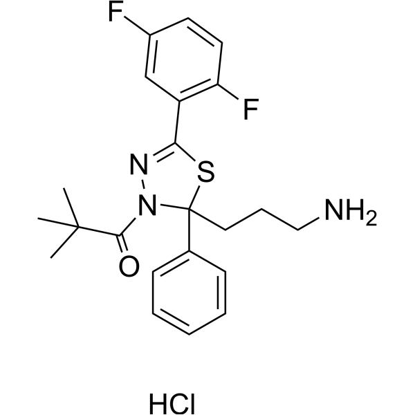
 Mitotic kinesin-IN-2 hydrochloride
InvivoChem的所有产品仅用于作科学研究,不面向患者销售
Copyright 2020 InvivoChem LLC | All Rights Reserved 粤ICP备20063088号-1


 COA
COA


 463611831
