| 规格 | 价格 | 库存 | 数量 |
|---|---|---|---|
| 5mg |
|
||
| 10mg |
|
||
| 25mg |
|
||
| 50mg |
|
||
| 100mg |
|
||
| 250mg |
|
||
| 500mg |
|
||
| Other Sizes |
|

| 靶点 |
dynamin (IC50 = 15 μM); HSV-1/2
Dynasore specifically targets dynamin GTPase (isoforms dynamin 1, 2, 3) with IC50 values of 15 μM (dynamin 1), 18 μM (dynamin 2), and 20 μM (dynamin 3) for inhibiting GTPase activity [1] Dynasore binds to the GTP-binding pocket of dynamin, blocking GTP hydrolysis without inhibiting other GTPases (e.g., Ras, Rho) at concentrations up to 100 μM [1] |
|---|---|
| 体外研究 (In Vitro) |
Dynasore 抑制 dynamin1、dynamin2 和线粒体 dynamin Drp1 GTPase 活性,但不抑制其他小 GTPase。添加后,dynasore 会迅速阻止包被囊泡的形成,从而充当已知的动力依赖性内吞途径的强大抑制剂。这种效果在几秒钟内就可以看到。 G 形半成形凹坑和 O 形完全成形凹坑(在夹断过程中截留)是 dynasore 加工过程中积累的两种类型的涂层凹坑中间体 [1]。人类胎儿神经元、星形胶质细胞、初级生殖道细胞以及上皮细胞和神经细胞都容易受到HSV-1和HSV-2感染。 Dynasore 可以预防这些感染。病毒进入后八小时给予 dynasore 可以防止新生成的病毒蛋白离开细胞核,并增加到达核孔的病毒衣壳数量 [2]。 Dynasore 抑制缺血或再灌注引起的左心室舒张末压升高。此外,Dynamicore 可减少再灌注时的梗塞面积和心肌肌钙蛋白 I 流出。 Dynasore 增加了氧化应激下生长的成年小鼠心肌细胞的活力和存活率 [3]。
20 μM Dynasore 抑制 HeLa 和 COS-7 细胞的网格蛋白介导的内吞作用,使转铁蛋白内化减少 85% [1] - 30 μM Dynasore 干扰 Vero 细胞中单纯疱疹病毒(HSV-1)包膜蛋白(gB、gD)的转运,使病毒颗粒释放减少 70% [2] - 10-25 μM Dynasore 在氧化应激下保护新生大鼠心肌细胞的线粒体形态和功能,减少 60% 的线粒体活性氧(ROS)产生,维持线粒体膜电位(ΔΨm)[3] - 15 μM Dynasore 抑制氧糖剥夺(OGD)后大鼠脊髓来源神经元的凋亡,使半胱天冬酶 -3 激活减少 55%,TUNEL 阳性细胞减少 48% [4] - 20 μM Dynasore 抑制原代大鼠星形胶质细胞增殖,使 BrdU 掺入率降低 62% [4] - 25 μM Dynasore 阻断 HT1080 纤维肉瘤细胞的迁移小体释放,使迁移小体数量减少 75%,且不影响细胞迁移速度 [5] - Dynasore(10-30 μM)对正常细胞(HeLa、心肌细胞、神经元)毒性极低,细胞活力 > 80%,但可抑制 HSV-1 感染的 Vero 细胞活力(IC50 = 45 μM)[1][2][3][4] - Western blot 分析显示,Dynasore(15-25 μM)使 OGD 处理的神经元中 Bcl-2/Bax 比值升高 2.3 倍,使增殖星形胶质细胞中磷酸化 ERK1/2 减少 40% [4] |
| 体内研究 (In Vivo) |
大鼠脊髓损伤 (SCI) 后,dynasore 在第 3、7 和 10 天显着减少了运动功能障碍。通过防止脊髓损伤后大鼠神经元中导致线粒体凋亡和星形胶质细胞增殖的途径激活,Dynasore 极大地改善了运动功能 [4]。
在 Langendorff 灌流小鼠心脏模型中,Dynasore(10 μM,灌流 30 分钟)改善心脏舒张功能,使左心室舒张末期容积增加 25%,舒张末期压力降低 30% [3] - 在大鼠脊髓损伤(SCI)模型中,Dynasore 腹腔给药(5 mg/kg,每日一次,连续 7 天)改善运动功能恢复,28 天后 BBB(Basso-Beattie-Bresnahan)评分从 4.2 提升至 8.6 [4] - Dynasore 处理的 SCI 大鼠损伤部位神经元凋亡减少(TUNEL 阳性细胞减少 35%),星形胶质细胞瘢痕形成减少(GFAP 阳性区域减少 40%)[4] - Dynasore 灌流的小鼠心脏线粒体形态得到保护(碎片化线粒体减少 65%),ATP 生成增加 32% [3] |
| 酶活实验 |
ATP测量[3]
发光分析仪用于定量心肌细胞和Hela细胞ATP含量。简而言之,在Dynasore治疗和H2O2暴露后,心肌细胞被裂解,细胞裂解物中的ATP含量被测量。同时,在遵循相同实验方案的另一组孔中,使用TBE测定法对存活的心肌细胞进行计数。然后计算每种治疗条件下每个活心肌细胞的细胞ATP。类似的程序也应用于用对照或Dynasore处理的培养的非应激Hela细胞。 动力蛋白 GTP 酶活性测定:重组动力蛋白(亚型 1/2/3)与 [γ-32P]GTP 底物共孵育。加入系列浓度的 Dynasore(5 μM 至 100 μM),37°C 孵育 60 分钟。通过检测释放的 32Pi 定量 GTP 水解,从抑制作用的剂量 - 反应曲线计算 IC50 值 [1] - 动力蛋白 -GTP 结合测定:荧光标记的 GTP 与重组 dynamin 2 共孵育。加入系列浓度的 Dynasore(10 μM 至 80 μM),25°C 下测量荧光偏振。从竞争曲线推导结合亲和力(Kd),显示 Dynasore 置换 GTP 的 IC50 为 16 μM [1] |
| 细胞实验 |
成年小鼠心肌细胞的分离与培养[3]
使用先前描述的方法用胶原酶II(2mg/ml)分离后,从雄性成年C6/黑小鼠(8-12周)中分离出小鼠心室肌细胞。分离后,将心肌细胞以约1500/mm2的密度铺在层粘连蛋白预涂的35mm2培养皿上,并在37°C下保持在5%CO2的加湿气氛中。接种1小时后,用新鲜培养基(补充或耗尽血清)补充心肌细胞,并进行2小时的药物治疗(Dynasore或赋形剂),然后进行氧化应激(30µM H2O2 35分钟)。对于ATP补充实验,细胞在暴露于H2O2之前用3 mM ATP处理30分钟。 旋转圆盘共聚焦显微镜活细胞线粒体成像[3] HeLa细胞在添加了10%FBS和100µg/ml Normocin的DMEM中维持。细胞在37°C、5%CO2的加湿气氛中保持。以7×10⁴个细胞/cm2的密度接种细胞,并使其粘附过夜。然后用Organille Lights™Mito RFPBacMam 1.0转导细胞。转导后24小时,细胞用对照或1µM Dynasore预处理1小时,然后暴露于正常条件或200µM H2O2 15分钟。在暴露于H2O2之前和之后,使用尼康Ti倒置显微镜、横河CSU-X1旋转盘共聚焦单元和568nm DPSS激光源以及高分辨率Cool SNAP HQ2相机对细胞进行成像。图像以每帧400毫秒的曝光量采集,并使用浅浮雕滤镜自动处理以突出边缘。 网格蛋白介导的内吞实验:HeLa 细胞与 Alexa Fluor 488 标记的转铁蛋白及 Dynasore(10-40 μM)共孵育 30 分钟。流式细胞术定量内化的转铁蛋白,相对于溶媒对照组计算抑制率 [1] - HSV-1 转运实验:Vero 细胞感染 HSV-1(MOI = 1)后,用 Dynasore(10-50 μM)处理 12 小时。免疫荧光检测病毒包膜蛋白(gB、gD),共聚焦显微镜评分细胞内转运缺陷 [2] - 线粒体功能检测:新生大鼠心肌细胞暴露于 H2O2(200 μM)及 Dynasore(10-25 μM)24 小时。DCFH-DA 染色检测线粒体 ROS,JC-1 染色检测 ΔΨm [3] - 神经元凋亡实验:大鼠脊髓神经元经 OGD(2 小时)处理后,用 Dynasore(10-20 μM)孵育 24 小时。TUNEL 染色和半胱天冬酶 -3 活性测定评估凋亡 [4] - 星形胶质细胞增殖实验:原代大鼠星形胶质细胞用 Dynasore(10-30 μM)处理 48 小时。BrdU 掺入法和细胞计数检测增殖 [4] - 迁移小体形成实验:HT1080 细胞接种于纤连蛋白包被的培养皿,用 Dynasore(15-30 μM)处理 16 小时。GFP 标记的四跨膜蛋白 4(TSPAN4)可视化迁移小体,荧光显微镜计数 [5] |
| 动物实验 |
在Dynasore组中,大鼠在脊髓损伤后立即通过腹腔注射给予1、10或30 mg/kg剂量的dynasore,而假手术组和脊髓损伤组的大鼠则通过腹腔注射给予DMSO(与dynasore组相同体积)。
大鼠Langendorff灌注心脏[3] 雄性C6/Black小鼠(8~12周龄)在麻醉箱中用异氟烷(流量3%)和100% O2麻醉,并用肝素(50 IU,腹腔注射)抗凝。颈椎脱位后,迅速取出心脏,将其安装在朗根多夫灌注装置上,并以2.6 ml/min的恒定流速进行逆行灌注。灌注液为含氧的克氏-亨氏缓冲液(mmol/L):NaCl 118、NaHCO3 24、CaCl2·2H2O 2.5、KCl 4.7、KH2PO4 1.2、MgSO4·7H2O 1.2、葡萄糖 11、EDTA 0.5,pH值调节至7.4。该装置采用水套式温控系统,以维持心脏核心温度在37°C。灌注液流经由医用级Silastic™管材制成的膜状“肺”,该“肺”持续通入95% O2-5% CO2混合气体。在右心房和左心室心尖处放置精细的铂电极,用于记录整个实验过程中的心电图和心率。将 Millar MIKRO-TIP 导管压力传感器从左心房插入左心室,以测量左心室压力。使用 PowerLab 系统连续监测并记录左心室舒张末期压力 (LVEDP)、左心室收缩末期压力 (LVESP) 和心率。左心室收缩末期压力 (LVDP) 通过 LVESP 减去 LVEDP 计算得出。使用刺激器提供的刺激,通过连接在右心房的双极电极以 360 bpm 的频率对心脏进行起搏。 经过最初 15 分钟的稳定期后,如果心脏出现以下一项或多项排除标准,则将其排除在后续研究之外:LVEDP 高于 20 mmHg;LVDP 低于 50 mmHg;固有心率低于 280 bpm 或不规则;或主动脉瓣反流。灌注液的体积减少至 200 ml 并使其循环。随后将心脏随机分为以下两个处理组:Dynasore 组(n = 8,在循环灌注液中逐步添加,在 120 分钟内达到 1 µM 的最终浓度)或 DMSO 对照组(n = 8,以与 Dynasore 类似的方式添加)。之后,心脏经历 30 分钟的全局缺血,随后进行 1 小时的再灌注。除缺血期间外,在稳定后开始起搏,并在再灌注 2 分钟后重新开始起搏。 Langendorff 灌注小鼠心脏模型:将雄性 C57BL/6 小鼠(8-10 周龄)安乐死,取出心脏,并用 Krebs-Henseleit 缓冲液灌注。稳定(20 分钟)后,将 Dynasore(10 μM)添加到灌注液中,持续 30 分钟。采用压力-容积导管测量心脏功能(左心室压力、容积)[3] - 大鼠脊髓损伤(SCI)模型:雌性Sprague-Dawley大鼠(200-250 g)在T9-T10节段进行脊髓挫伤。损伤后立即将大鼠随机分组(每组n=10),并分别进行以下治疗:(1)腹腔注射溶剂(DMSO + 生理盐水);(2)腹腔注射Dynasore(5 mg/kg),每日一次,连续7天。每周采用BBB评分评估运动功能,持续28天,并收集脊髓组织进行组织学检查[4] |
| 毒性/毒理 (Toxicokinetics/TK) |
Dynasore在正常细胞中显示出较低的体外细胞毒性:HeLa细胞(IC50 > 100 μM)、新生儿心肌细胞(IC50 > 80 μM)和大鼠脊髓神经元(IC50 > 75 μM)[1][3][4]
- 在脊髓损伤大鼠的重复给药毒性研究中(5 mg/kg,腹腔注射,连续7天),Dynasore未引起明显的体重减轻,组织学检查显示肝脏、肾脏或心脏均无异常[4] - Dynasore(30 μM)在体外不抑制人细胞色素P450酶(CYP1A2、CYP2C9、CYP3A4)[1] |
| 参考文献 | |
| 其他信息 |
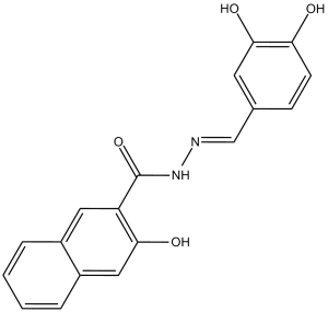
Dynasore 是一种碳酰肼,由 3,4-二羟基苯甲醛腙的腙部分与 3-羟基-2-萘甲酸的羧基缩合而成。它是一种细胞渗透性、可逆的非竞争性 GTP 酶抑制剂,可抑制动力蛋白 1 和 2 以及 Drp1(线粒体)的活性,而对另外两种小 GTP 酶 MxA 和 Cdc42 没有显著影响。它是一种 EC 3.6.5.5(动力蛋白 GTP 酶)抑制剂。它属于儿茶酚类、萘酚类、酰肼类和腙类化合物。其功能与 3-羟基-2-萘甲酸酯相关。动力蛋白是网格蛋白依赖性包被囊泡形成所必需的。在从完全形成的凹陷到收缩囊泡的转变过程中,膜出芽的后期阶段需要动力蛋白。动力蛋白在囊泡形成的早期阶段也可能发挥其他作用。我们筛选了约16,000种小分子,并鉴定出一种名为dynasore的化合物1,该化合物在体外可干扰动力蛋白1、动力蛋白2和线粒体动力蛋白Drp1的GTP酶活性,但不会干扰其他小GTP酶的活性。dynasore是一种强效的内吞途径抑制剂,已知该途径依赖于动力蛋白,其作用机制是在加入dynasore后数秒内迅速阻断包被囊泡的形成。在dynasore处理过程中,会积累两种类型的包被凹陷中间体:U形的半成形凹陷和O形的完全成形凹陷(在收缩过程中被捕获)。因此,动力蛋白在网格蛋白包被形成过程中的两个步骤中发挥作用。 GTP水解可能在这两个步骤中都是必需的。[1]
Dynasore是一种小分子动力蛋白GTP酶活性抑制剂,可抑制多种病毒(包括单纯疱疹病毒(HSV))的入侵,但其对病毒生命周期其他步骤的影响尚未明确。本研究旨在验证动力蛋白是否参与病毒蛋白转运,从而对HSV感染具有多效性抑制作用。Dynasore抑制了HSV-1和HSV-2对人上皮细胞和神经元细胞(包括原代生殖道细胞、人胎儿神经元和星形胶质细胞)的感染。当用表达显性负性动力蛋白的质粒转染细胞时,也获得了类似的结果。动力学研究表明,若在病毒进入细胞时加入dynasore,则可减少到达核孔的病毒衣壳数量;若在病毒进入细胞后8小时加入dynasore,则可阻断新合成的病毒蛋白从细胞核向细胞质的转运。邻近连接分析表明,dynasore处理可阻止VP5和动力蛋白的共定位。这导致从蔗糖梯度中分离出的病毒衣壳数量减少。与对照组相比,dynasore处理组细胞中通过电镜观察到的衣壳数量也更少。此外,释放到培养上清液中的感染性子代病毒数量以及细胞间传播也均有所减少。综上所述,这些发现提示靶向动力蛋白-HSV相互作用可能为HSV的治疗和预防提供一种新的策略。重要性:单纯疱疹病毒 (HSV) 感染仍然是一个全球性的健康问题,与显著的发病率相关,尤其是在新生儿和免疫功能低下者中,这凸显了开发新的治疗和预防方法的必要性。目前的研究表明,动力蛋白在病毒生命周期的多个步骤中发挥作用,并为抗病毒治疗提供了一个新的靶点。动力蛋白小分子抑制剂 Dynasore 对 HSV-1 和 HSV-2 感染具有多效性,并能抑制病毒进入细胞、病毒蛋白转运和衣壳形成。[2] 背景:舒张功能障碍引起的心力衰竭在美国造成了巨大的经济、发病率和死亡率负担。目前用于改善舒张功能障碍的治疗药物有限。最近发现,动力蛋白相关蛋白 1 (Drp1) 介导缺血/再灌注 (I/R) 损伤期间的线粒体分裂,而抑制 Drp1 可减少心肌梗死面积。我们假设,小分子非竞争性动力蛋白GTP酶抑制剂Dynasore可能对缺血/再灌注(I/R)损伤期间的心脏生理功能产生有益影响。方法和结果:在Langendorff灌注的小鼠心脏中,经受I/R(30分钟全局缺血,随后1小时再灌注)后,1 µM Dynasore预处理可预防I/R引起的左心室舒张末期压力(LVEDP)升高,表明其具有显著且特异性的舒张功能增强作用。Dynasore还降低了再灌注期间心肌肌钙蛋白I的释放,并缩小了梗死面积。在经受氧化应激的培养成年小鼠心肌细胞中,Dynasore可提高心肌细胞的存活率和活力(通过台盼蓝排除法测定),并降低细胞内三磷酸腺苷(ATP)的消耗。此外,在培养细胞中,Dynasore预处理可保护线粒体免受氧化应激诱导的断裂。结论:Dynasore通过维持受损细胞中线粒体形态和细胞内ATP水平的机制,保护心肌舒张功能并减少细胞损伤。Dynasore等药物对线粒体的保护作用可通过积极影响舒张功能障碍的能量代谢而产生临床获益。[3] 脊髓损伤(SCI)是一种常见且严重的神经系统损伤,目前缺乏有效的治疗方法。我们之前的实验结果表明,动力蛋白相关蛋白1(Drp1)介导SCI期间的线粒体分裂,抑制Drp1在SCI大鼠模型中具有显著的保护作用。Dynasore可抑制质膜(动力蛋白1、2)和线粒体膜(Drp1)上的GTP酶活性。本研究旨在探讨Dynasore对SCI大鼠模型的有益作用及其潜在机制。将Sprague-Dawley大鼠随机分为假手术组、脊髓损伤(SCI)组以及1 mg、10 mg和30 mg Dynasore组。采用Allen氏模型建立大鼠SCI模型。Dynasore立即通过腹腔注射给药。运动功能测试结果表明,Dynasore在SCI后3、7和10天显著改善了大鼠的运动功能障碍(P < 0.05)。Western blot结果显示,Dynasore在SCI后3天显著降低了Drp1、动力蛋白1和动力蛋白2的表达,并降低了Bax、细胞色素C和活性Caspase-3的表达,但增加了Bcl-2的表达(P < 0.05)。值得注意的是,脊髓损伤(SCI)后3天,dynasore可抑制增殖细胞核抗原(PCNA)和胶质纤维酸性蛋白(GAFP)的表达上调(P < 0.05)。免疫荧光双标记结果显示,与SCI组相比,dynasore组的凋亡神经元和增殖性星形胶质细胞数量均显著减少(P < 0.05)。此外,尼氏染色组织学评估表明,与SCI组相比,dynasore组的存活神经元数量显著增加(P < 0.05)。这种神经保护作用呈剂量依赖性(P < 0.05)。据我们所知,这是首项研究表明,Dynasore 能显著增强脊髓损伤(SCI)大鼠的运动功能,其机制可能是通过抑制神经元线粒体凋亡通路和星形胶质细胞增殖来实现的。[4] Dynasore 是一种细胞可渗透的、可逆的动力蛋白 GTP 酶抑制剂,动力蛋白 GTP 酶是膜裂变事件(例如,网格蛋白介导的内吞作用、线粒体裂变、病毒运输)的关键调节因子。[1] Dynasore 的作用机制是通过抑制 GTP 水解来阻断动力蛋白依赖的膜重塑,从而破坏需要动力蛋白介导裂变的过程。[1][5] Dynasore 具有多种生物活性:抗病毒(HSV-1)、心脏保护(线粒体保护)和神经保护(SCI 恢复)。 [2][3][4] Dynasore是一种用于研究动力蛋白依赖性细胞过程的宝贵工具化合物,在病毒感染、心血管疾病和神经损伤方面具有潜在的治疗应用价值[1][2][3][4][5] |
| 分子式 |
C18H14N2O4
|
|
|---|---|---|
| 分子量 |
322.31
|
|
| 精确质量 |
322.095
|
|
| 元素分析 |
C, 67.08; H, 4.38; N, 8.69; O, 19.85
|
|
| CAS号 |
304448-55-3
|
|
| 相关CAS号 |
|
|
| PubChem CID |
135533054
|
|
| 外观&性状 |
Light brown to brown solid powder
|
|
| 密度 |
1.36±0.1 g/cm3
|
|
| 折射率 |
1.665
|
|
| LogP |
4.06
|
|
| tPSA |
102.15
|
|
| 氢键供体(HBD)数目 |
4
|
|
| 氢键受体(HBA)数目 |
5
|
|
| 可旋转键数目(RBC) |
3
|
|
| 重原子数目 |
24
|
|
| 分子复杂度/Complexity |
470
|
|
| 定义原子立体中心数目 |
0
|
|
| SMILES |
C1=CC=C2C=C(C(=CC2=C1)C(=O)N/N=C/C3=CC(=C(C=C3)O)O)O
|
|
| InChi Key |
SYNDQCRDGGCQRZ-VXLYETTFSA-N
|
|
| InChi Code |
InChI=1S/C18H14N2O4/c21-15-6-5-11(7-17(15)23)10-19-20-18(24)14-8-12-3-1-2-4-13(12)9-16(14)22/h1-10,21-23H,(H,20,24)/b19-10+
|
|
| 化学名 |
(E)-N-(3,4-dihydroxybenzylidene)-3-hydroxy-2-naphthohydrazide
|
|
| 别名 |
|
|
| HS Tariff Code |
2934.99.9001
|
|
| 存储方式 |
Powder -20°C 3 years 4°C 2 years In solvent -80°C 6 months -20°C 1 month |
|
| 运输条件 |
Room temperature (This product is stable at ambient temperature for a few days during ordinary shipping and time spent in Customs)
|
| 溶解度 (体外实验) |
|
|||
|---|---|---|---|---|
| 溶解度 (体内实验) |
配方 1 中的溶解度: ≥ 2.5 mg/mL (7.76 mM) (饱和度未知) in 10% DMSO + 40% PEG300 + 5% Tween80 + 45% Saline (这些助溶剂从左到右依次添加,逐一添加), 澄清溶液。
例如,若需制备1 mL的工作液,可将100 μL 25.0 mg/mL澄清DMSO储备液加入到400 μL PEG300中,混匀;然后向上述溶液中加入50 μL Tween-80,混匀;加入450 μL生理盐水定容至1 mL。 *生理盐水的制备:将 0.9 g 氯化钠溶解在 100 mL ddH₂O中,得到澄清溶液。 配方 2 中的溶解度: ≥ 2.5 mg/mL (7.76 mM) (饱和度未知) in 10% DMSO + 90% (20% SBE-β-CD in Saline) (这些助溶剂从左到右依次添加,逐一添加), 澄清溶液。 例如,若需制备1 mL的工作液,可将 100 μL 25.0 mg/mL澄清DMSO储备液加入900 μL 20% SBE-β-CD生理盐水溶液中,混匀。 *20% SBE-β-CD 生理盐水溶液的制备(4°C,1 周):将 2 g SBE-β-CD 溶解于 10 mL 生理盐水中,得到澄清溶液。 请根据您的实验动物和给药方式选择适当的溶解配方/方案: 1、请先配制澄清的储备液(如:用DMSO配置50 或 100 mg/mL母液(储备液)); 2、取适量母液,按从左到右的顺序依次添加助溶剂,澄清后再加入下一助溶剂。以 下列配方为例说明 (注意此配方只用于说明,并不一定代表此产品 的实际溶解配方): 10% DMSO → 40% PEG300 → 5% Tween-80 → 45% ddH2O (或 saline); 假设最终工作液的体积为 1 mL, 浓度为5 mg/mL: 取 100 μL 50 mg/mL 的澄清 DMSO 储备液加到 400 μL PEG300 中,混合均匀/澄清;向上述体系中加入50 μL Tween-80,混合均匀/澄清;然后继续加入450 μL ddH2O (或 saline)定容至 1 mL; 3、溶剂前显示的百分比是指该溶剂在最终溶液/工作液中的体积所占比例; 4、 如产品在配制过程中出现沉淀/析出,可通过加热(≤50℃)或超声的方式助溶; 5、为保证最佳实验结果,工作液请现配现用! 6、如不确定怎么将母液配置成体内动物实验的工作液,请查看说明书或联系我们; 7、 以上所有助溶剂都可在 Invivochem.cn网站购买。 |
| 制备储备液 | 1 mg | 5 mg | 10 mg | |
| 1 mM | 3.1026 mL | 15.5130 mL | 31.0260 mL | |
| 5 mM | 0.6205 mL | 3.1026 mL | 6.2052 mL | |
| 10 mM | 0.3103 mL | 1.5513 mL | 3.1026 mL |
1、根据实验需要选择合适的溶剂配制储备液 (母液):对于大多数产品,InvivoChem推荐用DMSO配置母液 (比如:5、10、20mM或者10、20、50 mg/mL浓度),个别水溶性高的产品可直接溶于水。产品在DMSO 、水或其他溶剂中的具体溶解度详见上”溶解度 (体外)”部分;
2、如果您找不到您想要的溶解度信息,或者很难将产品溶解在溶液中,请联系我们;
3、建议使用下列计算器进行相关计算(摩尔浓度计算器、稀释计算器、分子量计算器、重组计算器等);
4、母液配好之后,将其分装到常规用量,并储存在-20°C或-80°C,尽量减少反复冻融循环。
计算结果:
工作液浓度: mg/mL;
DMSO母液配制方法: mg 药物溶于 μL DMSO溶液(母液浓度 mg/mL)。如该浓度超过该批次药物DMSO溶解度,请首先与我们联系。
体内配方配制方法:取 μL DMSO母液,加入 μL PEG300,混匀澄清后加入μL Tween 80,混匀澄清后加入 μL ddH2O,混匀澄清。
(1) 请确保溶液澄清之后,再加入下一种溶剂 (助溶剂) 。可利用涡旋、超声或水浴加热等方法助溶;
(2) 一定要按顺序加入溶剂 (助溶剂) 。
|
|---|
|
|
InvivoChem的所有产品仅用于作科学研究,不面向患者销售
Copyright 2020 InvivoChem LLC | All Rights Reserved 粤ICP备20063088号-1


 COA
COA


 463611831
