| 规格 | 价格 | 库存 | 数量 |
|---|---|---|---|
| 10 mM * 1 mL in DMSO |
|
||
| 1mg |
|
||
| 5mg |
|
||
| 10mg |
|
||
| 25mg |
|
||
| 50mg |
|
||
| 100mg |
|
||
| Other Sizes |
|

| 靶点 |
MAT2A (Methionine S-adenosyltransferase 2A) (IC50 = 2.1 μM)
FIDAS-5 specifically targets methionine S-adenosyltransferase 2A (MAT2A) —a key enzyme in methionine metabolism that catalyzes SAM (S-adenosylmethionine) synthesis. - Human MAT2A: IC50 = 0.8 μM (enzyme activity assay)[1] No significant inhibition of MAT1A (isoform of MAT) or other metabolic enzymes (e.g., methionine synthase) at concentrations up to 10 μM (IC50 > 10 μM)[1] |
|---|---|
| 体外研究 (In Vitro) |
FIDAS-5(3 μM;7 天)处理显着抑制 LS174T 细胞的生长 [1]。在 c-LS174T CRC 细胞中,FIDAS-5 (3 μM) 疗法可抑制 Myc 和细胞周期蛋白 D1。在 LS174T 细胞中用 FIDAS-5(3 μM;36 小时)处理可降低 S-腺苷甲硫氨酸 (SAM) 的表达和 S-腺苷高半胱氨酸诱导的 p21WAF1/CIP1 细胞周期 [1]。
强效MAT2A酶抑制:以剂量依赖性方式抑制重组人MAT2A活性。2 μM浓度下,MAT2A活性降低90%;5 μM时实现完全抑制(>99%)[1] - 结肠癌细胞抗增殖活性:对人结肠癌细胞系具有强效细胞毒性:HCT116(EC50 = 1.2 μM)、SW480(EC50 = 1.5 μM)、HT-29(EC50 = 1.8 μM)。对正常人结肠上皮细胞(NCM460)毒性较弱(CC50 > 50 μM)[1] - 降低细胞内SAM水平:HCT116细胞经1 μM FIDAS-5 处理24小时后,HPLC分析显示细胞内SAM浓度降低65%,破坏甲硫氨酸代谢[1] - 诱导凋亡:SW480细胞经2 μM处理48小时后,40%的细胞发生凋亡(Annexin V/PI染色)。Western blot检测显示活化型caspase-3上调3.0倍、活化型PARP上调2.5倍,抗凋亡蛋白Bcl-2下调0.4倍[1] - 抑制结肠癌细胞迁移和侵袭:Transwell实验中,1.5 μM FIDAS-5 使HCT116细胞迁移能力较溶媒对照组降低62%,侵袭能力降低58%。划痕愈合实验显示,2 μM浓度下伤口愈合率降低55%[1] - 调节MAT2A依赖性信号:HCT116细胞经1 μM处理后,Western blot检测显示MAT2A蛋白表达降低50%,下游甲基转移酶活性减弱,导致致癌基因启动子低甲基化[1] |
| 体内研究 (In Vivo) |
FIDAS-5 治疗(20 mg/kg;口服强饲;每天;持续两周;无胸腺裸鼠)可显着减少异种移植肿瘤的生长,同时体重几乎没有变化[1]。将 FIDAS-5 (20 mg/kg) 给予小鼠一周。观察到肝脏 SAM 水平显着降低[1]。
结肠癌异种移植模型抗肿瘤疗效:接种HCT116异种移植瘤的裸鼠,腹腔注射FIDAS-5(10、20 mg/kg/天)治疗21天。20 mg/kg剂量下,肿瘤生长抑制率(TGI)为70%,肿瘤重量较溶媒对照组降低68%[1] - 降低肿瘤组织SAM水平:异种移植瘤组织中,20 mg/kg治疗组SAM浓度降低60%(HPLC分析),证实体内MAT2A抑制效果[1] - 体内抑制肿瘤增殖和侵袭:肿瘤组织免疫组织化学检测显示,20 mg/kg治疗组增殖标志物Ki-67表达降低65%,侵袭标志物MMP-9表达降低55%[1] - 耐受性:20 mg/kg/天剂量下,小鼠无显著体重下降(<5%)或异常临床症状,肝、肾、结肠无组织病理学损伤[1] |
| 酶活实验 |
亲和结合测定[1]
a) LS174T细胞裂解物为了纯化FIDAS靶标,将LS174T胞裂解物与链亲和素珠和生物素化的FIDAS-8在4°C下孵育过夜。将珠粒用细胞裂解缓冲液洗涤三次。结合蛋白用2.5mM D-生物素洗脱。纯化的样品通过4.12%梯度SDS-PAGE分离,并通过银染色或Sypro Ruby荧光染色进行分析。如前所述,切除FIDAS-8样品中特异性存在的蛋白带,并通过MALDI-TOF/TOF和LC-MS/MS进行分析 b) 将重组MAT2A、MAT2A和MAT2B克隆到pGEX-6P-3载体中。将构建体转染到大肠杆菌BL21中。GST融合蛋白通过IPTG诱导并通过谷胱甘肽珠纯化,如前所述。对于结合测定,将纯化的蛋白质与链亲和素珠和上述生物素化的FIDAS-8化合物一起孵育。用抗GST、MAT2A或MAT2B的抗体通过蛋白质印迹分析洗脱的蛋白质。他标记的MAT2A由pETDuet载体表达,用于SAM合成和诱变研究。根据生产说明使用HIS Select树脂纯化蛋白质,并使用补充有300mM咪唑的缓冲液洗脱 各向异性分析[1] 将FIDAS-3(2.5μM)与DMSO或MAT2A在96孔板中的100μL PBS缓冲液中混合。为了进行竞争测定,将SAM或L-甲硫氨酸加入到混合物中。使用SpectraMax M5在23°C下测量荧光各向异性,激发波长为358nm,发射波长为454nm,发射滤光片波长为420nm。样品在总体积为100μL的彩色96孔板中进行测量。[1] 孔雀绿磷酸盐(Pi)含量测定[1] 将L-蛋氨酸(1 mM)和ATP(1 mM)与纯化的His标记的MAT2A(5μg)在0.5 mL反应缓冲液(50 mM Tris pH8.0,50 mM KCl,10 mL MgCl2)中孵育30分钟。用SensoLyte MG磷酸盐测定试剂盒测量反应释放的无机磷酸盐。在酶标板读数器上在620nm处测量吸光度。对于抑制测定,将MAT2A与FIDAS试剂在室温下孵育20分钟,然后在0.5mL反应缓冲液中与L-甲硫氨酸和ATP混合。加入冷去离子水(2mL)以停止反应并稀释样品。 MAT2A活性抑制实验:将重组人MAT2A与L-甲硫氨酸(底物)、ATP及系列稀释的FIDAS-5(0.01-10 μM)在反应缓冲液(pH 7.5)中混合,37°C孵育60分钟后,反相HPLC结合紫外检测量化SAM生成量,从剂量-反应曲线计算IC50值[1] - MAT2A结合实验:将纯化的重组MAT2A固定在传感芯片上,FIDAS-5(0.05-50 μM)以恒定流速注入,通过表面等离子体共振(SPR)分析测定结合亲和力(KD = 0.5 μM)[1] - 酶选择性实验:采用特异性底物和检测方法,评估FIDAS-5(0.01-10 μM)对MAT1A及其他代谢酶(甲硫氨酸合酶、腺苷同型半胱氨酸酶)的抑制作用,非靶点酶的抑制率<10%[1] |
| 细胞实验 |
细胞活力测定 [1]
细胞类型: LS174T 结直肠癌 (CRC) 细胞 测试浓度: 3 μM 孵育时间: 7天 实验结果:显着抑制LS174T细胞的增殖。 (SAH)水平[1]。 抗增殖实验:结肠癌细胞(HCT116、SW480、HT-29)和正常NCM460细胞以5×10³个细胞/孔接种到96孔板,过夜培养。用FIDAS-5(0.01-50 μM)处理72小时,MTT法检测细胞活力,从剂量-反应曲线推导EC50/CC50值[1] - 细胞内SAM水平检测实验:HCT116细胞经FIDAS-5(0.5-2 μM)处理24小时后裂解,提取SAM并通过反相HPLC结合紫外检测量化,与溶媒对照组对比[1] - 凋亡实验:SW480细胞以2×10⁵个细胞/孔接种到6孔板,用FIDAS-5(0.5-2 μM)处理48小时后,Annexin V-FITC/PI染色,流式细胞术量化凋亡细胞。Western blot检测活化型caspase-3、活化型PARP和Bcl-2的表达[1] - 细胞迁移和侵袭实验:HCT116细胞(2×10⁴个细胞/孔)接种到Transwell小室(迁移实验无包被,侵袭实验包被基质胶),上室加入FIDAS-5(0.5-2 μM)。孵育24小时后,对迁移/侵袭细胞进行染色计数,计算相对于对照组的抑制百分比[1] - MAT2A信号Western blot实验:HCT116细胞经FIDAS-5(0.1-2 μM)处理24小时后,制备细胞裂解液,Western blot检测MAT2A蛋白水平,密度分析法量化条带强度[1] |
| 动物实验 |
动物/疾病模型: 16只无胸腺裸鼠注射HT29 CRC细胞[1]。
剂量: 20 mg/kg。 给药途径: 口服(灌胃);kg)。肝脏SAM水平显著升高。显著降低[1]。常规;两周。 实验结果: 异种移植瘤生长显著抑制。 结肠癌异种移植疗效研究:雌性BALB/c-nu小鼠(6-8周龄,18-22 g)皮下接种5×10⁶个HCT116细胞。当肿瘤体积达到 100-150 mm³ 时,将小鼠随机分为 3 组(每组 n=8):1)载体对照组(10% DMSO + 90% 生理盐水);2)FIDAS-5 组(10 mg/kg/天,腹腔注射);3)FIDAS-5 组(20 mg/kg/天,腹腔注射)。治疗持续 21 天。每 3 天测量一次肿瘤体积,每周记录一次体重。于第 21 天处死小鼠,收集肿瘤组织进行 SAM 定量、免疫组织化学和组织病理学分析[1] |
| 毒性/毒理 (Toxicokinetics/TK) |
体外细胞毒性:对正常人结肠上皮细胞(NCM460,CC50 > 50 μM)毒性低,治疗指数(EC50 结肠癌/EC50 NCM460)> 40[1]
- 体内急性毒性:小鼠单次腹腔注射剂量高达 50 mg/kg 未引起死亡或显著毒性(例如嗜睡、摄食行为异常)[1] - 亚慢性毒性:小鼠连续 21 天以 20 mg/kg/天的剂量给药,血液学参数(红细胞、白细胞、血小板)或肝肾功能(ALT、AST、BUN、肌酐)均未见显著变化。主要器官(肝脏、肾脏、结肠、心脏)未观察到组织病理学病变[1] |
| 参考文献 | |
| 其他信息 |
甲硫氨酸S-腺苷转移酶2A (MAT2A) 是S-腺苷甲硫氨酸 (SAM) 合成的催化亚基,SAM是许多生物过程中的主要甲基供体。MAT2A在多种癌症中表达上调,包括肝癌和结直肠癌 (CRC),因此是一个潜在的重要药物靶点。我们开发了一类氟化N,N-二烷基氨基芪类化合物,称为FIDAS类化合物,它们在体外和体内均能抑制CRC细胞的增殖。利用生物素标记的FIDAS类似物,我们鉴定出MAT2A的催化亚基是这些FIDAS类化合物的直接且唯一的结合靶点。MAT2B是MAT2A的关联调节亚基,它通过与MAT2A的结合间接与FIDAS类化合物结合。FIDAS类化合物抑制了MAT2A在SAM合成中的活性,而shRNA介导的MAT2A敲低则抑制了CRC细胞的生长。一种新型的FIDAS类药物经口服给药后,在无胸腺裸鼠体内抑制了CRC异种移植瘤的生长。这些发现表明,靶向MAT2A的FIDAS类似物代表了一类新型且可能用于癌症治疗的药物。[1]
总之,研究人员通过优化其抗癌活性,开发了一类新型的芪类类似物——FIDAS类药物。正如本文所述,我们的研究提供了令人信服的证据,表明FIDAS类似物的直接靶点是MAT2A,特别是负责SAM合成的催化亚基。MAT2A在CRC和肝癌中显著升高,表明MAT2A是这些癌症的一个潜在靶点。我们还分析了FIDAS类药物对其他癌细胞的影响,发现这些药物可以抑制多种人类癌细胞系的生长,包括乳腺癌、前列腺癌、肺癌、肝癌、类癌和头颈癌细胞。我们发现 FIDAS 类药物会影响 SAM 的合成,而 SAM 在众多生物过程中发挥着核心作用,这提示了一种可用于癌症治疗的重要机制。FIDAS 类药物能够特异性地阻断 MAT2A 催化亚基的活性,是极具潜力的先导化合物,也是研究 MAT2A 抑制在癌症治疗中作用的优秀实验工具。 背景:FIDAS-5 是一种合成的氟化 N,N-二烷基氨基芪衍生物,已被证实是一种选择性 MAT2A 抑制剂,可用于结肠癌治疗[1] - 作用机制:与 MAT2A 的活性位点结合,抑制其酶活性,从而减少 SAM 的合成。这会扰乱蛋氨酸代谢和表观遗传调控(DNA/蛋白质甲基化),导致结肠癌细胞增殖停滞、凋亡和侵袭能力降低[1] - 治疗适应症:拟用于治疗结肠癌,靶向MAT2A过表达的结肠癌亚型[1] - 结构特征:含有氟化芪骨架和N,N-二烷基氨基部分,这对MAT2A的结合亲和力和选择性至关重要。氟取代增强了代谢稳定性和靶向结合[1] - 主要优势:对MAT2A的选择性高于MAT1A和其他代谢酶;对结肠癌细胞具有强大的抗增殖和抗侵袭活性;安全性良好,全身毒性低[1] |
| 分子式 |
C15H13CLFN
|
|---|---|
| 分子量 |
261.721826314926
|
| 精确质量 |
261.07205
|
| CAS号 |
1391934-98-7
|
| PubChem CID |
57521314
|
| 外观&性状 |
Light yellow to yellow solid
|
| LogP |
4.9
|
| tPSA |
12Ų
|
| 氢键供体(HBD)数目 |
1
|
| 氢键受体(HBA)数目 |
2
|
| 可旋转键数目(RBC) |
3
|
| 重原子数目 |
18
|
| 分子复杂度/Complexity |
274
|
| 定义原子立体中心数目 |
0
|
| SMILES |
ClC1C=CC=C(C=1/C=C/C1C=CC(=CC=1)NC)F
|
| InChi Key |
KXVXICBOMOGFMH-JXMROGBWSA-N
|
| InChi Code |
InChI=1S/C15H13ClFN/c1-18-12-8-5-11(6-9-12)7-10-13-14(16)3-2-4-15(13)17/h2-10,18H,1H3/b10-7+
|
| 化学名 |
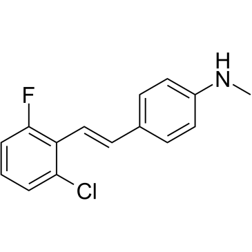
4-[(E)-2-(2-chloro-6-fluorophenyl)ethenyl]-N-methylaniline
|
| 别名 |
FIDAS-5; 1391934-98-7; (E)-4-(2-chloro-6-fluorostyryl)-N-methylaniline; 4-[(E)-2-(2-chloro-6-fluorophenyl)ethenyl]-N-methylaniline; 4-[(1E)-2-(2-chloro-6-fluorophenyl)ethenyl]-N-methylaniline; CHEMBL3314420; SCHEMBL11895362; SCHEMBL11895364;
|
| HS Tariff Code |
2934.99.9001
|
| 存储方式 |
Powder -20°C 3 years 4°C 2 years In solvent -80°C 6 months -20°C 1 month 注意: 该产品在溶液状态不稳定,请现配现用。 |
| 运输条件 |
Room temperature (This product is stable at ambient temperature for a few days during ordinary shipping and time spent in Customs)
|
| 溶解度 (体外实验) |
DMSO : ~125 mg/mL (~477.61 mM)
|
|---|---|
| 溶解度 (体内实验) |
配方 1 中的溶解度: ≥ 2.08 mg/mL (7.95 mM) (饱和度未知) in 10% DMSO + 40% PEG300 + 5% Tween80 + 45% Saline (这些助溶剂从左到右依次添加,逐一添加), 澄清溶液。
例如,若需制备1 mL的工作液,可将100 μL 20.8 mg/mL澄清DMSO储备液加入400 μL PEG300中,混匀;然后向上述溶液中加入50 μL Tween-80,混匀;加入450 μL生理盐水定容至1 mL。 *生理盐水的制备:将 0.9 g 氯化钠溶解在 100 mL ddH₂O中,得到澄清溶液。 配方 2 中的溶解度: ≥ 2.08 mg/mL (7.95 mM) (饱和度未知) in 10% DMSO + 90% (20% SBE-β-CD in Saline) (这些助溶剂从左到右依次添加,逐一添加), 澄清溶液。 例如,若需制备1 mL的工作液,可将 100 μL 20.8 mg/mL澄清DMSO储备液加入900 μL 20% SBE-β-CD生理盐水溶液中,混匀。 *20% SBE-β-CD 生理盐水溶液的制备(4°C,1 周):将 2 g SBE-β-CD 溶解于 10 mL 生理盐水中,得到澄清溶液。 请根据您的实验动物和给药方式选择适当的溶解配方/方案: 1、请先配制澄清的储备液(如:用DMSO配置50 或 100 mg/mL母液(储备液)); 2、取适量母液,按从左到右的顺序依次添加助溶剂,澄清后再加入下一助溶剂。以 下列配方为例说明 (注意此配方只用于说明,并不一定代表此产品 的实际溶解配方): 10% DMSO → 40% PEG300 → 5% Tween-80 → 45% ddH2O (或 saline); 假设最终工作液的体积为 1 mL, 浓度为5 mg/mL: 取 100 μL 50 mg/mL 的澄清 DMSO 储备液加到 400 μL PEG300 中,混合均匀/澄清;向上述体系中加入50 μL Tween-80,混合均匀/澄清;然后继续加入450 μL ddH2O (或 saline)定容至 1 mL; 3、溶剂前显示的百分比是指该溶剂在最终溶液/工作液中的体积所占比例; 4、 如产品在配制过程中出现沉淀/析出,可通过加热(≤50℃)或超声的方式助溶; 5、为保证最佳实验结果,工作液请现配现用! 6、如不确定怎么将母液配置成体内动物实验的工作液,请查看说明书或联系我们; 7、 以上所有助溶剂都可在 Invivochem.cn网站购买。 |
| 制备储备液 | 1 mg | 5 mg | 10 mg | |
| 1 mM | 3.8209 mL | 19.1044 mL | 38.2088 mL | |
| 5 mM | 0.7642 mL | 3.8209 mL | 7.6418 mL | |
| 10 mM | 0.3821 mL | 1.9104 mL | 3.8209 mL |
1、根据实验需要选择合适的溶剂配制储备液 (母液):对于大多数产品,InvivoChem推荐用DMSO配置母液 (比如:5、10、20mM或者10、20、50 mg/mL浓度),个别水溶性高的产品可直接溶于水。产品在DMSO 、水或其他溶剂中的具体溶解度详见上”溶解度 (体外)”部分;
2、如果您找不到您想要的溶解度信息,或者很难将产品溶解在溶液中,请联系我们;
3、建议使用下列计算器进行相关计算(摩尔浓度计算器、稀释计算器、分子量计算器、重组计算器等);
4、母液配好之后,将其分装到常规用量,并储存在-20°C或-80°C,尽量减少反复冻融循环。
计算结果:
工作液浓度: mg/mL;
DMSO母液配制方法: mg 药物溶于 μL DMSO溶液(母液浓度 mg/mL)。如该浓度超过该批次药物DMSO溶解度,请首先与我们联系。
体内配方配制方法:取 μL DMSO母液,加入 μL PEG300,混匀澄清后加入μL Tween 80,混匀澄清后加入 μL ddH2O,混匀澄清。
(1) 请确保溶液澄清之后,再加入下一种溶剂 (助溶剂) 。可利用涡旋、超声或水浴加热等方法助溶;
(2) 一定要按顺序加入溶剂 (助溶剂) 。
InvivoChem的所有产品仅用于作科学研究,不面向患者销售
Copyright 2020 InvivoChem LLC | All Rights Reserved 粤ICP备20063088号-1


 COA
COA
 463611831
