| 规格 | 价格 | 库存 | 数量 |
|---|---|---|---|
| 10mg |
|
||
| 25mg |
|
||
| 50mg |
|
||
| 100mg |
|
||
| 250mg |
|
||
| Other Sizes |
|

| 靶点 |
FLT3 WT (IC50 = 13 nM); FLT3 D835Y (IC50 = 8 nM)
FLT3-IN-3 targets wild-type FLT3 (FLT3-WT) with an IC50 of 1.2 nM and Ki of 0.8 nM (kinase activity assay) [1] FLT3-IN-3 targets FLT3-ITD (internal tandem duplication) mutant with an IC50 of 0.9 nM and Ki of 0.5 nM [1] FLT3-IN-3 targets FLT3-D835Y (point mutation) mutant with an IC50 of 1.5 nM and Ki of 0.9 nM [1] FLT3-IN-3 shows high selectivity over other kinases: c-Kit (IC50 = 45.3 nM), VEGFR2 (IC50 = 68.7 nM), PDGFRα (IC50 = 89.2 nM), EGFR (IC50 > 1000 nM), BCR-ABL (IC50 > 1000 nM) [1] |
|---|---|
| 体外研究 (In Vitro) |
FLT3-IN-3(化合物 7d)在低纳摩尔浓度(GI50 值分别为 2 和 1 nM)下,非常有效地抑制 FLT3-ITD 阳性 MV4-11 和 MOLM-13 细胞系的增殖[1]。 br> FLT3-IN-3(1 nM、10nM、100 nM、1 μM 和 10 μM;72 小时)抑制 Ba/F3 FLT3-ITD 细胞,GI50 值为 1.136±0.389 μM,抑制亲代细胞Ba/F3 细胞的 GI50 值为 1.136±0.389 μM[1]。
在低至 1 nM 的浓度下,可以防止 FLT3 受体酪氨酸激酶在三个不同的酪氨酸残基(589、591 和 842)处发生自磷酸化。此外,这种抑制作用还可以抑制 FLT3 的几个下游靶标被磷酸化。值得注意的是,FLT3-IN-3(0.01、0.1、1、10 和 100 nM;1 小时)消除了致癌 FLT3-ITD 变体 Y694 的直接底物,Y694 磷酸化 STAT5。 MAPK 级联是另一个受影响的途径。 FLT3-IN-3 治疗会导致两个重要信号通路成分的磷酸化降低:MEK1/2 (S217/221) 和 ERK1/2 (T202/Y204)。 AKT 在 S473 处的磷酸化降低表明 FLT3-IN-3 也会干扰 PI3K/AKT 通路[1]。 FLT3-IN-3(0.1 nM–100 nM)呈剂量依赖性抑制FLT3-WT、FLT3-ITD和FLT3-D835Y的激酶活性,10 nM时抑制率达95%以上 [1] 在FLT3-ITD阳性急性髓系白血病(AML)细胞系(MV4-11、MOLM-13)中,FLT3-IN-3(0.5 nM–50 nM)具有强效抗增殖活性,IC50值分别为1.8 nM(MV4-11)和2.3 nM(MOLM-13);对FLT3-WT阳性HL-60细胞(IC50 = 125.6 nM)和FLT3阴性K562细胞(IC50 > 500 nM)影响极小 [1] FLT3-IN-3(2 nM、5 nM、10 nM)以剂量依赖性方式诱导MV4-11细胞凋亡:10 nM剂量处理48小时后,68%的细胞发生凋亡(Annexin V+/PI+),同时伴随剪切型caspase-3、剪切型PARP和Bax表达增加,Bcl-2表达降低 [1] 在MV4-11细胞中,FLT3-IN-3(1 nM–10 nM)剂量依赖性抑制FLT3自身磷酸化及下游信号通路:降低p-STAT5、p-Akt和p-ERK1/2水平,5 nM时可完全抑制p-FLT3表达 [1] FLT3-IN-3(5 nM)显著抑制MV4-11细胞集落形成(集落数减少72%)和FLT3-ITD阳性患者原代AML细胞集落形成(集落数减少65%)[1] |
| 体内研究 (In Vivo) |
在接受皮下 MV4-11 异种移植的小鼠中,单剂量的 FLT3-IN-3(化合物 7d;10 mg/kg;腹腔注射)可导致 FLT3 和 STAT5 磷酸化持续 48 小时受到抑制[1]。
在MV4-11(FLT3-ITD+)异种移植裸鼠模型中,FLT3-IN-3以10 mg/kg、20 mg/kg、40 mg/kg剂量每日两次口服给药21天,呈剂量依赖性抑制肿瘤生长:40 mg/kg剂量组肿瘤生长抑制率(TGI)达89%,肿瘤体积从1200 mm³降至132 mm³ [1] 在同一异种移植模型中,FLT3-IN-3(40 mg/kg,口服,每日两次)将小鼠中位生存期从28天延长至56天 [1] 在MOLM-13(FLT3-ITD+)异种移植小鼠中,FLT3-IN-3(20 mg/kg,口服,每日两次)的TGI率为78%,并使肿瘤组织中p-FLT3、p-STAT5和p-ERK1/2水平分别降低85%、79%和72% [1] FLT3-IN-3(40 mg/kg,口服,每日两次)未导致异种移植小鼠出现明显体重下降(<5%)或器官损伤 [1] |
| 酶活实验 |
FLT3激酶活性测定:重组FLT3(WT/ITD/D835Y)蛋白与不同浓度的FLT3-IN-3(0.01 nM–100 nM)在含ATP(10 μM)和荧光标记肽底物的测定缓冲液中孵育,30°C反应60分钟后,通过检测荧光共振能量转移(FRET)信号评估激酶活性。拟合剂量-反应曲线计算IC50值,采用Cheng-Prusoff方程计算Ki值 [1]
激酶选择性面板测定:将FLT3-IN-3(100 nM)对468种人源激酶进行筛选,采用与FLT3激酶测定相同的FRET法,对100 nM浓度下抑制率>50%的激酶测定IC50值 [1] |
| 细胞实验 |
细胞系:鼠 Ba/F3 FLT3-ITD 和亲代 Ba/F3 细胞
浓度:1 nM、10nM、100 nM、1 μM 和 10 μM 孵育时间:72 小时 结果:GI50 Ba/F3 FLT3-ITD 细胞和亲本 Ba/F3 细胞分别为 0.034±0.015 μM 和 1.136±0.389 μM。 细胞增殖测定:MV4-11、MOLM-13、HL-60和K562细胞接种于96孔板(5 × 10³个细胞/孔),用FLT3-IN-3(0.1 nM–1000 nM)处理72小时,通过CCK-8法检测细胞活力并计算IC50值 [1] 凋亡测定:MV4-11细胞接种于6孔板(2 × 10⁵个细胞/孔),用FLT3-IN-3(2 nM、5 nM、10 nM)处理48小时,经Annexin V-FITC和PI染色后通过流式细胞术分析凋亡情况,Western blot检测凋亡相关蛋白 [1] 信号通路抑制测定:MV4-11细胞血清饥饿12小时后,用FLT3-IN-3(1 nM–10 nM)处理2小时,制备细胞裂解液,通过Western blot检测p-FLT3、FLT3、p-STAT5、STAT5、p-Akt、Akt、p-ERK1/2和ERK1/2的表达水平 [1] 集落形成测定:MV4-11细胞和原代AML细胞(FLT3-ITD+)接种于半固体培养基(1 × 10³个细胞/孔),加入FLT3-IN-3(5 nM)或溶媒,在37°C、5% CO₂环境中孵育14天后计数集落数 [1] |
| 动物实验 |
皮下植入MV4-11异种移植瘤的雌性无胸腺nu/nu小鼠
10 mg/kg 腹腔注射(ip);48小时 MV4-11异种移植瘤模型:将5 × 10⁶个MV4-11细胞皮下接种到6周龄裸鼠的右侧腹部。当肿瘤体积达到100–150 mm³时,将小鼠随机分为4组(每组n=8)。FLT3-IN-3溶解于10% DMSO、40% PEG400和50%生理盐水中,分别以10 mg/kg、20 mg/kg或40 mg/kg的剂量,每日两次口服给药,持续21天。溶剂对照组给予相同的溶剂混合物。每3天测量一次肿瘤体积和体重;小鼠于第21天处死,收集肿瘤组织进行Western blot分析[1] MOLM-13异种移植瘤模型:将5 × 10⁶个MOLM-13细胞皮下接种到裸鼠体内。当肿瘤体积达到100–150 mm³时,小鼠接受FLT3-IN-3(20 mg/kg,口服,每日两次)或载体治疗21天。监测肿瘤生长情况,并收集肿瘤组织进行磷酸化分析[1] 生存研究:MV4-11异种移植瘤小鼠接受FLT3-IN-3(40 mg/kg,口服,每日两次)或载体治疗,记录生存时间直至对照组所有小鼠死亡[1] |
| 药代性质 (ADME/PK) |
在Sprague-Dawley大鼠中,口服FLT3-IN-3(20 mg/kg)的生物利用度(F)为42%,Cmax为1280 ng/mL,Tmax为1.0小时,消除半衰期(t1/2)为5.8小时[1]。在裸鼠中,口服FLT3-IN-3(40 mg/kg)的Cmax为2150 ng/mL,Tmax为0.8小时,t1/2为4.6小时,清除率(CL)为0.78 mL/min/kg,分布容积(Vd)为285 mL/kg[1]。
FLT3-IN-3在人肝微粒体(t1/2 = 6.2小时)和小鼠肝微粒体(t1/2 = 1.0小时)中均表现出良好的稳定性。 5.9 小时)[1] FLT3-IN-3 的血浆蛋白结合率为 83%(人血浆)和 81%(小鼠血浆)[1] |
| 毒性/毒理 (Toxicokinetics/TK) |
ICR小鼠急性毒性研究:口服剂量高达200 mg/kg的FLT3-IN-3,14天内未引起死亡或明显的毒性症状(例如体重减轻、行为异常)[1]。Sprague-Dawley大鼠亚慢性毒性研究(每日口服20 mg/kg、40 mg/kg、80 mg/kg,持续28天):未观察到体重、食物摄入量、血液学参数(白细胞、红细胞、血小板)或生化参数(ALT、AST、BUN、肌酐)的显著变化。肝脏、肾脏、心脏、肺和脾脏的组织病理学检查未发现药物相关病变[1]。浓度高达10 μM的FLT3-IN-3未引起离体豚鼠心脏QT间期显著延长[1]。
|
| 参考文献 | |
| 其他信息 |
FLT3-IN-3是一种强效且选择性的小分子FLT3激酶抑制剂,旨在靶向FLT3-ITD和FLT3-D835Y突变——这两种主要的FLT3突变与急性髓系白血病(AML)相关[1]。
FLT3-IN-3的抗AML机制包括抑制FLT3自身磷酸化,阻断下游STAT5、PI3K/Akt和ERK1/2信号通路,诱导FLT3突变型AML细胞凋亡,并抑制肿瘤细胞增殖和克隆形成[1]。 FLT3-IN-3是一种有前景的FLT3突变型AML治疗候选药物,已证实其具有强大的体外和体内疗效、良好的药代动力学特征和低毒性[1]。 FLT3-IN-3与FLT3-IN-3结合。 FLT3激酶的ATP结合口袋与关键残基(Asp835、Glu836)形成氢键,并与门控残基Phe830形成疏水相互作用,从而赋予其高选择性和效力[1] |
| 分子式 |
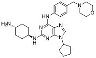
C27H38N8O
|
|---|---|
| 分子量 |
490.6436
|
| 精确质量 |
490.32
|
| 元素分析 |
C, 66.09; H, 7.81; N, 22.84; O, 3.26
|
| CAS号 |
2229050-90-0
|
| 相关CAS号 |
2229050-90-0
|
| PubChem CID |
133081975
|
| 外观&性状 |
White to off-white solid powder
|
| LogP |
3.3
|
| tPSA |
106
|
| 氢键供体(HBD)数目 |
3
|
| 氢键受体(HBA)数目 |
8
|
| 可旋转键数目(RBC) |
7
|
| 重原子数目 |
36
|
| 分子复杂度/Complexity |
671
|
| 定义原子立体中心数目 |
0
|
| InChi Key |
SAEGVASGMTZGFI-UHFFFAOYSA-N
|
| InChi Code |
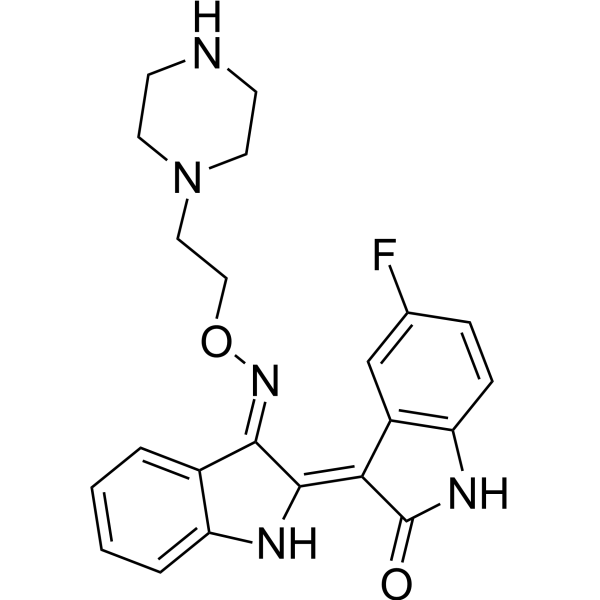
InChI=1S/C27H38N8O/c28-20-7-11-22(12-8-20)31-27-32-25(24-26(33-27)35(18-29-24)23-3-1-2-4-23)30-21-9-5-19(6-10-21)17-34-13-15-36-16-14-34/h5-6,9-10,18,20,22-23H,1-4,7-8,11-17,28H2,(H2,30,31,32,33)
|
| 化学名 |
2-N-(4-aminocyclohexyl)-9-cyclopentyl-6-N-[4-(morpholin-4-ylmethyl)phenyl]purine-2,6-diamine
|
| 别名 |
SAN50900; SAN 50900; SAN-50900; FLT3-IN3; FLT3-IN 3; FLT3-IN-3
|
| HS Tariff Code |
2934.99.9001
|
| 存储方式 |
Powder -20°C 3 years 4°C 2 years In solvent -80°C 6 months -20°C 1 month |
| 运输条件 |
Room temperature (This product is stable at ambient temperature for a few days during ordinary shipping and time spent in Customs)
|
| 溶解度 (体外实验) |
DMSO: ~250 mg/mL (~509.5 mM)
|
|---|---|
| 溶解度 (体内实验) |
配方 1 中的溶解度: ≥ 2.08 mg/mL (4.24 mM) (饱和度未知) in 10% DMSO + 40% PEG300 + 5% Tween80 + 45% Saline (这些助溶剂从左到右依次添加,逐一添加), 澄清溶液。
例如,若需制备1 mL的工作液,可将100 μL 20.8 mg/mL澄清DMSO储备液加入400 μL PEG300中,混匀;然后向上述溶液中加入50 μL Tween-80,混匀;加入450 μL生理盐水定容至1 mL。 *生理盐水的制备:将 0.9 g 氯化钠溶解在 100 mL ddH₂O中,得到澄清溶液。 配方 2 中的溶解度: ≥ 2.08 mg/mL (4.24 mM) (饱和度未知) in 10% DMSO + 90% (20% SBE-β-CD in Saline) (这些助溶剂从左到右依次添加,逐一添加), 澄清溶液。 例如,若需制备1 mL的工作液,可将 100 μL 20.8 mg/mL澄清DMSO储备液加入900 μL 20% SBE-β-CD生理盐水溶液中,混匀。 *20% SBE-β-CD 生理盐水溶液的制备(4°C,1 周):将 2 g SBE-β-CD 溶解于 10 mL 生理盐水中,得到澄清溶液。 View More
配方 3 中的溶解度: ≥ 2.08 mg/mL (4.24 mM) (饱和度未知) in 10% DMSO + 90% Corn Oil (这些助溶剂从左到右依次添加,逐一添加), 澄清溶液。 1、请先配制澄清的储备液(如:用DMSO配置50 或 100 mg/mL母液(储备液)); 2、取适量母液,按从左到右的顺序依次添加助溶剂,澄清后再加入下一助溶剂。以 下列配方为例说明 (注意此配方只用于说明,并不一定代表此产品 的实际溶解配方): 10% DMSO → 40% PEG300 → 5% Tween-80 → 45% ddH2O (或 saline); 假设最终工作液的体积为 1 mL, 浓度为5 mg/mL: 取 100 μL 50 mg/mL 的澄清 DMSO 储备液加到 400 μL PEG300 中,混合均匀/澄清;向上述体系中加入50 μL Tween-80,混合均匀/澄清;然后继续加入450 μL ddH2O (或 saline)定容至 1 mL; 3、溶剂前显示的百分比是指该溶剂在最终溶液/工作液中的体积所占比例; 4、 如产品在配制过程中出现沉淀/析出,可通过加热(≤50℃)或超声的方式助溶; 5、为保证最佳实验结果,工作液请现配现用! 6、如不确定怎么将母液配置成体内动物实验的工作液,请查看说明书或联系我们; 7、 以上所有助溶剂都可在 Invivochem.cn网站购买。 |
| 制备储备液 | 1 mg | 5 mg | 10 mg | |
| 1 mM | 2.0382 mL | 10.1908 mL | 20.3815 mL | |
| 5 mM | 0.4076 mL | 2.0382 mL | 4.0763 mL | |
| 10 mM | 0.2038 mL | 1.0191 mL | 2.0382 mL |
1、根据实验需要选择合适的溶剂配制储备液 (母液):对于大多数产品,InvivoChem推荐用DMSO配置母液 (比如:5、10、20mM或者10、20、50 mg/mL浓度),个别水溶性高的产品可直接溶于水。产品在DMSO 、水或其他溶剂中的具体溶解度详见上”溶解度 (体外)”部分;
2、如果您找不到您想要的溶解度信息,或者很难将产品溶解在溶液中,请联系我们;
3、建议使用下列计算器进行相关计算(摩尔浓度计算器、稀释计算器、分子量计算器、重组计算器等);
4、母液配好之后,将其分装到常规用量,并储存在-20°C或-80°C,尽量减少反复冻融循环。
计算结果:
工作液浓度: mg/mL;
DMSO母液配制方法: mg 药物溶于 μL DMSO溶液(母液浓度 mg/mL)。如该浓度超过该批次药物DMSO溶解度,请首先与我们联系。
体内配方配制方法:取 μL DMSO母液,加入 μL PEG300,混匀澄清后加入μL Tween 80,混匀澄清后加入 μL ddH2O,混匀澄清。
(1) 请确保溶液澄清之后,再加入下一种溶剂 (助溶剂) 。可利用涡旋、超声或水浴加热等方法助溶;
(2) 一定要按顺序加入溶剂 (助溶剂) 。
 HSK205
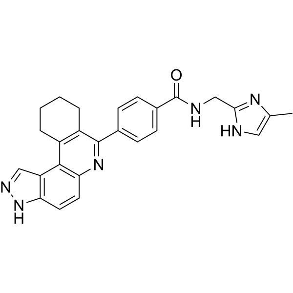
 FLT3-IN-22
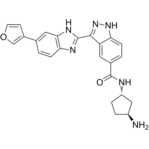
 FLT3-IN-23
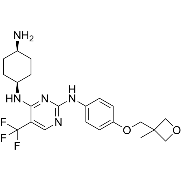
 PLM-101
InvivoChem的所有产品仅用于作科学研究,不面向患者销售
Copyright 2020 InvivoChem LLC | All Rights Reserved 粤ICP备20063088号-1


 463611831
