| 规格 | 价格 | 库存 | 数量 |
|---|---|---|---|
| 10 mM * 1 mL in DMSO |
|
||
| 5mg |
|
||
| 10mg |
|
||
| 50mg |
|
||
| 100mg |
|
||
| 250mg |
|
||
| 500mg |
|
||
| 1g |
|
||
| 2g |
|
||
| 5g |
|
||
| Other Sizes |
|

| 靶点 |
DNA synthesis (Capan2 cells) ( IC50 = 12 nM ); DNA synthesis (BxPC3 cells) ( IC50 = 18 nM ); DNA synthesis (MIAPaCa2 cells) ( IC50 = 40 nM ); DNA synthesis (PANC1 cells) ( IC50 = 50 nM )
Ribonucleotide reductase (RR) [3] Human equilibrative nucleoside transporter 1 (hENT1) (functional target for cellular uptake) [1] |
|---|---|
| 体外研究 (In Vitro) |
吉西他滨诱导 BxPC-3、PANC-1 和 MIA PaCa-2 细胞中的 NF-κB 活性,并降低 BxPC-3 和 PANC-1 细胞中 NF-κB 抑制剂 IκBα 的水平。用低剂量吉西他滨处理 BxPC-3 细胞 48 小时会导致 NF-κB 结合呈剂量依赖性增加。相比之下,用较高吉西他滨剂量处理 48 小时的 BxPC-3 细胞中 NF-κB DNA 结合减少;然而,用这些较高剂量进行 24 小时处理会增加 BxPC-3 细胞中 NF-κB 的结合。细胞测定:将 BxPC-3、MIA PaCa-2 和 PANC-1 细胞接种到 96 孔板中。 24小时后,用媒介物、DMAPT和/或吉西他滨再处理细胞24小时或48小时。使用细胞死亡检测 ELISA 来定量细胞凋亡,以检测细胞质组蛋白相关 DNA 片段的量并相对于载体处理的细胞进行表达。
针对人胰腺癌细胞系(PANC-1、MiaPaCa-2),盐酸吉西他滨表现出浓度依赖性抗增殖活性,IC50值分别为0.15 μM(PANC-1)和0.22 μM(MiaPaCa-2)。与吲哚-3-甲醇(I3C)联合处理可通过上调hENT1表达,将IC50值降低40-50%,增强药效[1] - 在人结直肠癌细胞(HCT-116)中,盐酸吉西他滨(0.1-1 μM)通过靶向核糖核苷酸还原酶(RR)抑制细胞增殖并诱导S期细胞周期阻滞。当RR大亚基(RRM1)与硫氧还蛋白(Trx)相互作用时,RR活性增强,药物敏感性降低[3] - 胰腺癌细胞中药物细胞毒性与hENT1表达呈正相关:hENT1沉默细胞的IC50值是亲本细胞的3倍[1] - 诱导胰腺癌细胞凋亡,表现为caspase-3激活和PARP切割增加,I3C联合给药可增强该效应[1] |
| 体内研究 (In Vivo) |
本研究的目的是评估吉西他滨肺部给药的安全性,并在动物模型中确定每周肺给药的最大耐受剂量。五组八只Wistar大鼠接受2、4、6或8mg/kg剂量的吉西他滨或载体溶液,通过气管内喷雾进行肺沉积闪烁显像。为了记录消化暴露的安全性,五组八只大鼠接受相同剂量的吉西他滨或灌胃载体溶液。计划每周一次,并在第64天对活体动物进行血细胞计数和组织学检查。闪烁显像证实316次喷雾给药中有310次(98%)出现肺部沉积,沉积模式均匀。通过肺部给药的吉西他滨最大耐受剂量为4 mg/kg。在该剂量下,连续9周每周给药一次,除血小板和红细胞计数减少外,没有与化疗相关的死亡,也没有临床、组织学或血液学毒性症状,没有临床意义。在2、4和6 mg/kg的剂量下,吉西他滨经口给药的毒性在体重减轻和白细胞毒性方面高于经肺给药。吉西他滨的最大耐受剂量为4 mg/kg,对大鼠进行肺部给药是安全的,每周一次,持续9周。在同等剂量下,吉西他滨的肺部毒性低于口服给药。[2]
与 PBS 治疗的小鼠相比,吉西他滨治疗的小鼠瘤内 NF-κB 活性显着升高(1.3 至 1.8 倍),表明吉西他滨也诱导 NF-κB 激活。 在胰腺癌转基因小鼠模型(KrasG12D; Trp53R172H; Pdx1-Cre)中,以120 mg/kg的剂量每周两次腹腔注射盐酸吉西他滨,连续3周,中位存活时间较对照组延长28%。与二甲基氨基小白菊内酯(DMAPT)联合使用,中位存活时间进一步延长52%[4] - 大鼠肺部给药盐酸吉西他滨(10、20 mg/kg)后,药物分布至肺组织并达到治疗浓度,无显著全身毒性。[2] |
| 酶活实验 |
核糖核苷酸还原酶(RR)活性检测:将重组人RR(RRM1/RRM2复合物)与ADP底物在反应缓冲液中于37°C孵育。加入系列浓度(0.05-2 μM)的盐酸吉西他滨,混合物孵育60分钟。加入高氯酸终止反应,通过高效液相色谱(HPLC)定量dADP(产物)生成量,评估RR抑制效果[3]
- hENT1介导的转运检测:PANC-1细胞与盐酸吉西他滨(0.5 μM)和[3H]标记吉西他滨在37°C孵育30分钟。洗涤细胞去除未结合药物,通过液体闪烁计数法测量放射性,定量hENT1依赖的摄取量。在hENT1沉默细胞中重复实验以确认特异性[1] |
| 细胞实验 |
在 96 孔板中,接种 BxPC-3、MIA PaCa-2 和 PANC-1 细胞。 24小时后用媒介物、DMAPT和/或吉西他滨进一步处理细胞24或48小时。使用细胞死亡检测 ELISA,通过计算细胞质组蛋白相关 DNA 片段的数量来测量与载体处理的细胞相关的细胞凋亡。
接受吉西他滨(2',2'-二氟脱氧胞苷)治疗的癌症患者最终可能产生耐药性。最近,我们实验室公布的数据表明,吉西他滨与膳食药物吲哚-3-甲醇(I3C)的疗效增强。目前的研究探讨了这种I3C增强疗效的可能机制。检测了几种胰腺细胞系(BxPC-3、Mia Paca-2、PL-45、AsPC-1和PANC-1)单独使用I3C和与吉西他滨联合使用对人平衡核苷转运蛋白1(hENT1)表达的调节,hENT1是吉西他滨的主要转运蛋白。I3C显著(p<0.01)上调了几种细胞系中hENT1的表达。单独使用吉西他滨对hENT1表达没有影响。然而,吉西他滨与I3C联合使用进一步增加了hENT1的表达。细胞活力测定显示I3C对正常细胞hTERT-HPNE没有影响。hENT1特异性抑制剂硝基苄硫肌苷显著消除了I3C诱导的吉西他滨细胞毒性,进一步证明了其特异性。本研究表明,hENT1表达的上调可能是I3C和吉西他滨加性作用的一种新机制。[1] 胰腺癌细胞抗增殖及联合用药检测:将PANC-1和MiaPaCa-2细胞以4×10³个细胞/孔接种到96孔板中,用0.01-1 μM的盐酸吉西他滨单独或与25 μM I3C联合处理72小时。采用四唑盐比色法检测细胞活力。蛋白质印迹法检测hENT1表达,分析caspase-3/PARP水平评估凋亡[1] - 结直肠癌细胞RR相互作用检测:HCT-116细胞转染RRM1和Trx表达质粒后,用0.5 μM的盐酸吉西他滨处理48小时。计数存活细胞评估增殖情况。免疫共沉淀(Co-IP)验证RRM1-Trx相互作用,通过ADP向dADP转化实验检测RR活性[3] - 细胞周期检测:用0.2 μM的盐酸吉西他滨处理PANC-1细胞24小时。乙醇固定细胞,碘化丙啶染色后通过流式细胞术检测S期阻滞[1] |
| 动物实验 |
溶于PBS;50或100 mg/kg;腹腔注射
无胸腺裸鼠携带MIA PaCa-2细胞。本研究采用表达突变型Kras和p53的LSL-KrasG12D/+;LSL-Trp53R172H;Pdx-1-Cre小鼠胰腺癌模型,评估DMAPT和吉西他滨在化学预防试验中的疗效。小鼠随机分为四组(安慰剂组、DMAPT组[40 mg/kg/天]、吉西他滨组[50 mg/kg,每周两次]以及DMAPT/吉西他滨联合组)。持续治疗直至小鼠出现健康问题,此时处死小鼠。采用Bio-Plex免疫分析法测定血浆细胞因子水平。所采用的统计检验方法包括对数秩检验、Dunnett事后检验的方差分析、学生t检验和Fisher精确检验。[4] 结果:吉西他滨或DMAPT/吉西他滨联合用药显著延长了中位生存期,并降低了胰腺腺癌的发生率和数量。DMAPT/吉西他滨联合用药还显著缩小了肿瘤体积,并降低了肝转移的发生率。各治疗组间正常胰管或癌前胰腺病变的百分比无显著差异。对未形成肿瘤的胰腺组织进行分析,以确定癌前病变的程度;结果显示,大部分为正常胰管或低级别胰腺病变,表明该联合用药可预防这些动物发生更高级别的病变。吉西他滨治疗可增加小鼠血浆中炎症细胞因子白细胞介素 1α (IL-1α)、IL-1β 和 IL-17 的水平,而 DMAPT 和 DMAPT/吉西他滨则可降低炎症细胞因子 IL-12p40、单核细胞趋化蛋白-1 (MCP-1)、巨噬细胞炎症蛋白-1β (MIP-1β)、嗜酸性粒细胞趋化因子和肿瘤坏死因子-α (TNF-α) 的水平,这些都是 NF-κB 的靶基因。[4] 胰腺癌小鼠模型:将自发性胰腺癌的转基因小鼠 (KrasG12D; Trp53R172H; Pdx1-Cre) 随机分为对照组、吉西他滨单药组和吉西他滨 + DMAPT 组(每组 n=10)。将盐酸吉西他滨溶于无菌生理盐水中,以120 mg/kg的剂量腹腔注射,每周两次,持续3周。DMAPT以20 mg/kg的剂量每日口服。记录生存时间,并收集肿瘤组织进行组织病理学分析[4]。 - 肺毒性大鼠模型:将雄性Sprague-Dawley大鼠随机分为对照组和吉西他滨组(每组n=6)。将盐酸吉西他滨制成气雾剂,以10或20 mg/kg的剂量单次经肺部吸入给药。72小时后处死大鼠;收集肺、肝、肾组织进行组织病理学检查,并收集血清进行肝肾功能检测[2]。 |
| 药代性质 (ADME/PK) |
吸收:吉西他滨静脉输注30分钟后,血浆峰浓度范围为10至40 mg/L,并在15至30分钟达到。一项研究表明,在53至1000 mg/m²的剂量范围内,吉西他滨的稳态浓度与剂量呈线性关系。吉西他滨的活性代谢物吉西他滨三磷酸盐可在循环外周血单核细胞中蓄积。一项研究显示,外周血单核细胞中吉西他滨三磷酸盐的Cmax出现在输注结束后30分钟内,并随吉西他滨剂量(最高达350 mg/m²)的增加而呈比例增加。
消除途径:吉西他滨主要经肾脏排泄。单次静脉输注1000 mg/m²吉西他滨后一周内,约92-98%的剂量从尿液中回收,其中89%以二氟脱氧尿苷(dFdU)形式排出,不足10%以吉西他滨形式排出。尿液中检测不到吉西他滨的单磷酸、二磷酸或三磷酸代谢物。在一项单剂量研究中,约1%的给药剂量从粪便中回收。分布容积:在患有各种实体瘤的患者中,分布容积随输注时间的延长而增加。输注时间少于70分钟时,吉西他滨的分布容积为50 L/m²。长时间输注后,分布容积增加至370 L/m²。吉西他滨三磷酸盐是吉西他滨的活性代谢产物,在体外和体内均可在实体瘤细胞中蓄积和滞留。短时输注(持续时间少于70分钟)后,其在组织中的分布并不广泛。目前尚不清楚吉西他滨是否能穿过血脑屏障,但吉西他滨可广泛分布于包括腹水在内的各种组织中。在大鼠中,给药后5至15分钟内即可迅速通过胎盘和乳糜管转运。 清除率:静脉输注(持续时间少于70分钟)后,雄性大鼠的清除率为41至92 L/h/m²,雌性大鼠的清除率为31至69 L/h/m²。清除率随年龄增长而降低。女性患者的药物清除率比男性患者低约30%。 代谢/代谢产物:吉西他滨给药后被癌细胞吸收,首先被脱氧胞苷激酶(dCK)磷酸化,少量被线粒体外胸苷激酶2磷酸化,生成吉西他滨单磷酸(dFdCMP)。dFdCMP随后被核苷激酶磷酸化,生成活性代谢产物吉西他滨二磷酸(dFdCDP)和吉西他滨三磷酸(dFdCTP)。吉西他滨还可在细胞内和细胞外被胞苷脱氨酶脱氨,生成其无活性代谢产物2′,2′-二氟脱氧尿苷或2′-脱氧-2′,2′-二氟尿苷(dFdU)。脱氨基作用发生在血液、肝脏、肾脏和其他组织中,该代谢途径是药物清除的主要途径。 生物半衰期:静脉输注时间少于70分钟后,终末半衰期为0.7至1.6小时。输注时间在70至285分钟之间时,终末半衰期为4.1至10.6小时。女性患者的半衰期通常比男性患者长。吉西他滨的活性代谢物吉西他滨三磷酸盐可在循环外周血单核细胞中蓄积。吉西他滨三磷酸盐(活性代谢物)在单核细胞中的终末半衰期为 1.7 至 19.4 小时。 吸收:大鼠肺部给药吉西他滨盐酸盐后,肺部生物利用度约为 45-55%,吸入后 1 小时内即可达到肺部峰浓度[2] - 分布:肺部给药后,药物主要分布于肺组织,全身暴露量极低(血浆浓度为肺部浓度的 10-15%)[2] - 血浆蛋白结合率:吉西他滨盐酸盐与人血浆蛋白的结合率约为 10-15%[2] |
| 毒性/毒理 (Toxicokinetics/TK) |
妊娠期和哺乳期影响
◉ 哺乳期用药概述 大多数资料认为,母亲在接受抗肿瘤药物治疗期间不宜进行母乳喂养。在间歇性吉西他滨治疗期间,如果采取适当的哺乳间隔期,或许可以安全地进行母乳喂养;生产商建议在最后一次给药后至少停止哺乳 1 周。化疗可能会对母乳的正常微生物群和化学成分产生不利影响。妊娠期接受化疗的女性更有可能出现哺乳困难。 ◉ 对母乳喂养婴儿的影响 截至修订日期,未找到相关的已发表信息。 ◉ 对哺乳和母乳的影响 一项电话随访研究对 74 名在妊娠中期或晚期于同一中心接受癌症化疗的女性进行了调查,以确定她们产后是否成功进行母乳喂养。仅有34%的女性能够纯母乳喂养婴儿,66%的女性报告在母乳喂养过程中遇到困难。相比之下,在22名孕期确诊但未接受化疗的母亲中,母乳喂养成功率高达91%。其他具有统计学意义的相关性包括:1. 存在母乳喂养困难的母亲平均接受了5.5个疗程的化疗,而没有母乳喂养困难的母亲平均接受了3.8个疗程的化疗;2. 存在母乳喂养困难的母亲首次接受化疗的时间平均比没有母乳喂养困难的母亲提前3.4周。接受含氟尿嘧啶方案治疗的9名女性中,8名出现哺乳困难。 肺毒性:大鼠肺部给予吉西他滨盐酸盐(20 mg/kg)可引起轻度肺部炎症(中性粒细胞浸润)和上皮细胞增生,这些症状在14天内可逆[2] -全身毒性:小鼠接受治疗剂量(120 mg/kg,腹腔注射)后,该药物可引起轻度骨髓抑制(白细胞计数降低20-25%)和血清转氨酶水平短暂升高(1.3倍),但未见明显的肾毒性[4] |
| 参考文献 |
|
| 其他信息 |
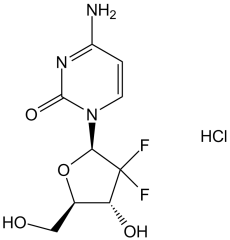
吉西他滨盐酸盐是抗代谢核苷脱氧胞苷类似物的盐酸盐,具有抗肿瘤活性。吉西他滨在细胞内转化为活性代谢物二氟脱氧胞苷二磷酸和三磷酸(dFdCDP、dFdCTP)。dFdCDP抑制核糖核苷酸还原酶,从而减少可用于DNA合成的脱氧核苷酸池; dFdCTP 可掺入 DNA,导致 DNA 链终止和细胞凋亡。
一种脱氧胞苷抗代谢物,用作抗肿瘤药物。 药物适应症 治疗尿路上皮癌 盐酸吉西他滨是一种合成的嘧啶核苷类似物,是一种前药,需要磷酸化生成活性代谢物 (dFdCTP) 才能发挥抗肿瘤作用[1][3] - 作用机制:活性 dFdCTP 抑制核糖核苷酸还原酶 (RR) 以减少脱氧核糖核苷酸池,并掺入 DNA 以阻断复制/转录。细胞摄取依赖于hENT1,疗效与hENT1表达相关[1][3] - 临床适应症:已获批用于治疗胰腺癌、非小细胞肺癌、结直肠癌和乳腺癌[1][4] - 耐药机制:hENT1表达降低、RR活性增强(通过RRM1-Trx相互作用)或药物外排增强均会导致耐药[1][3] - 联合用药优势:与靶向hENT1(例如I3C)或RR调节蛋白(例如DMAPT)的药物联合用药可增强其在临床前模型中的抗肿瘤疗效[1][4] |
| 分子式 |
C9H11F2N3O4.HCI
|
|
|---|---|---|
| 分子量 |
299.66
|
|
| 精确质量 |
299.048
|
|
| 元素分析 |
C, 36.07; H, 4.04; Cl, 11.83; F, 12.68; N, 14.02; O, 21.36
|
|
| CAS号 |
122111-03-9
|
|
| 相关CAS号 |
|
|
| PubChem CID |
60749
|
|
| 外观&性状 |
White solid powder
|
|
| 沸点 |
482.7ºC at 760 mmHg
|
|
| 熔点 |
>250°C dec.
|
|
| 蒸汽压 |
2.41E-11mmHg at 25°C
|
|
| LogP |
0.094
|
|
| tPSA |
110.6
|
|
| 氢键供体(HBD)数目 |
4
|
|
| 氢键受体(HBA)数目 |
6
|
|
| 可旋转键数目(RBC) |
2
|
|
| 重原子数目 |
19
|
|
| 分子复杂度/Complexity |
426
|
|
| 定义原子立体中心数目 |
3
|
|
| SMILES |
Cl[H].FC1([C@]([H])(N2C(N=C(C([H])=C2[H])N([H])[H])=O)O[C@]([H])(C([H])([H])O[H])[C@@]1([H])O[H])F
|
|
| InChi Key |
OKKDEIYWILRZIA-OSZBKLCCSA-N
|
|
| InChi Code |
InChI=1S/C9H11F2N3O4.ClH/c10-9(11)6(16)4(3-15)18-7(9)14-2-1-5(12)13-8(14)17;/h1-2,4,6-7,15-16H,3H2,(H2,12,13,17);1H/t4-,6-,7-;/m1./s1
|
|
| 化学名 |
4-amino-1-[(2R,4R,5R)-3,3-difluoro-4-hydroxy-5-(hydroxymethyl)oxolan-2-yl]pyrimidin-2-one;hydrochloride
|
|
| 别名 |
Abbreviations: dFdC; dFdCyd; LY188011; LY-188011; LY 188011; gemcitabine; Gemzar
|
|
| HS Tariff Code |
2934.99.9001
|
|
| 存储方式 |
Powder -20°C 3 years 4°C 2 years In solvent -80°C 6 months -20°C 1 month 注意: 请将本产品存放在密封且受保护的环境中(例如氮气保护),避免吸湿/受潮和光照。 |
|
| 运输条件 |
Room temperature (This product is stable at ambient temperature for a few days during ordinary shipping and time spent in Customs)
|
| 溶解度 (体外实验) |
|
|||
|---|---|---|---|---|
| 溶解度 (体内实验) |
配方 1 中的溶解度: ≥ 2.08 mg/mL (6.94 mM) (饱和度未知) in 10% DMSO + 40% PEG300 + 5% Tween80 + 45% Saline (这些助溶剂从左到右依次添加,逐一添加), 澄清溶液。
例如,若需制备1 mL的工作液,可将100 μL 20.8 mg/mL澄清DMSO储备液加入400 μL PEG300中,混匀;然后向上述溶液中加入50 μL Tween-80,混匀;加入450 μL生理盐水定容至1 mL。 *生理盐水的制备:将 0.9 g 氯化钠溶解在 100 mL ddH₂O中,得到澄清溶液。 配方 2 中的溶解度: ≥ 2.08 mg/mL (6.94 mM) (饱和度未知) in 10% DMSO + 90% (20% SBE-β-CD in Saline) (这些助溶剂从左到右依次添加,逐一添加), 澄清溶液。 例如,若需制备1 mL的工作液,可将 100 μL 20.8 mg/mL澄清DMSO储备液加入900 μL 20% SBE-β-CD生理盐水溶液中,混匀。 *20% SBE-β-CD 生理盐水溶液的制备(4°C,1 周):将 2 g SBE-β-CD 溶解于 10 mL 生理盐水中,得到澄清溶液。 View More
配方 3 中的溶解度: ≥ 2.08 mg/mL (6.94 mM) (饱和度未知) in 10% DMSO + 90% Corn Oil (这些助溶剂从左到右依次添加,逐一添加), 澄清溶液。 配方 4 中的溶解度: Saline: 20 mg/mL 配方 5 中的溶解度: 60 mg/mL (200.23 mM) in PBS (这些助溶剂从左到右依次添加,逐一添加), 澄清溶液; 超声助溶. 1、请先配制澄清的储备液(如:用DMSO配置50 或 100 mg/mL母液(储备液)); 2、取适量母液,按从左到右的顺序依次添加助溶剂,澄清后再加入下一助溶剂。以 下列配方为例说明 (注意此配方只用于说明,并不一定代表此产品 的实际溶解配方): 10% DMSO → 40% PEG300 → 5% Tween-80 → 45% ddH2O (或 saline); 假设最终工作液的体积为 1 mL, 浓度为5 mg/mL: 取 100 μL 50 mg/mL 的澄清 DMSO 储备液加到 400 μL PEG300 中,混合均匀/澄清;向上述体系中加入50 μL Tween-80,混合均匀/澄清;然后继续加入450 μL ddH2O (或 saline)定容至 1 mL; 3、溶剂前显示的百分比是指该溶剂在最终溶液/工作液中的体积所占比例; 4、 如产品在配制过程中出现沉淀/析出,可通过加热(≤50℃)或超声的方式助溶; 5、为保证最佳实验结果,工作液请现配现用! 6、如不确定怎么将母液配置成体内动物实验的工作液,请查看说明书或联系我们; 7、 以上所有助溶剂都可在 Invivochem.cn网站购买。 |
| 制备储备液 | 1 mg | 5 mg | 10 mg | |
| 1 mM | 3.3371 mL | 16.6856 mL | 33.3712 mL | |
| 5 mM | 0.6674 mL | 3.3371 mL | 6.6742 mL | |
| 10 mM | 0.3337 mL | 1.6686 mL | 3.3371 mL |
1、根据实验需要选择合适的溶剂配制储备液 (母液):对于大多数产品,InvivoChem推荐用DMSO配置母液 (比如:5、10、20mM或者10、20、50 mg/mL浓度),个别水溶性高的产品可直接溶于水。产品在DMSO 、水或其他溶剂中的具体溶解度详见上”溶解度 (体外)”部分;
2、如果您找不到您想要的溶解度信息,或者很难将产品溶解在溶液中,请联系我们;
3、建议使用下列计算器进行相关计算(摩尔浓度计算器、稀释计算器、分子量计算器、重组计算器等);
4、母液配好之后,将其分装到常规用量,并储存在-20°C或-80°C,尽量减少反复冻融循环。
计算结果:
工作液浓度: mg/mL;
DMSO母液配制方法: mg 药物溶于 μL DMSO溶液(母液浓度 mg/mL)。如该浓度超过该批次药物DMSO溶解度,请首先与我们联系。
体内配方配制方法:取 μL DMSO母液,加入 μL PEG300,混匀澄清后加入μL Tween 80,混匀澄清后加入 μL ddH2O,混匀澄清。
(1) 请确保溶液澄清之后,再加入下一种溶剂 (助溶剂) 。可利用涡旋、超声或水浴加热等方法助溶;
(2) 一定要按顺序加入溶剂 (助溶剂) 。
Testing the Addition of an Anti-cancer Drug, Pembrolizumab, to the Usual Intravesical Chemotherapy Treatment (Gemcitabine) for the Treatment of BCG-Unresponsive Non-muscle Invasive Bladder Cancer
CTID: NCT04164082
Phase: Phase 2 Status: Recruiting
Date: 2024-11-25
|
|
|
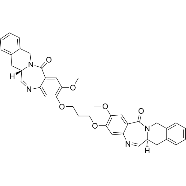 (S,S)-D211
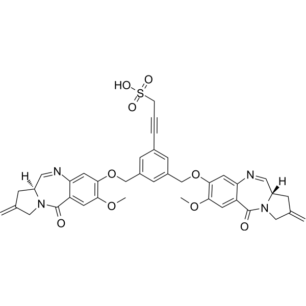 Di(MMY-SJG)-Ph-prop-2-yne-1-sulfonic acid
 MOMA-341
 Morpholino G phosphoramidite
InvivoChem的所有产品仅用于作科学研究,不面向患者销售
Copyright 2020 InvivoChem LLC | All Rights Reserved 粤ICP备20063088号-1


 COA
COA


 463611831
