| 规格 | 价格 | 库存 | 数量 |
|---|---|---|---|
| 5mg |
|
||
| 10mg |
|
||
| 25mg |
|
||
| 50mg |
|
||
| 100mg |
|
||
| Other Sizes |
|
| 靶点 |
Endogenous metabolite; glycine-conjugated secondary bile acid
|
|---|---|
| 体外研究 (In Vitro) |
石胆酸(LCA)是一种与胆汁淤积等不良反应相关的胆汁酸,在体内主要以Glycolithocholic acid(GLCA)和tauro-LCA(TLCA)的结合物存在。三苯氧胺与胆汁淤积的发展有关,它抑制磺基转移酶2A1(SULT2A1)催化的脱氢表雄酮(DHEA)磺化。本研究旨在表征LCA、GLCA和TLCA的磺化作用,并研究选择性雌激素受体调节剂(SERM)类的三苯乙烯(氯米芬、三苯氧胺、托瑞米芬、奥赛美芬、屈洛昔芬)、苯并噻吩(雷洛昔芬、阿唑昔芬)、四氢萘(拉索昔芬、萘呋辛)、吲哚(巴多昔芬)和苯并吡喃(acolbifene)是否抑制LCA、GLPA和TLCA磺化。人重组SULT2A1,但不是SULT2B1b或SULT1E1,催化LCA、GLCA和TLCA磺化,而这些酶中的每一种都催化DHEA磺化。LCA、GLCA和TLCA磺化由人肝细胞溶质催化,SULT2A1遵循底物抑制模型,具有相当的表观Km值(≤1µm)。每种SERM都以不同的效力和酶抑制模式抑制LCA、GLCA和TLCA磺化。通过对托瑞米芬、巴多昔芬和拉索菲芬进行结构修饰,LCA磺化的效力和抑制程度减弱或增加。在HepG2人肝细胞癌细胞中也观察到雷洛昔芬、巴多昔芬和阿可联苯对LCA磺化的抑制作用。总体而言,在所研究的SERM中,巴多昔芬和雷洛昔芬是LCA、GLCA和TLCA磺化最有效的抑制剂。这些发现为特定SERM的结构特征提供了见解,这些特征有助于抑制SULT2A1催化的LCA磺化。SERM对LCA、GLCA和TLCA解毒的抑制可能为SERM相关的不良反应提供生化基础[3]。
|
| 体内研究 (In Vivo) |
在杂合FH IIa型患者中,我们观察到与对照组的相应值相比,甘鹅脱氧胆酸、甘熊脱氧胆酸和Glycolithocholic acid的摩尔百分比显著降低,牛磺脱氧胆酸显著增加。在普罗布考治疗16周后,对6名患者的胆汁分析进行了复查。普罗布考显著降低了血清胆固醇水平。然而,治疗没有改变胆汁脂质组成和单个胆汁酸比例。结果表明,大多数杂合性FH患者胆汁过饱和,易形成胆固醇结石。此外,普罗布考降低血清胆固醇的机制似乎与胆汁脂质代谢的任何变化无关。[1]
结果:本研究纳入了32名UC患者和23名HC患者。研究发现,与HC相比,UC患者的肠道微生物群多样性降低。UC患者的厚壁菌门、梭菌IV、丁球菌、梭菌XlVa、粪杆菌和Roseburia显著降低(分别为P=0.75E-05、P=0.28E-07、P=0.0002、P=0.003、P=0.0003和P=0.0004)。UC组中变形杆菌、大肠杆菌、肠球菌、克雷伯氏菌和链球菌显著富集(分别为P=2.99E-09、P=3.63E-05、P=8.59E-05、P=0.003和P=0.016)。UC患者粪便中的继发性BAs,如石胆酸、脱氧胆酸、糖脱氧胆酸、Glycolithocholic acid和牛磺胆酸的浓度显著低于HCs(分别为P=8.1E-08、P=1.2E-07、P=3.5E-04、P=1.9E-03和P=1.8E-02),并与丁球菌、玫瑰孢菌、梭菌IV、粪杆菌和梭菌XlVb呈正相关(P<0.01)。UC患者的原发性BA,如牛磺胆酸、胆酸、牛磺脱氧胆酸和甘鹅脱氧胆酸的浓度显著高于HC患者(分别为P=5.3E-03、P=4E-02、P=0.042和P=0.045),并且与肠球菌、克雷伯氏菌、链球菌、乳杆菌和促炎细胞因子呈正相关(P<0.01)。UC患者TGR5的表达显著升高(0.019±0.013 vs 0.006±0.003,P=0.0003)。UC患者结肠黏膜标本中VDR表达显著降低(0.011±0.007 vs 0.016±0.004,P=0.033)。 结论:粪便BA谱与肠道微生物群和血清炎性细胞因子密切相关。肠道微生物群失调和粪便BAs结构改变可能通过BA受体TGR5和VDR参与调节炎症反应[2]。 |
| 酶活实验 |
LCA、GLCA/Glycolithocholic acid、TLCA和DHEA磺化试验的优化[3]
之前已经优化了人肝细胞质中的LCA磺化试验(Bansal和Lau,2016a)。LCA和GLCA/Glycolithocholic acid磺化在100µg细胞质蛋白时线性增加,而TLCA磺化在80µg细胞质蛋白质时线性增加(补充图S1,A-C)。重组SULT2A1催化的LCA、Glycolithocholic acid/GLCA和TLCA磺化在5µg酶下呈线性(补充图S1,D-F)。人肝细胞溶质催化LCA磺化反应在45分钟内呈线性,而GLCA在45分钟后呈线性。。。 |
| 动物实验 |
背景:肠道菌群及其代谢产物可能参与炎症性肠病的发生发展。近期多项临床研究表明,溃疡性结肠炎(UC)患者的粪便胆汁酸(BA)谱发生改变。研究发现,胆汁酸受体Takeda G蛋白偶联受体5(TGR5)和维生素D受体(VDR)通过调节NF-κB信号通路参与肠道炎症反应。我们假设,UC患者粪便胆汁酸谱的改变可能与肠道菌群和炎症反应相关。目的:探讨UC患者粪便胆汁酸的变化,并分析胆汁酸与肠道菌群和炎症之间的关系。方法:本研究采用16S rDNA测序技术检测UC患者和健康对照组(HCs)肠道菌群的差异。采用靶向代谢组学方法检测粪便胆汁酸水平。采用免疫组织化学方法分析粘膜TGR5和VDR的表达,并采用ELISA方法检测血清炎症细胞因子水平。[2]
|
| 参考文献 |
|
| 其他信息 |
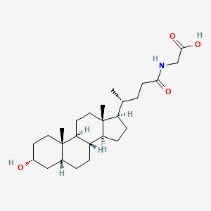
甘氨胆酸是石胆酸的甘氨酸结合物。它是一种胆汁酸甘氨酸结合物,也是一种N-酰基甘氨酸。它在功能上与石胆酸相关。它是甘氨胆酸盐的结合酸。
已有报道称,在智人和牛体内均存在甘氨胆酸,并有相关数据可供参考。 另见:甘氨胆酸盐(注释已移至此处)。 背景与目的:血清胆汁酸水平升高与肝硬化以及肝脏相关疾病和死亡风险增加相关。本文报告了在代谢性或胆汁淤积性肝病患者的前瞻性II期研究中,对肠道激素成纤维细胞生长因子19的工程化类似物aldafermin对循环胆汁酸谱的二次分析。方法:176例经活检确诊的非酒精性脂肪性肝炎(NASH)伴纤维化和肝脏脂肪含量升高(磁共振成像质子密度脂肪分数≥8%)的患者,分别接受0.3 mg(n = 23)、1 mg(n = 49)、3 mg(n = 49)、6 mg(n = 28)的阿达菲明或安慰剂(n = 27)治疗12周。62例原发性硬化性胆管炎(PSC)伴碱性磷酸酶升高(>正常值上限的1.5倍)的患者,分别接受1 mg(n = 21)、3 mg(n = 21)的阿达菲明或安慰剂(n = 20)治疗12周。分别于第1天和第12周采集血清样本,用于测定胆汁酸谱和III型胶原蛋白新表位特异性N端前肽(Pro-C3),后者是纤维化发生的直接指标。结果:阿达弗明治疗可显著降低血清胆汁酸水平,且呈剂量依赖性。尤其值得注意的是,在非酒精性脂肪性肝炎(NASH)和原发性硬化性胆管炎(PSC)患者中,阿达弗明均能显著降低具有较高疏水性指数的胆汁酸,例如脱氧胆酸、石胆酸、甘氨脱氧胆酸、甘氨鹅脱氧胆酸和甘氨胆酸。此外,阿达弗明主要抑制甘氨酸结合胆汁酸,而非牛磺酸结合胆汁酸。在合并的非酒精性脂肪性肝炎(NASH)和原发性硬化性胆管炎(PSC)人群中,胆汁酸水平的变化与新型纤维化标志物Pro-C3的变化相关。Pro-C3可检测III型胶原蛋白形成过程中的新表位。结论:Aldafermin显著降低了具有更强去污活性和细胞毒性的主要疏水性胆汁酸。我们的数据表明,胆汁酸可能有助于维持代谢性和胆汁淤积性肝病中肝脏的促纤维化微环境。简而言之:Aldafermin是一种肠道激素类似物,目前正在开发用于治疗慢性肝病。本文表明,无论病因如何,Aldafermin都能有效且稳定地抑制有毒的疏水性胆汁酸。利用Aldafermin的治疗策略可能广泛适用于其他慢性胃肠道和肝脏疾病。临床试验注册:该研究已在 Clinicaltrials.gov 注册,注册号为 NCT02443116 和 NCT02704364。[4] |
| 分子式 |
C26H43NO4
|
|---|---|
| 分子量 |
433.63
|
| 精确质量 |
433.319
|
| 元素分析 |
C, 72.02; H, 10.00; N, 3.23; O, 14.76
|
| CAS号 |
474-74-8
|
| 相关CAS号 |
24404-83-9 (mono-hydrochloride salt)
|
| PubChem CID |
115245
|
| 外观&性状 |
White to off-white solid powder
|
| 密度 |
1.112g/cm3
|
| 沸点 |
619.7ºC at 760mmHg
|
| 熔点 |
212-214°C (lit.)
|
| 闪点 |
328.6ºC
|
| LogP |
5.014
|
| tPSA |
86.63
|
| 氢键供体(HBD)数目 |
3
|
| 氢键受体(HBA)数目 |
4
|
| 可旋转键数目(RBC) |
6
|
| 重原子数目 |
31
|
| 分子复杂度/Complexity |
695
|
| 定义原子立体中心数目 |
9
|
| SMILES |
C[C@H](CCC(=O)NCC(=O)O)[C@H]1CC[C@@H]2[C@@]1(CC[C@H]3[C@H]2CC[C@H]4[C@@]3(CC[C@H](C4)O)C)C
|
| InChi Key |
XBSQTYHEGZTYJE-OETIFKLTSA-N
|
| InChi Code |
InChI=1S/C26H43NO4/c1-16(4-9-23(29)27-15-24(30)31)20-7-8-21-19-6-5-17-14-18(28)10-12-25(17,2)22(19)11-13-26(20,21)3/h16-22,28H,4-15H2,1-3H3,(H,27,29)(H,30,31)/t16-,17-,18-,19+,20-,21+,22+,25+,26-/m1/s1
|
| 化学名 |
2-[[(4R)-4-[(3R,5R,8R,9S,10S,13R,14S,17R)-3-hydroxy-10,13-dimethyl-2,3,4,5,6,7,8,9,11,12,14,15,16,17-tetradecahydro-1H-cyclopenta[a]phenanthren-17-yl]pentanoyl]amino]acetic acid
|
| 别名 |
Lithocholic acid glycine conjugate; Lithocholylglycine; Glycolithocholic acid; Lithocholylglycine; 474-74-8; Lithocholic acid glycine conjugate; Glycine, N-[(3a,5b)-3-hydroxy-24-oxocholan-24-yl]-; Q53GV75CJG; CHEMBL258818; CHEBI:37998; Glycolithocholic acid
|
| HS Tariff Code |
2934.99.9001
|
| 存储方式 |
Powder -20°C 3 years 4°C 2 years In solvent -80°C 6 months -20°C 1 month |
| 运输条件 |
Room temperature (This product is stable at ambient temperature for a few days during ordinary shipping and time spent in Customs)
|
| 溶解度 (体外实验) |
DMSO : ~50 mg/mL (~115.31 mM)
|
|---|---|
| 溶解度 (体内实验) |
配方 1 中的溶解度: ≥ 1.25 mg/mL (2.88 mM) (饱和度未知) in 10% DMSO + 90% (20% SBE-β-CD in Saline) (这些助溶剂从左到右依次添加,逐一添加), 澄清溶液。
例如,若需制备1 mL的工作液,可将100 μL 12.5mg/mL澄清的DMSO储备液加入到900μL 20%SBE-β-CD生理盐水中,混匀。 *20% SBE-β-CD 生理盐水溶液的制备(4°C,1 周):将 2 g SBE-β-CD 溶解于 10 mL 生理盐水中,得到澄清溶液。 配方 2 中的溶解度: ≥ 1.25 mg/mL (2.88 mM) (饱和度未知) in 10% DMSO + 90% Corn Oil (这些助溶剂从左到右依次添加,逐一添加), 澄清溶液。 例如,若需制备1 mL的工作液,可将 100 μL 12.5 mg/mL 澄清 DMSO 储备液添加到 900 μL 玉米油中并混合均匀。 请根据您的实验动物和给药方式选择适当的溶解配方/方案: 1、请先配制澄清的储备液(如:用DMSO配置50 或 100 mg/mL母液(储备液)); 2、取适量母液,按从左到右的顺序依次添加助溶剂,澄清后再加入下一助溶剂。以 下列配方为例说明 (注意此配方只用于说明,并不一定代表此产品 的实际溶解配方): 10% DMSO → 40% PEG300 → 5% Tween-80 → 45% ddH2O (或 saline); 假设最终工作液的体积为 1 mL, 浓度为5 mg/mL: 取 100 μL 50 mg/mL 的澄清 DMSO 储备液加到 400 μL PEG300 中,混合均匀/澄清;向上述体系中加入50 μL Tween-80,混合均匀/澄清;然后继续加入450 μL ddH2O (或 saline)定容至 1 mL; 3、溶剂前显示的百分比是指该溶剂在最终溶液/工作液中的体积所占比例; 4、 如产品在配制过程中出现沉淀/析出,可通过加热(≤50℃)或超声的方式助溶; 5、为保证最佳实验结果,工作液请现配现用! 6、如不确定怎么将母液配置成体内动物实验的工作液,请查看说明书或联系我们; 7、 以上所有助溶剂都可在 Invivochem.cn网站购买。 |
| 制备储备液 | 1 mg | 5 mg | 10 mg | |
| 1 mM | 2.3061 mL | 11.5306 mL | 23.0611 mL | |
| 5 mM | 0.4612 mL | 2.3061 mL | 4.6122 mL | |
| 10 mM | 0.2306 mL | 1.1531 mL | 2.3061 mL |
1、根据实验需要选择合适的溶剂配制储备液 (母液):对于大多数产品,InvivoChem推荐用DMSO配置母液 (比如:5、10、20mM或者10、20、50 mg/mL浓度),个别水溶性高的产品可直接溶于水。产品在DMSO 、水或其他溶剂中的具体溶解度详见上”溶解度 (体外)”部分;
2、如果您找不到您想要的溶解度信息,或者很难将产品溶解在溶液中,请联系我们;
3、建议使用下列计算器进行相关计算(摩尔浓度计算器、稀释计算器、分子量计算器、重组计算器等);
4、母液配好之后,将其分装到常规用量,并储存在-20°C或-80°C,尽量减少反复冻融循环。
计算结果:
工作液浓度: mg/mL;
DMSO母液配制方法: mg 药物溶于 μL DMSO溶液(母液浓度 mg/mL)。如该浓度超过该批次药物DMSO溶解度,请首先与我们联系。
体内配方配制方法:取 μL DMSO母液,加入 μL PEG300,混匀澄清后加入μL Tween 80,混匀澄清后加入 μL ddH2O,混匀澄清。
(1) 请确保溶液澄清之后,再加入下一种溶剂 (助溶剂) 。可利用涡旋、超声或水浴加热等方法助溶;
(2) 一定要按顺序加入溶剂 (助溶剂) 。
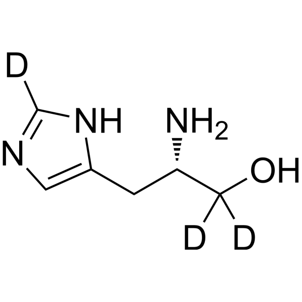 L-Histidinol-d3
 D-myo-Inositol-3-phosphate sodium
 (±)19(20)-DiHDPA
 4-Hydroxy-2-butanone
InvivoChem的所有产品仅用于作科学研究,不面向患者销售
Copyright 2020 InvivoChem LLC | All Rights Reserved 粤ICP备20063088号-1


 463611831
