| 规格 | 价格 | 库存 | 数量 |
|---|---|---|---|
| 5mg |
|
||
| 10mg |
|
||
| 25mg |
|
||
| 50mg |
|
||
| 100mg |
|
||
| 250mg |
|
||
| 500mg |
|
||
| Other Sizes |
|

| 靶点 |
STING (stimulator of interferon genes)
|
|---|---|
| 体外研究 (In Vitro) |
在 HEK293T 细胞中,H-151 (0.02-2 μM) 降低 IFNβ 荧光素酶报告基因测量值 [1]。在 THP-1 细胞中,H-151(0.5 μM;2 小时)抑制 TBK1 磷酸化 [1]。 hsSTING 掌化被 H-151(1 μM;3 小时)抑制 [1]。
|
| 体内研究 (In Vivo) |
在 CMA 治疗的小鼠中,H-151(每只小鼠 750 nmol;单次腹腔注射)可显着降低全身细胞因子反应 [1]。在 CMA 治疗的小鼠中,H-151(每只小鼠 750 nmol;单次腹腔注射)可显着降低全身细胞因子反应 [1]。 d) Trex1®/?表达生物发光 IFNβ 报告基因(750 nmol/小鼠;单次腹腔注射)的 H-151 小鼠达到很强的全身水平,显示出显着的血清半衰期,并且功能与野生型小鼠相似。在小鼠中表现出显着的效果[1]。 mmSTING 加合物形成 [1]。
|
| 酶活实验 |
高通量化合物筛选
使用GeneJuice 转染表达具有N-末端mCherry标签的小鼠STING的HEK293T细胞,该细胞具有编码环状二GMP合酶的构建体和IFN-β萤火虫萤光素酶报告质粒。三小时后,将转染的细胞接种在涂有文库化合物的384孔板 中。对于每种化合物,选择40nl的体积以在测定板中获得10μM的浓度。将每个孔中DMSO的量标准化为0.1%。过夜处理后,将细胞在裂解缓冲液(25mM磷酸三钠(pH 7.8),2mM DTT,2mM 1,2-二氨基环己烷-N,N,N′,N′-四乙酸,10%甘油,1%Triton X-100)中裂解20分钟,然后加入萤火虫萤光素酶底物。使用Tecan Infinite平板读数器测量报告活性。对来自EPFL BSF核心设施的约20000种化学多样化合物进行了筛选。为了鉴定hsSTING特异性化合物,用编码小鼠环状GMP–AMP合酶的构建体和IFN-β萤火虫萤光素酶报告质粒转染表达人STING构建体的HEK293T细胞。如上所述进行了进一步的分析。对来自EPFL BSF核心设施的约30000种化学多样化合物进行了筛选。[1] 竞争分析 将表达Flag–STING的HEK293T细胞与所示化合物孵育,1小时后,加入C-176-AL 1小时。将细胞收集在PBS中,并通过C-176-AL-介导的STING标记的凝胶内分析进行分析(见“化合物与STING结合的基于凝胶的分析”)。[1] |
| 细胞实验 |
免疫沉淀
Flag–STING在HEK293T细胞中通过多西环素诱导表达过夜。将细胞与或不与C-178或C-176(1μM)一起孵育1小时,并用DMSO或CMA(250μg ml−1)处理2小时。将细胞在PBS中洗涤,并在裂解缓冲液(50mM HEPES、150mM NaCl、10%甘油、1mM MgCl、1mM CaCl、1%Brij-58和蛋白酶抑制剂混合物(Sigma P8340))中裂解30分钟。使用抗Flag M2亲和凝胶琼脂糖凝胶(Sigma)在4 °C。在裂解缓冲液和PBS中严格洗涤后,完全去除上清液,并在进行SDS–PAGE之前在样品缓冲液中煮沸树脂。对于内源性STING的免疫沉淀,将脾细胞在上述裂解缓冲液中裂解,并与抗STING(RD System AF6516)和G琼脂糖珠(GE Healthcare,17-0618-01)孵育过夜。在PBS中洗涤珠,并对C-176-AL与STING的结合进行基于凝胶的分析 基于凝胶的化合物与STING结合的分析 将表达Flag–STING的HEK293T细胞与C-176-AL、C-175-AZ、碘代叠氮化物或H-151-AL在无血清培养基中孵育,收集在PBS中,并通过重复冷冻和解冻裂解。用新制备的“点击试剂”混合物处理43微升裂解的细胞,该混合物含有三(苄基三唑基甲基)胺(TBTA)(每个样品3μl,在1:4 DMSO:t-ButOH中3 mM)、四甲基罗丹明(TAMRA)叠氮化物(Thermo Fisher)、SiR叠氮化物(Spichrochrome)或SiR炔烃(Spichrome)(每个样本2μl,DMSO中1.25 mM)和新制备CuSO4(每个样本1μl)和三-(2-羧乙基)膦盐酸盐(TCEP)通过加入还原性样品缓冲液而淬灭。使用Fusion FX(Vilber-Lourmat)对凝胶内荧光进行可视化,并通过Fusion-capt高级采集软件进行分析 与二琥珀酰亚胺亚油酸酯交联 将表达Flag–mmSTING的HEK293T细胞与C-176(1μM)一起或不与C-176一起孵育1小时,并用DMSO或CMA(250μg ml−1)处理2小时。在PBS中与室温下在DMSO中新制备的1 mM二琥珀酰亚胺基亚油酸酯(DSS)(赛默飞世尔)进行交联1小时。 |
| 动物实验 |
小鼠及体内研究
C57BL/6J小鼠(品系编号000664)购自Jackson Laboratories。TREX1缺陷小鼠由T. Lindahl惠赠,并已回交至C57BL/6NJ品系超过10代。小鼠饲养于瑞士洛桑联邦理工学院(EPFL)的特定病原体清除(SPF)条件下。在药代动力学研究中,每只野生型小鼠腹腔注射750 nmol C-176(溶于200 μl玉米油中)。分别于注射后30分钟、2小时和4小时采集血液样本,并采用液相色谱-高分辨率质谱法(LC-HRMS)测定血清中C-176的浓度。为评估C-176的体内抑制作用,将8-12周龄的野生型小鼠分别注射载体或C-176。分别在1小时或4小时后,以224 mg kg⁻¹的浓度给予CMA。4小时后,处死小鼠并收集血清,以检测CMA诱导的细胞因子水平。为了评估H-151的体内抑制作用,将750 nmol H-151(溶于200 μl含10% Tween-80的PBS溶液中)腹腔注射到野生型小鼠体内。1小时后给予CMA(112 mg kg⁻¹),4小时后处死小鼠并收集血清。Trex1⁻/⁻小鼠的疗效研究如下:将7.5 μl C-176或DMSO(溶于85 μl玉米油中)注射到2-5周龄的小鼠体内,连续11天,每天两次。小鼠在CO₂麻醉箱中麻醉后,通过颈椎脱臼处死。在毒理学研究中,8周龄小鼠每日注射562.5 nmol C-176,持续2周。第14天,用肝素锂抗凝管采集血样,4℃离心后分离血浆,并储存于-80℃。使用DimensionXpand Plus测定血浆参数。外周血细胞分析中,采集100 μl血液于EDTA-K抗凝管中。使用ADVIA120血液分析仪进行全血细胞计数。为检测荧光素酶活性,将Trex1−/−Ifnb1Δβ-luc/Δβ-luc报告基因小鼠(4-7周龄)腹腔注射750 nmol H-151或DMSO(溶于200 μl含0.1% Tween-80的PBS缓冲液中),持续7天。对于体内成像,小鼠用异氟烷麻醉,并经静脉注射15 mg kg−1 XenoLight D-荧光素(溶于等渗氯化钠溶液)。注射两分钟后,使用In-vivo Xtreme II成像设备定量光子通量,像素合并设置为8 × 8,积分时间为3分钟。动物实验已获得沃州消费和兽医事务服务处或德累斯顿州政府的批准,并按照相关法律法规进行。 制剂和剂量:腹腔注射;每只小鼠腹腔注射750 nmol H-151,溶于200 μl含10% Tween-80的PBS溶液中。 |
| 参考文献 | |
| 其他信息 |
先天免疫通路异常激活与多种疾病相关。对先天免疫通路分子机制的深入理解为靶向治疗方法带来了希望,但开发能够特异性作用于目标分子的药物仍然面临挑战。本文报道了高效且选择性的小分子干扰素基因刺激因子(STING)拮抗剂的发现和表征。STING是细胞内DNA感应通路的核心信号组分1,2。从机制上讲,这些化合物共价靶向预测的跨膜半胱氨酸残基91,从而阻断STING的激活诱导棕榈酰化。利用这些抑制剂,我们发现STING的棕榈酰化对于其在Golgi体组装成多聚体复合物至关重要,进而影响下游信号因子的募集。这些化合物及其衍生物能够降低人和小鼠细胞中STING介导的炎症细胞因子的产生。此外,我们还发现这些小分子拮抗剂能够减轻小鼠自身炎症性疾病的病理特征。总之,我们的研究揭示了STING可被药物抑制的机制,并证明了靶向STING的疗法在治疗自身炎症性疾病方面的潜力。[1]
|
| 分子式 |
C17H17N3O
|
|---|---|
| 分子量 |
279.336383581162
|
| 精确质量 |
279.137
|
| 元素分析 |
C, 73.10; H, 6.13; N, 15.04; O, 5.73
|
| CAS号 |
941987-60-6
|
| 相关CAS号 |
941987-60-6;
|
| PubChem CID |
7616033
|
| 外观&性状 |
White to off-white solid powder
|
| LogP |
3.4
|
| tPSA |
56.9Ų
|
| 氢键供体(HBD)数目 |
3
|
| 氢键受体(HBA)数目 |
1
|
| 可旋转键数目(RBC) |
3
|
| 重原子数目 |
21
|
| 分子复杂度/Complexity |
352
|
| 定义原子立体中心数目 |
0
|
| InChi Key |
UJZDIKVQFMCLBE-UHFFFAOYSA-N
|
| InChi Code |
InChI=1S/C17H17N3O/c1-2-12-7-9-13(10-8-12)19-17(21)20-16-11-18-15-6-4-3-5-14(15)16/h3-11,18H,2H2,1H3,(H2,19,20,21)
|
| 化学名 |
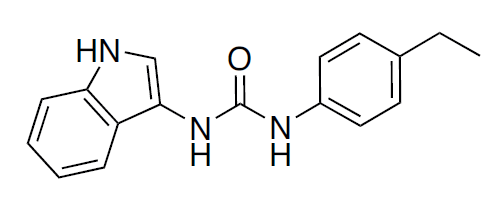
N-(4-Ethylphenyl)-N'-1H-indol-3-yl-urea
|
| 别名 |
H 151; H-151; 1-(4-ethylphenyl)-3-(1H-indol-3-yl)urea; H-151; N-(4-ethylphenyl)-N'-1H-indol-3-yl-urea; H 151; MFCD08026581; H151
|
| HS Tariff Code |
2934999001
|
| 存储方式 |
Powder -20°C 3 years 4°C 2 years In solvent -80°C 6 months -20°C 1 month |
| 运输条件 |
Room temperature (This product is stable at ambient temperature for a few days during ordinary shipping and time spent in Customs)
|
| 溶解度 (体外实验) |
DMSO : ~100 mg/mL (~357.99 mM)
|
|---|---|
| 溶解度 (体内实验) |
配方 1 中的溶解度: 2.5 mg/mL (8.95 mM) in 5% DMSO 5% Tween80 + 90% PBS (这些助溶剂从左到右依次添加,逐一添加), 悬浮液;超声助溶。
配方 2 中的溶解度: ≥ 2.08 mg/mL (7.45 mM) (饱和度未知) in 10% DMSO + 40% PEG300 + 5% Tween80 + 45% Saline (这些助溶剂从左到右依次添加,逐一添加), 澄清溶液。 例如,若需制备1 mL的工作液,可将 100 μL 20.8 mg/mL澄清的DMSO储备液加入到400 μL PEG300中,混匀;再向上述溶液中加入50 μL Tween-80,混匀;然后加入450 μL生理盐水定容至1 mL。 *生理盐水的制备:将 0.9 g 氯化钠溶解在 100 mL ddH₂O中,得到澄清溶液。 View More
配方 3 中的溶解度: 2.08 mg/mL (7.45 mM) in 10% DMSO + 90% (20% SBE-β-CD in Saline) (这些助溶剂从左到右依次添加,逐一添加), 悬浊液; 超声助溶。 配方 4 中的溶解度: ≥ 2.08 mg/mL (7.45 mM) in 10% DMSO + 40% PEG300 + 5% Tween80 + + 45% Saline ≥ 2.08 mg/mL (7.45 mM) in 10% DMSO + 90% (20% SBE-β-CD in saline) ≥ 2.08 mg/mL (7.45 mM) in 10% DMSO + 90% Corn oil 1、请先配制澄清的储备液(如:用DMSO配置50 或 100 mg/mL母液(储备液)); 2、取适量母液,按从左到右的顺序依次添加助溶剂,澄清后再加入下一助溶剂。以 下列配方为例说明 (注意此配方只用于说明,并不一定代表此产品 的实际溶解配方): 10% DMSO → 40% PEG300 → 5% Tween-80 → 45% ddH2O (或 saline); 假设最终工作液的体积为 1 mL, 浓度为5 mg/mL: 取 100 μL 50 mg/mL 的澄清 DMSO 储备液加到 400 μL PEG300 中,混合均匀/澄清;向上述体系中加入50 μL Tween-80,混合均匀/澄清;然后继续加入450 μL ddH2O (或 saline)定容至 1 mL; 3、溶剂前显示的百分比是指该溶剂在最终溶液/工作液中的体积所占比例; 4、 如产品在配制过程中出现沉淀/析出,可通过加热(≤50℃)或超声的方式助溶; 5、为保证最佳实验结果,工作液请现配现用! 6、如不确定怎么将母液配置成体内动物实验的工作液,请查看说明书或联系我们; 7、 以上所有助溶剂都可在 Invivochem.cn网站购买。 |
| 制备储备液 | 1 mg | 5 mg | 10 mg | |
| 1 mM | 3.5799 mL | 17.8993 mL | 35.7987 mL | |
| 5 mM | 0.7160 mL | 3.5799 mL | 7.1597 mL | |
| 10 mM | 0.3580 mL | 1.7899 mL | 3.5799 mL |
1、根据实验需要选择合适的溶剂配制储备液 (母液):对于大多数产品,InvivoChem推荐用DMSO配置母液 (比如:5、10、20mM或者10、20、50 mg/mL浓度),个别水溶性高的产品可直接溶于水。产品在DMSO 、水或其他溶剂中的具体溶解度详见上”溶解度 (体外)”部分;
2、如果您找不到您想要的溶解度信息,或者很难将产品溶解在溶液中,请联系我们;
3、建议使用下列计算器进行相关计算(摩尔浓度计算器、稀释计算器、分子量计算器、重组计算器等);
4、母液配好之后,将其分装到常规用量,并储存在-20°C或-80°C,尽量减少反复冻融循环。
计算结果:
工作液浓度: mg/mL;
DMSO母液配制方法: mg 药物溶于 μL DMSO溶液(母液浓度 mg/mL)。如该浓度超过该批次药物DMSO溶解度,请首先与我们联系。
体内配方配制方法:取 μL DMSO母液,加入 μL PEG300,混匀澄清后加入μL Tween 80,混匀澄清后加入 μL ddH2O,混匀澄清。
(1) 请确保溶液澄清之后,再加入下一种溶剂 (助溶剂) 。可利用涡旋、超声或水浴加热等方法助溶;
(2) 一定要按顺序加入溶剂 (助溶剂) 。
 STING Degrader-2
 ZSA-215
 STING agonist-44
 Sim-9
Sim-9
InvivoChem的所有产品仅用于作科学研究,不面向患者销售
Copyright 2020 InvivoChem LLC | All Rights Reserved 粤ICP备20063088号-1


 COA
COA
 463611831
