| 规格 | 价格 | 库存 | 数量 |
|---|---|---|---|
| 5mg |
|
||
| 10mg |
|
||
| 25mg |
|
||
| 50mg |
|
||
| 100mg |
|
||
| 250mg |
|
||
| 500mg |
|
||
| Other Sizes |
|

| 靶点 |
NF-κB; TNF-α; IKKβ; IL-6
|
|---|---|
| 体外研究 (In Vitro) |
Homoplantaginin 还原 DPPH 自由基的 IC50 为 0.35 μg/mL。Homoplantaginin(0.1-100μg/mL)除了增加暴露于 H2O2 的人肝细胞 HL-7702 细胞上清液中的谷胱甘肽、谷胱甘肽过氧化物酶和超氧化物歧化酶外,还显着减少乳酸脱氢酶的渗漏[1]。棕榈酸 (100 μM) 诱导的 Toll 样受体 4 表达会被Homoplantaginin (0.1、1、10 μM) 剂量依赖性地降低。通过抑制 NLRP3 和 caspase-1 蛋白(活性氧敏感的硫氧还蛋白相互作用蛋白),Homoplantaginin严格调节棕榈酸诱导的活性氧,以防止 NLRP3 炎症小体激活 [2]。 Homoplantaginin 预处理显着降低棕榈酸诱导的人脐静脉内皮细胞中 TNF-α 和 IL-6 的 mRNA 表达以及 IKKβ 和 NF-κB p65 磷酸化。 Homoplantaginin 显着改变 IRS-1 的 Ser/Thr 磷酸化,增强 Akt 和内皮一氧化氮合酶磷酸化,并在胰岛素存在下增加 NO 产生 [3]。
Homoplantaginin [1] 的抗氧化作用 为了评价Homoplantaginin对自由基的直接清除作用,我们测定了一种稳定的自由基DPPH的清除活性。如图2所示,当浓度达到1.6 μg/ml时,Homoplantaginin降低DPPH自由基水平,显示出清除自由基的作用,IC50值为0.35 μg/ml。l-抗坏血酸具有清除自由基的作用,IC50值为0.43 μg/ml。 细胞活力暴露于Homoplantaginin体外[1] 将HL-7702细胞分别暴露于Homoplantaginin(0.1、1、10、50、100、200 μg/ml)中72 h,通过MTT法测定活细胞数,评估细胞生长抑制率。如图3所示,这些浓度的Homoplantaginin均未引起任何明显的细胞毒性(与未处理的细胞相比P < 0.05)。 LDH释放抑制[1] 采用H2O2暴露HL-7702细胞模型,研究Homoplantaginin对肝细胞损伤的保护作用。暴露于H2O2中18小时显著增加LDH向介质的泄漏。如图4所示,对照细胞上清液中LDH水平为21 U/ml, H2O2胁迫下LDH水平升高至122 U/ml。Homoplantaginin预处理HL-7702细胞可显著抑制LDH渗漏,且呈剂量依赖性。在浓度为0.1、1、10、50和100 μg/ml时,LDH水平分别降至73.5±4.4、65.2±5.1、58.4±3.2、52.1±2.6和41.6±4.3 U/ml(图4)。 GSH、GSH- px、SOD升高[1] 测定h2o2损伤HL-7702细胞中GSH、GSH- px和SOD的活性。结果见表1。h2o2损伤细胞的GSH水平从58.4±4.5 nmol/mg蛋白的正常值降至40.5±5.7 nmol/mg蛋白。Homoplantaginin阻止了H2O2对GSH的消耗。预处理24 h后,在0.1 ~ 100 μg/ml浓度范围内,GSH呈剂量依赖性升高(表1)。 然后我们检测了Homoplantaginin对肝细胞GSH-Px和SOD的影响。HL-7702细胞暴露于H2O2后,GSH-Px和SOD活性降低(表1)。Homoplantaginin增加两种抗氧化酶的活性呈剂量依赖性(表1)。 棕榈酸(PA)诱导的血管内皮炎症在血管疾病的发生发展中起着至关重要的作用。本文研究了中药鼠尾草中主要类黄酮Homoplantaginin的作用。研究了pa处理的人脐静脉内皮细胞炎症及其分子机制。首先,我们发现Homoplantaginin(0.1,1,10 μM)剂量依赖性地降低了PA (100 μM)诱导的toll样受体-4的表达。在脂多糖刺激下,进一步证实了Homoplantaginin的抑制作用。此外,在PA处理下,下游适应蛋白包括髓样分化初级反应基因88、toll/白细胞介素-1受体结构域含有适配器诱导干扰素-β和肿瘤坏死因子受体相关因子-6被Homoplantaginin成功抑制。此外,我们发现Homoplantaginin通过抑制活性氧物种敏感的硫氧还蛋白相互作用蛋白NLRP3和caspase-1,严格控制pa诱导的活性氧来阻止核苷酸结合结构域样受体3 (NLRP3)炎性体的激活。同时,也降低了炎症介质(白细胞介素-1β、细胞间黏附分子-1和单核细胞趋化蛋白-1)的蛋白和mRNA水平。此外,Homoplantaginin还能恢复pa损伤的一氧化氮生成。综上所述,这些结果表明,Homoplantaginin通过抑制toll样受体-4和NLRP3通路,保护内皮细胞免受pa诱导的内皮炎症的改善,并恢复一氧化氮的产生,这表明它可能是一个潜在的候选物,可以进一步开发用于预防和治疗血管疾病。[2] 最近的数据表明,炎症在胰岛素抵抗的发展中起着重要作用。本研究旨在研究中药鼠尾草中的类黄酮Homoplantaginin的活性。内皮细胞中棕榈酸(PA)诱导的胰岛素敏感性及其抗炎特性的潜在机制。前处理的Homoplantaginin对人脐静脉内皮细胞(HUVECs)有显著抑制PA诱导的肿瘤坏死因子-α (TNF-α)和白细胞介素-6 (IL-6) mRNA表达,抑制κB激酶β (IKKβ)和核因子-κB (NF-κB) p65磷酸化。对于PA损伤胰岛素受体底物-1 (IRS-1)的胰岛素依赖性酪氨酸磷酸化和一氧化氮(NO)的产生,前处理的Homoplantaginin可以有效逆转PA的作用。此外,Homoplantaginin显著调节IRS-1的丝氨酸/苏氨酸磷酸化,改善Akt和内皮型一氧化氮合酶(eNOS)的磷酸化,并在胰岛素存在下增加NO的产生。综上所述,我们的研究结果表明,Homoplantaginin通过抑制炎症和通过IKKβ/IRS-1/pAkt/peNOS通路调节细胞信号传导来改善内皮胰岛素抵抗,这表明它可能用于预防和治疗与胰岛素抵抗相关的内皮功能障碍。[3] |
| 体内研究 (In Vivo) |
Homoplantaginin (25-100mg/kg) 显着降低血清丙氨酸和天冬氨酸转氨酶水平的升高,并降低 TNF-α 和 IL-1 水平。相同的过程还增加了肝匀浆中 GSH、GSH-Px 和 SOD 的水平,并降低了硫代巴比妥酸反应物质的含量[1]。Homoplantaginin吸收迅速(Tmax=16.00±8.94min),其平均Cmax范围在0.77至1.27 nmol/mL之间。据估计,只有 0.75% 的口服生物利用度是绝对的。
Homoplantaginin对BCG/LPS诱导的ILI的治疗作用 采用卡介苗/LPS诱导小鼠ILI的方法,评价了Homoplantaginin的治疗作用。在该模型中,小鼠表现出严重的肝损伤,血清转氨酶(ALT、AST)升高,促炎细胞因子如TNF-α、IL-1升高,多种抗氧化酶如GSH、GSH- px、SOD降低(表2、表3)。Homoplantaginin明显抑制ILI的发生。如表2所示,给药10 d后,小鼠血清中谷丙转氨酶和谷丙转氨酶水平显著降低。ALT和AST的降低呈剂量依赖性(25、50、100 mg/kg/d)。另一方面,在连续给药期间没有观察到明显的体重减轻(数据未显示)。同样,与BCG + lps处理的小鼠相比,联苯酯(100 mg/kg)降低了ALT和AST的水平(表2)。 抗氧化酶升高[1] 测定小鼠肝组织中GSH、GSH- px、SOD水平。如表3所示,Homoplantaginin(25、50、100 mg/kg)显著提高GSH、GSH- px活性(P < 0.01),微弱提高SOD活性(25、50 mg/kg, P < 0.05)。100 mg/kg,与未处理模型相比P < 0.05,表3)。 TBARS下降[1] 小鼠BCG启动后注射LPS导致肝组织TBARS水平显著升高(表3)。Homoplantaginin显著降低TBARS升高(25 mg/kg, P > 0.05);50和100 mg/kg,与未处理模型相比P < 0.05,表3)。 TNF-α和IL-1 [1] 如表4所示,BCG/ lps诱导小鼠血清中TNF-α和IL-1水平明显高于对照组。Homoplantaginin可明显抵消ILI小鼠血清中TNF-α和IL-1水平升高(25 mg/kg, P < 0.05);50和100 mg/kg,与未处理模型相比P < 0.01,表4)。 Homoplantaginin对肝脏组织学的影响[1] 正常小鼠肝脏组织病理学检查未见组织学异常(图5A)。LPS注射bcg小鼠,可见肝窦充血,炎性细胞散在浸润,出现点状坏死、碎片状坏死、桥状坏死,炎性细胞在坏死组织周围排列(图5B)。在Homoplantaginin(25、50、100 mg/kg/d)处理的小鼠中,坏死的面积和程度减少,炎症细胞的迁移减少(图5D-F)。表2显示了各组小鼠的肝损伤类别。Homoplantaginin治疗组(50、100 mg/kg/d, P < 0.05)大鼠肝损伤程度明显改善(表2)。 本研究旨在研究鼠尾草主要活性成分 的药动学特性。本研究测定了Homoplantaginin的有效分配系数、大鼠肠段的原位吸收以及大鼠肠道细菌对Homoplantaginin的体外生物转化。此外,大鼠还分别通过静脉、腹腔和口服给药给药Homoplantaginin。采用高效液相色谱法测定Homoplantaginin和Homoplantaginin代谢物hispidulin的浓度。经静脉、腹膜注射后,无法测定海鞘磷脂的浓度。口服后同时定量hispidulin和homintaginin, homintaginin吸收迅速(Tmax=16.00±8.94min),平均Cmax在0.77 ~ 1.27nmol/mL之间。计算出其绝对口服生物利用度仅为0.75%,曲线下面积(AUC)约为Homoplantaginin的5.4倍。大鼠肠道细菌对Homoplantaginin的生物转化可能是其口服生物利用度较差的原因。[4] |
| 酶活实验 |
抗氧化活性[1]
Homoplantaginin的抗氧化活性通过dpph清除试验进行了评估(Watjen et al., 2007, Yang et al., 2005)。将含Homoplantaginin(实验化合物)和40 μl DPPH(1,1-二苯基-2-picrylhydrazyl自由基溶解于MeOH)的反应混合物160微升置于96孔板中,暗孵育30 min。对照组用蒸馏水代替实验化合物溶液。反应后,用517 nm比色法测定剩余的DPPH。从反应混合物的吸光度中减去Homoplantaginin的吸光度作为空白。自由基清除活性计算为100 × (ODcontrol−ODtest)/ODcontrol。IC50值定义为抑制DPPH自由基形成50%所需的测试化合物浓度。l-抗坏血酸,一种众所周知的抗氧化剂,被用作阳性对照(Yang et al., 2005)。 Homoplantaginin在体外的生物转化[4] 采用大鼠肠道菌群和含2.16 μMHomoplantaginin的GAM溶液(1:9,v:v)组成完整的培养体系进行肠道细菌培养。然后将混合物在37℃厌氧培养中孵育。在设计时间(5、10、20、30、45、60、90、120、180、300、480、720 min),取出反应混合物,用甲醇(1:1,v:v)萃取。旋流3min, 12000 rpm离心10min,最后取20 μL上清液入HPLC系统分析。条件与2.3相同。 |
| 细胞实验 |
MTT 测定用于评估培养细胞的活力。Homoplantaginin以不同浓度(0.1、1、3、10、30、100 M)作用于人脐静脉内皮细胞 48 小时。然后,每孔加入 20 μL MTT (5 mg/mL),在 37°C 下再培养 4 小时。除去上清液后,添加 DMSO 以帮助溶解甲臜晶体。在 540 nm[3] 处测量吸光度。
细胞培养和活力[1] 人肝细胞系HL-7702在rmi -1640中维持,rmi -1640中添加10% (v/v)热灭活胎牛血清、青霉素-链霉素(100 IU/ml - 100 μg/ml)、2 mM谷氨酰胺和10 mM Hepes缓冲液,在37℃的潮湿环境中(5% CO2, 95%空气)。采用3-[4,5-二甲基噻唑-2-基]-2,5-二苯基溴化四氮唑(MTT)测定法评估细胞生长和活力,详见其他文献(Zhang et al., 2008a)。 H2O2诱导的氧化应激[1] HL-7702细胞以3 × 10⁵ / ml的密度在6孔板中培养,并使其生长到所需的合流度。用不同浓度(0.1 ~ 100 μg/ml)的Homoplantaginin预处理细胞24 h,然后暴露于750 μM的H2O2中18 h (Wang et al., 2005a)。未经H2O2处理的细胞在与实验方案中使用的H2O2相同的条件下孵育。 细胞内ROS水平测定[2] 将HUVECs (5 × 10⁴细胞/孔)接种于6孔板中,在5% CO2培养箱中37℃保存48小时。然后将培养基替换为无血清培养基,使细胞饥饿2.5小时。细胞分别用PA (100 μM)处理1.5、3和6小时,或在PA (100 μM)处理3小时之前,用不同浓度的Homoplantaginin(0.1、1和10 μM)或Sal (500 μM)预处理0.5小时。收获细胞,用10 μM DCFH-DA在37℃下暗室孵育20分钟。用冷磷酸盐缓冲盐水洗涤两次后,用FACS Calibur流式细胞仪 在激发波长为488 nm,发射波长为525 nm.24下对细胞进行分析利用Cell Quest软件计算二氯荧光素(DCF)的荧光分布。 Western Blotting [2] 将HUVECs (5 × 10⁴细胞/孔)接种于6孔板中,在5% CO2培养箱中37℃保存48小时。然后将培养基替换为无血清培养基,使细胞饥饿2.5小时。分别用不同浓度Homoplantaginin(0.1、1、10 μM)或Sal (500 μM)预处理细胞0.5 h,再用PA (100 μM)处理细胞3 h。此外,如上所述,对HUVECs进行播种和饥饿。然后,用Homoplantaginin(10 μM)或水杨酸盐(500 μM)预处理细胞0.5 h,再加脂多糖(1 μg/mL)预处理1 h。根据制造商推荐的膜和细胞质蛋白提取试剂盒获得相关蛋白裂解物,并用BCA蛋白测定试剂盒检测蛋白浓度。等量的蛋白质(30 μg)通过10%十二烷基硫酸钠-聚丙烯酰胺凝胶电泳分离,随后电转移到聚偏二氟乙烯膜上。在含有5%脱脂牛奶和0.05% Tween 20的tris缓冲盐水中37℃阻断2小时后,将膜与指定的抗体孵育。使用ChemiDoc XRS系统显示蛋白条带,并使用Image J软件(National Institutes of Health)进行分析。 ELISA法检测炎症介质[2] 将HUVECs (5 × 10⁴细胞/孔)接种于6孔板中,在5% CO2培养箱中37℃保存48小时。然后将培养基替换为无血清培养基,使细胞饥饿2.5小时。分别用不同浓度的Homoplantaginin(0.1、1、10 μM)或Sal (500 μM)预处理细胞0.5 h,再用PA (100 μM)预处理细胞3 h。使用相应的ELISA试剂盒,按照制造商的说明,定量细胞上清液中IL-1β、ICAM-1和MCP-1的浓度。 细胞内一氧化氮水平测定[2] 将HUVECs (2 × 10⁴个细胞/孔)接种于48孔板中,在5% CO2培养箱中37°C保存48小时。然后将培养基替换为无血清培养基,使细胞饥饿2.5小时。分别用不同浓度的Homoplantaginin(0.1、1、10 μM)或Sal (500 μM)预处理细胞0.5 h,再用PA (100 μM)预处理细胞3 h。然后用PBS洗涤细胞2次,并用5 μM, DAF-FM DA在37℃下孵育25分钟。用PBS洗涤2次后,在激发波长为495nm、发射波长为515nm的Olympus荧光显微镜下分析细胞。用Image J软件分析荧光强度。 |
| 动物实验 |
大鼠:将高车前苷溶解于DMSO、PEG 400、乙醇和生理盐水(体积比2:2:3:3)的混合溶液中,配制成浓度为10 mg/mL的溶液。将大鼠随机分为三组,分别进行口服(150 mg/kg)、尾静脉注射(15 mg/kg)和腹腔注射(15 mg/kg)。给药后5、10、20、30、45、60、90、120和180分钟,从眼眶后静脉丛抽取约0.5 mL血液样本至肝素化微量离心管中。将血液样本以10,000 rpm离心[4]提取血浆。
小鼠:将高车前苷溶解于5%淀粉溶液中。在实验期间,分别以 25、50 和 100 mg/kg/d 的剂量,通过胃管灌注给予小鼠高车前苷。注射脂多糖 (LPS) 8 小时后,用乙醚麻醉小鼠,并从下静脉放血取血。为了便于组织学检查,取出肝脏并用福尔马林固定[1]。 急性肝损伤模型和高车前苷治疗[1] 在卡介苗/脂多糖 (BCG/LPS) 诱导的小鼠免疫性肝损伤 (ILI) 模型中评估了高车前苷的保肝作用。雄性 Balb/c 小鼠 (20 ± 2 g) 饲养于塑料笼中,可自由获取食物和水。将2.5 mg卡介苗悬浮于0.2 ml生理盐水中,经尾静脉注射入小鼠体内。10天后,注射7.5 μg脂多糖(LPS),该物质溶解于0.2 ml生理盐水中。实验期间,分别以25、50和100 mg/kg/d的剂量,通过胃管灌注给予小鼠高车前苷。目前临床上用于治疗肝炎的联苯酯作为阳性对照,经口给予(Pan et al., 2006)。注射LPS 8小时后,用乙醚麻醉小鼠,并通过下腹静脉放血采集血样。取出肝脏并用福尔马林固定,用于组织学分析(Lu et al., 2008; Wang et al., 2005b)。 表观分配系数的测定[4] 采用摇瓶法测定高车前苷和毛蕊花素的表观分配系数。将高车前苷和毛蕊花素溶解于不同pH值的溶液中。将所得高车前苷和毛蕊花素溶液与等体积的正辛醇(预先用水饱和)混合,摇匀,并在37℃水浴中孵育24小时。离心后,采用高效液相色谱法(HPLC)测定水相和辛醇相中高车前苷和毛蕊花素的浓度。然后,通过辛醇相中高车前苷和毛蕊花素的浓度与水相中浓度的比值,得到表观分配系数。 大鼠原位单次肠灌注(SPIP)研究[4] 灌注缓冲液由以下成分组成:1.4 g/L 葡萄糖、20 mg/L 酚红、7.8 g/L NaCl、0.35 g/L KCl、1.37 g/L NaHCO3、0.32 g/L NaH2PO4、0.02 g/L MgCl2、0.37 g/L CaCl2。高车前苷的一级标准储备液用二甲基亚砜配制,浓度为10 g/L。将高车前苷工作标准溶液用肠道空白灌注缓冲液稀释至所需浓度,制备高车前苷工作标准溶液。 简而言之,雄性SD大鼠禁食16-18小时,期间可自由饮水,然后腹腔注射10%水合氯醛(3.4 mL/kg)进行麻醉,并置于加热垫上以维持正常体温。确认痛觉反射消失后,沿腹部正中线做纵切口,分别取10 cm长的十二指肠、空肠、回肠和结肠,用预热至37℃的生理盐水轻轻冲洗,然后连接到灌注装置上。最后,用封口膜覆盖整个手术区域以减少水分蒸发。使用注射泵以0.2 mL/min的恒定流速灌注空白灌注缓冲液30分钟,随后以0.2 mL/min的恒定流速灌注高车前苷工作标准溶液120分钟。每隔15分钟用微量离心管收集出口灌注液样品。实验结束时,测量节段长度(不拉伸),最后对动物实施安乐死[12]。研究样品储存于-20 °C直至分析。渗透性研究产生的样品采用已验证的方法进行分析。样品以12,000 rpm离心10分钟,上清液直接注入高效液相色谱柱,无需预先制备。在分析过程中,除了渗透性样品外,还分发了质控样品。数据是基于使用大鼠肠灌注空白制备的质控样品的性能而接受的。 药代动力学研究[4] 将高车前苷溶解于二甲基亚砜、PEG 400、乙醇和生理盐水(体积比2:2:3:3)的混合溶液中,浓度为10 mg/mL。将大鼠随机分为三组,分别接受口服(150 mg/kg)、尾静脉注射(15 mg/kg)和腹腔注射(15 mg/kg)。分别于给药后5、10、20、30、45、60、90、120和180分钟,从眼眶后静脉丛采集血样(约0.5 mL)至肝素化微量离心管中。将血液样本在 4 °C 下以 10,000 rpm 离心 1 分钟分离血浆样本,并将血浆样本储存于 −20 °C 直至进一步分析。使用 Phoenix_1.1 药代动力学软件,根据非房室模型计算以下药代动力学参数:半衰期 (T1/2)、浓度-时间曲线下面积 (AUC)、平均滞留时间 (MRT)、清除率 (Cl) 和稳态表观分布容积 (Vss)。数据以均值 ± 标准差 (SD) 表示。最大浓度 (Cmax) 和达峰时间 (Tmax) 由浓度-时间曲线获得。口服生物利用度 (F) 和腹腔注射生物利用度 (Fi.p.) 使用以下公式计算: |
| 药代性质 (ADME/PK) |
表观分配系数的测定[4]
毛蕊花素和高车前苷在pH 1.2至pH 8.0范围内稳定,因为孵育24小时后,油相和水相中药物的总浓度均未发生变化,表明毛蕊花素和高车前苷在胃肠道pH条件下未发生降解。毛蕊花素和高车前苷在不同pH溶液中的表观分配系数结果如图2所示。可以看出,毛蕊花素和高车前苷的表观分配系数分别为2.4~2.8和1.0~1.2。结果表明,毛蕊花素和高车前苷在胃肠道pH条件下未发生电离,其吸收部位为胃和肠道。结果还表明,毛蕊花素和高车前苷的胃肠道吸收与pH值无关。 大鼠肠段中高车前苷的原位吸收[4] 据报道,酚红会干扰大鼠肠灌注中难溶性药物的吸收和转运,因为酚红在肠道中会被部分吸收[13]。采用重量法校准灌注液的体积变化。表1列出了高车前苷在三种浓度下于四个肠段的有效渗透率。高车前苷的吸收取决于肠道部位,在每个测试浓度下均可在十二指肠、空肠、回肠和结肠中发现吸收。三种浓度下的有效渗透率顺序均为回肠 > 十二指肠 > 空肠 > 结肠。在大鼠肠段中测得的最小和最大有效渗透系数(Peff)分别为0.259 × 10⁻⁴(结肠)和0.687 × 10⁻⁴(回肠)。单次肠灌注实验获得的同型车前草素的渗透系数与普萘洛尔的报道值(0.30–0.75 × 10⁻⁴ cm/s)[14]、[15]、[16]相当,普萘洛尔常被用作跨细胞被动吸收的参考化合物。此外,在每个同型车前草素浓度下,四个肠段的Peff值均无统计学差异,提示同型车前草素在四个肠段中的转运机制可能主要为被动转运。高车前苷的吸收率(Ka)在空肠最高,在十二指肠和回肠明显降低,然后在结肠显著降低。 大鼠肠道细菌体外生物转化高车前苷的时间进程[4] 图3显示了正常大鼠肠道细菌生物转化基质中高车前苷的平均时间-浓度曲线。如图3所示,高车前苷代谢迅速,在180分钟内几乎完全生物转化,最终约有75%转化为毛蕊花素。因此,我们知道,在肠道细菌存在的情况下,高车前苷很容易转化为毛蕊花素,从而影响其口服生物利用度。 药代动力学结果[4] 经验证的方法已成功应用于高车前苷的药代动力学研究。图4显示了静脉和腹膜给药后高车前苷的平均血浆浓度-时间曲线。本研究同时定量了口服给药后的毛蕊花素和高车前苷。图5显示了口服给药后高车前苷和毛蕊花素的平均血浆浓度-时间曲线。基于高车前苷和毛蕊花素的血浆浓度,计算了基本药代动力学参数,并汇总于表2。 正如我们所见,静脉给药后,血浆中高车前苷的浓度在50分钟内迅速消除。腹膜给药后,可以明显观察到血浆药物浓度在20分钟内迅速达到峰值,平均Cmax为15.45至21.99 nmol/mL,并在接下来的100分钟内迅速下降。计算得出Fi.p.为102.85%。这表明,如有效分配系数测定和原位吸收大鼠肠段研究所述,高车前苷在肠道内被完全吸收。经灌胃给药后,由于吸收迅速,高车前苷在给药后血浆中迅速达到Cmax,平均Tmax为7至25分钟。计算得出绝对口服生物利用度为0.75%。相比之下,毛蕊花素吸收迅速(Tmax = 5–23 分钟),平均 Cmax 介于 4.73 和 6.87 nmol/mL 之间,绝对生物利用度为 4.02%,约为高车前苷的 5 倍。研究表明,大部分高车前苷在大鼠肠道细菌的作用下转化为毛蕊花素。如图 5 所示,口服给药的浓度-时间曲线呈现双峰。多种机制可能解释这一现象,例如肠肝循环、胃排空分阶段以及不同的“吸收窗口”。肠肝循环可能是造成这一现象的原因,因为在静脉注射给药的浓度-时间曲线中未观察到双峰。此外,代谢产物毛蕊花素也呈现出相同的现象。[4] |
| 参考文献 |
|
| 其他信息 |
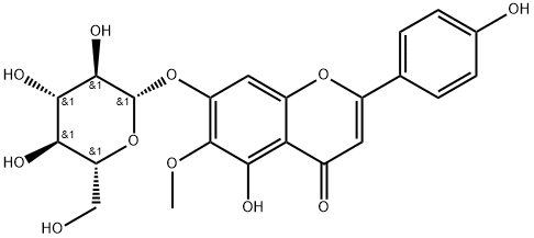
高车前苷是一种糖苷,属于黄酮类化合物。
据报道,高车前苷存在于鼠尾草(Salvia plebeia)、药用鼠尾草(Salvia officinalis)以及其他有相关数据的生物体中。 鼠尾草(Salvia plebeia R. Br)是一种传统中药,被认为是一种抗炎介质,用于治疗包括肝炎在内的多种感染性疾病。此前,我们课题组已分离得到高车前苷。因此,我们评估了高车前苷对肝细胞损伤的保护作用。高车前苷在无细胞体系中表现出抗氧化活性,对DPPH自由基的清除IC50值为0.35 μg/ml。在暴露于H₂O₂的人肝细胞HL-7702中,添加0.1-100 μg/ml的同型车前子苷(对细胞活力无毒性作用)可显著降低乳酸脱氢酶(LDH)的泄漏,并增加上清液中谷胱甘肽(GSH)、谷胱甘肽过氧化物酶(GSH-Px)和超氧化物歧化酶(SOD)的含量。体内实验中,我们采用卡介苗(BCG)/脂多糖(LPS)诱导的小鼠肝损伤模型来评估同型车前子苷的疗效。同型车前子苷(25-100 mg/kg)显著降低了血清丙氨酸氨基转移酶(ALT)和天冬氨酸氨基转移酶(AST)的升高,并降低了肿瘤坏死因子-α(TNF-α)和白细胞介素-1(IL-1)的水平。同样的治疗方法也降低了硫代巴比妥酸反应物质(TBARS)的含量,并提高了肝匀浆中谷胱甘肽(GSH)、谷胱甘肽过氧化物酶(GSH-Px)和超氧化物歧化酶(SOD)的水平。组织病理学分析显示,高车前苷治疗小鼠的肝损伤程度有所减轻,炎症细胞减少,肝细胞坏死也减少。这些结果表明,高车前苷对肝细胞损伤具有保护和治疗作用,这可能与其抗氧化特性有关。[1] 作为一种血管舒张剂和血管稳态分子,一氧化氮(NO)在调节生理性内皮功能中发挥着至关重要的作用。然而,在炎症内皮细胞中,NO的生物活性和生成通常会受损。[49] 我们的数据表明,在棕榈酸(PA)刺激下,NO的生成受损。正如预期的那样,我们发现高车前苷可以恢复NO的生成(图8)。研究表明,高车前苷可通过恢复受损的一氧化氮(NO)生成来保护内皮细胞免受棕榈酸(PA)损伤。结论:总之,我们的研究阐明了高车前苷可下调TLR4和NLRP3炎症小体通路,进而抑制相关炎症介质,从而恢复PA诱导的内皮细胞中受损的一氧化氮生成。这些发现提示高车前苷可能是一种具有开发潜力的血管疾病预防和治疗候选药物。[2] 总之,本研究首次报道了高车前苷在静脉注射、腹腔注射和口服给药后在大鼠体内的药代动力学。首次在口服给药后同时定量分析了毛蕊花素和高车前苷。毛蕊花素和高车前苷的表观分配系数在 pH 1.2 至 pH 8.0 范围内保持稳定,高车前苷在肠段中的转运可能主要通过被动转运进行。肠道是高车前苷的最佳吸收部位,但它几乎完全是经大鼠肠道细菌生物转化的中间体。口服后,高车前苷被迅速吸收,绝对口服生物利用度为 0.75%。[4] |
| 分子式 |
C22H22O11
|
|---|---|
| 分子量 |
462.4035
|
| 精确质量 |
462.116
|
| CAS号 |
17680-84-1
|
| 相关CAS号 |
17680-84-1
|
| PubChem CID |
5318083
|
| 外观&性状 |
Light yellow to yellow solid
|
| 密度 |
1.6±0.1 g/cm3
|
| 沸点 |
798.1±60.0 °C at 760 mmHg
|
| 熔点 |
256-258℃
|
| 闪点 |
279.7±26.4 °C
|
| 蒸汽压 |
0.0±3.0 mmHg at 25°C
|
| 折射率 |
1.695
|
| LogP |
-0.97
|
| tPSA |
179.28
|
| 氢键供体(HBD)数目 |
6
|
| 氢键受体(HBA)数目 |
11
|
| 可旋转键数目(RBC) |
5
|
| 重原子数目 |
33
|
| 分子复杂度/Complexity |
721
|
| 定义原子立体中心数目 |
5
|
| SMILES |
O1[C@]([H])([C@@]([H])([C@]([H])([C@@]([H])([C@@]1([H])C([H])([H])O[H])O[H])O[H])O[H])OC1C([H])=C2C(C(C([H])=C(C3C([H])=C([H])C(=C([H])C=3[H])O[H])O2)=O)=C(C=1OC([H])([H])[H])O[H]
|
| InChi Key |
GCLAFEGUXXHIFT-IWLDQSELSA-N
|
| InChi Code |
InChI=1S/C22H22O11/c1-30-21-14(32-22-20(29)19(28)17(26)15(8-23)33-22)7-13-16(18(21)27)11(25)6-12(31-13)9-2-4-10(24)5-3-9/h2-7,15,17,19-20,22-24,26-
|
| 化学名 |
5-hydroxy-2-(4-hydroxyphenyl)-6-methoxy-7-[(2S,3R,4S,5S,6R)-3,4,5-trihydroxy-6-(hydroxymethyl)oxan-2-yl]oxychromen-4-one
|
| 别名 |
Homoplantaginin; 17680-84-1; HISPIDULOSIDE; Dinatin 7-glucoside; (-)-Homoplantaginin; hispidulin-7-glucoside; 4H-1-Benzopyran-4-one, 7-(beta-D-glucopyranosyloxy)-5-hydroxy-2-(4-hydroxyphenyl)-6-methoxy-; 6-Methoxyapigenin 7-O-glucoside;
|
| HS Tariff Code |
2934.99.9001
|
| 存储方式 |
Powder -20°C 3 years 4°C 2 years In solvent -80°C 6 months -20°C 1 month |
| 运输条件 |
Room temperature (This product is stable at ambient temperature for a few days during ordinary shipping and time spent in Customs)
|
| 溶解度 (体外实验) |
DMSO: 50~92 mg/mL (108.1~199.0 mM)
|
|---|---|
| 溶解度 (体内实验) |
配方 1 中的溶解度: ≥ 2.08 mg/mL (4.50 mM) (饱和度未知) in 10% DMSO + 40% PEG300 + 5% Tween80 + 45% Saline (这些助溶剂从左到右依次添加,逐一添加), 澄清溶液。
例如,若需制备1 mL的工作液,可将100 μL 20.8 mg/mL澄清DMSO储备液加入400 μL PEG300中,混匀;然后向上述溶液中加入50 μL Tween-80,混匀;加入450 μL生理盐水定容至1 mL。 *生理盐水的制备:将 0.9 g 氯化钠溶解在 100 mL ddH₂O中,得到澄清溶液。 配方 2 中的溶解度: 2.08 mg/mL (4.50 mM) in 10% DMSO + 90% (20% SBE-β-CD in Saline) (这些助溶剂从左到右依次添加,逐一添加), 悬浊液; 超声助溶。 例如,若需制备1 mL的工作液,可将 100 μL 20.8 mg/mL澄清DMSO储备液加入900 μL 20% SBE-β-CD生理盐水溶液中,混匀。 *20% SBE-β-CD 生理盐水溶液的制备(4°C,1 周):将 2 g SBE-β-CD 溶解于 10 mL 生理盐水中,得到澄清溶液。 View More
配方 3 中的溶解度: ≥ 2.08 mg/mL (4.50 mM) (饱和度未知) in 10% DMSO + 90% Corn Oil (这些助溶剂从左到右依次添加,逐一添加), 澄清溶液。 1、请先配制澄清的储备液(如:用DMSO配置50 或 100 mg/mL母液(储备液)); 2、取适量母液,按从左到右的顺序依次添加助溶剂,澄清后再加入下一助溶剂。以 下列配方为例说明 (注意此配方只用于说明,并不一定代表此产品 的实际溶解配方): 10% DMSO → 40% PEG300 → 5% Tween-80 → 45% ddH2O (或 saline); 假设最终工作液的体积为 1 mL, 浓度为5 mg/mL: 取 100 μL 50 mg/mL 的澄清 DMSO 储备液加到 400 μL PEG300 中,混合均匀/澄清;向上述体系中加入50 μL Tween-80,混合均匀/澄清;然后继续加入450 μL ddH2O (或 saline)定容至 1 mL; 3、溶剂前显示的百分比是指该溶剂在最终溶液/工作液中的体积所占比例; 4、 如产品在配制过程中出现沉淀/析出,可通过加热(≤50℃)或超声的方式助溶; 5、为保证最佳实验结果,工作液请现配现用! 6、如不确定怎么将母液配置成体内动物实验的工作液,请查看说明书或联系我们; 7、 以上所有助溶剂都可在 Invivochem.cn网站购买。 |
| 制备储备液 | 1 mg | 5 mg | 10 mg | |
| 1 mM | 2.1626 mL | 10.8131 mL | 21.6263 mL | |
| 5 mM | 0.4325 mL | 2.1626 mL | 4.3253 mL | |
| 10 mM | 0.2163 mL | 1.0813 mL | 2.1626 mL |
1、根据实验需要选择合适的溶剂配制储备液 (母液):对于大多数产品,InvivoChem推荐用DMSO配置母液 (比如:5、10、20mM或者10、20、50 mg/mL浓度),个别水溶性高的产品可直接溶于水。产品在DMSO 、水或其他溶剂中的具体溶解度详见上”溶解度 (体外)”部分;
2、如果您找不到您想要的溶解度信息,或者很难将产品溶解在溶液中,请联系我们;
3、建议使用下列计算器进行相关计算(摩尔浓度计算器、稀释计算器、分子量计算器、重组计算器等);
4、母液配好之后,将其分装到常规用量,并储存在-20°C或-80°C,尽量减少反复冻融循环。
计算结果:
工作液浓度: mg/mL;
DMSO母液配制方法: mg 药物溶于 μL DMSO溶液(母液浓度 mg/mL)。如该浓度超过该批次药物DMSO溶解度,请首先与我们联系。
体内配方配制方法:取 μL DMSO母液,加入 μL PEG300,混匀澄清后加入μL Tween 80,混匀澄清后加入 μL ddH2O,混匀澄清。
(1) 请确保溶液澄清之后,再加入下一种溶剂 (助溶剂) 。可利用涡旋、超声或水浴加热等方法助溶;
(2) 一定要按顺序加入溶剂 (助溶剂) 。
|
InvivoChem的所有产品仅用于作科学研究,不面向患者销售
Copyright 2020 InvivoChem LLC | All Rights Reserved 粤ICP备20063088号-1


 463611831
