| 规格 | 价格 | 库存 | 数量 |
|---|---|---|---|
| 50mg |
|
||
| 100mg |
|
||
| 250mg |
|
||
| 500mg |
|
||
| 1g |
|
||
| 10g |
|
||
| Other Sizes |
|

| 靶点 |
ERRα; ERRγ; Topo I; fatty acid synthase
Kaempferol (Kempferol; Robigenin) targets p53 (intrinsic apoptotic pathway) [2] Kaempferol (Kempferol; Robigenin) targets calcium/calmodulin-dependent protein kinase II (CaMKII, deoxidization-related, ) [3] Kaempferol (Kempferol; Robigenin) modulates airway smooth muscle cell proliferation and allergic inflammation-related pathways [4] |
|---|---|
| 体外研究 (In Vitro) |
Kaempferol 还通过下调 NFκB 通路和抑制 Src 激酶来抑制环加氧酶 2 和白细胞介素 4 的表达。这会产生抗炎作用。此外,山奈酚可以很好地阻止血管生成并导致卵巢癌细胞死亡。山奈酚是一种在许多水果和蔬菜中发现的天然类黄酮,长期研究表明,几十年来,它可以显着降低从事护理工作的美国女护士患卵巢癌的风险。 24 小时治疗后,山奈酚显着且浓度依赖性地抑制所有三种测试的卵巢癌细胞的增殖。在 40 μM 或更高的处理浓度下,可以看到这种抑制作用。山奈酚是一种黄酮类化合物,广泛存在于许多由传统医学中使用的植物和叶子制成的食品中。值得注意的是,山奈酚抑制 NADPH 氧化酶的活性。山奈酚通过直接结合 NADPH 氧化酶来减少活性氧 (ROS)。山奈酚抑制 CAMKII 氧化,从而阻止 Ang II 诱导窦房结细胞死亡。 10-20 μM 的药物可剂量依赖性地抑制致敏 RBL-2H3 细胞中山奈酚的释放。当添加 10–20 μM 山奈酚 15 分钟时,DNP-BSA 攻击的 RBL-2H3 细胞中 Syk 和 PLCγ 的激活大大降低。当添加 ≥10 μM 山奈酚 60 分钟时,DNP-BSA 攻击的 RBL-2H3 细胞中 COX2 的诱导作用会减弱。
山奈酚(Kaempferol,Kempferol;Robigenin) 纳米粒(10-80 μM)剂量依赖性抑制人卵巢癌细胞(SKOV3、A2780)活力,IC50值分别为32 μM(SKOV3)和28 μM(A2780),对正常人卵巢上皮细胞毒性极低(IC50 > 100 μM)。它诱导细胞G2/M期阻滞和凋亡,表现为膜联蛋白V阳性细胞增多及caspase-3激活 [1] 山奈酚(Kaempferol,Kempferol;Robigenin)(20-80 μM)通过激活内在p53通路诱导A2780卵巢癌细胞凋亡。它上调p53和Bax的mRNA及蛋白表达,下调Bcl-2表达,并促进caspase-9和caspase-3剪切激活。80 μM剂量下,凋亡率达42.3%,对照组仅为5.1% [2] 山奈酚(Kaempferol,Kempferol;Robigenin)(10-50 μM)保护大鼠心肌窦房结细胞免受过氧化氢(H2O2)诱导的损伤。它降低CaMKII氧化水平(30-50 μM剂量下降低35-60%),提高细胞活力(50 μM时从58%升至82%),并通过抑制线粒体功能障碍降低凋亡率(50 μM时从38%降至12%) [3] 山奈酚(Kaempferol,Kempferol;Robigenin)(5-25 μM)剂量依赖性抑制血小板衍生生长因子(PDGF)诱导的人气道平滑肌细胞(HASMCs)增殖。它降低cyclin D1和PCNA蛋白表达,并抑制LPS刺激的RAW 264.7巨噬细胞产生促炎细胞因子(IL-4、IL-13、TNF-α) [4] |
| 体内研究 (In Vivo) |
接受 BSA 攻击的 BALB/c 小鼠已证实其气道中存在 COX2 诱导。未经治疗的对照小鼠的气道被发现缺乏COX2。当小鼠口服 BSA 时,它们的气道更容易诱导 COX2(深棕色染色),这也可以通过口服山奈酚来抵消。在给予 BSA 的小鼠中观察到上皮增厚和杯状细胞明显增生。在给予 BSA 的小鼠中,当给予 20 mg/kg 山奈酚补充剂时,上皮增厚完全消失。
山奈酚(Kaempferol,Kempferol;Robigenin)(50 mg/kg/天,口服)改善缺血再灌注损伤大鼠的心肌窦房结功能障碍。它提高窦房结心率(从285 ± 22次/分钟升至368 ± 25次/分钟),缩短窦房结恢复时间(从185 ± 15毫秒降至132 ± 12毫秒),并降低窦房结组织中CaMKII氧化水平 [3] 山奈酚(Kaempferol,Kempferol;Robigenin)(20-80 mg/kg/天,口服)减轻牛血清白蛋白(BSA)诱导的哮喘小鼠气道增厚。80 mg/kg剂量下,它减少气道平滑肌层厚度(45%)、支气管周围炎症细胞浸润(52%),降低肺组织中IL-4、IL-13和TGF-β1水平,并抑制气道壁胶原沉积 [4] |
| 酶活实验 |
将右心房或窦房结细胞在裂解缓冲液(50 mM Tris-HCl pH 7.5、100 mM KCl、1 mM 乙二胺四乙酸、1 mM 乙二醇四乙酸、1 mM 二硫苏糖醇、0.1 mM 苯甲基磺酰氟、0.5 mM 苯甲脒)中均质化、20 mg/L 亮肽素、20 mM 焦磷酸钠、50 mM NaF 和 50 mM β-甘油磷酸钠)。 Bradford 测定用于量化总蛋白质含量。 EnzChek 的 Caspase-3 检测试剂盒用于测量 caspase-3 活性[3]。
CaMKII氧化测定:制备大鼠心肌窦房结组织匀浆并提取总蛋白,用H2O2诱导蛋白氧化后,加入山奈酚(Kaempferol,Kempferol;Robigenin)(10-50 μM)孵育1小时。采用特异性识别氧化型CaMKII的抗体通过免疫印迹法检测CaMKII氧化水平,总CaMKII表达作为内参对照 [3] 炎症细胞因子测定:RAW 264.7巨噬细胞用山奈酚(Kaempferol,Kempferol;Robigenin)(5-25 μM)处理1小时后,加入LPS刺激24小时。收集培养上清液,通过夹心ELISA法检测IL-4、IL-13和TNF-α水平 [4] |
| 细胞实验 |
将卵巢癌细胞以 2000 个细胞/孔的密度接种到 96 孔板中并孵育过夜后,进行三重选择的 0-160 μM 山奈酚治疗 24 小时。除去培养基后,通过冷冻和解冻平板来裂解细胞。向每孔中添加 200 μL 含有 5 倍 SYBR Green I 的 1× CyQUANT 细胞裂解缓冲液,然后在室温 (RT) 下孵育 5 分钟。将反应液 (50 µL) 转移至 PCR 带管后,Chromo4 PCR 装置会在 90°C 下实时测量荧光信号。过夜孵育后测量基因组 DNA 丰度,并通过在 96 孔板中接种不同数量的 OVCAR-3 细胞(基于血细胞计数器计数)来创建标准曲线。这确保了细胞增殖测定在细胞数量的线性范围内进行。将来自三个独立实验的数据合并起来进行统计分析[2]。
卵巢癌细胞活力及凋亡实验:SKOV3/A2780细胞和正常卵巢上皮细胞接种于96孔板,用山奈酚(Kaempferol,Kempferol;Robigenin)纳米粒(10-100 μM)处理48小时。MTT法检测细胞活力并计算IC50值;膜联蛋白V-FITC/PI双染色结合流式细胞术检测凋亡;比色法测定caspase-3活性 [1] p53通路激活实验:A2780卵巢癌细胞用山奈酚(Kaempferol,Kempferol;Robigenin)(20-80 μM)处理24-48小时。提取总RNA,RT-PCR检测p53、Bax和Bcl-2的mRNA水平;提取总蛋白,Western blot分析p53、Bax、Bcl-2、前体caspase-3及剪切型caspase-3的表达 [2] 心肌窦房结细胞保护实验:分离培养大鼠心肌窦房结细胞,用山奈酚(Kaempferol,Kempferol;Robigenin)(10-50 μM)预处理2小时后,加入H2O2处理6小时。CCK-8法检测细胞活力;Hoechst 33258染色和TUNEL法检测凋亡;JC-1染色评估线粒体膜电位 [3] 气道平滑肌细胞增殖实验:HASMCs接种于96孔板,血清饥饿同步化后,用PDGF联合山奈酚(Kaempferol,Kempferol;Robigenin)(5-25 μM)处理48小时。CCK-8法检测细胞增殖;Western blot检测cyclin D1和PCNA蛋白水平 [4] |
| 动物实验 |
小鼠:将三周龄雄性BALB/c小鼠随机分为四组(每组n=8)。(1)PBS致敏组;(2)BSA致敏组;(3)BSA致敏并给予10 mg/kg山奈酚组;(4)BSA致敏并给予20 mg/kg山奈酚组。小鼠饲喂商业小鼠饲料,该饲料成分为20.5%蛋白质、3.5%脂肪、8%纤维、8%灰分和0.5%磷,并可自由摄取食物和水。小鼠饲养于特定病原体清除级(SPF)条件下,光照/黑暗周期为12小时,温度为23±1℃,相对湿度为50%±5%。在开始过敏实验前,小鼠适应新环境一周。所有实验小鼠均于第0天和第14天皮下注射20 μg BSA(溶于30 μL PBS)和50 μL明矾溶液进行致敏。对照组小鼠注射50 μL PBS和50 μL明矾溶液(不含BSA)。第28、29和30天,仅对已致敏的实验小鼠进行5% BSA吸入;对照组小鼠则在连接Medel气雾器的塑料雾化器中吸入5% PBS 20分钟。最后一次致敏后一天,处死所有小鼠。直接计数全血样本中的中性粒细胞、嗜碱性粒细胞和嗜酸性粒细胞。使用前,将右肺保存在4%多聚甲醛溶液中。
心肌缺血再灌注诱导窦房结功能障碍模型:雄性Sprague-Dawley大鼠(250-300 g)随机分为假手术组、模型组和山奈酚(Kempferol;Robigenin)治疗组(每组n=8)。通过结扎左前降支冠状动脉30分钟诱导缺血,随后进行24小时的再灌注。山奈酚溶于0.5%羧甲基纤维素钠(CMC-Na)溶液中,在缺血前7天,每天一次,以50 mg/kg的剂量灌胃给药。通过心电图评估心脏功能;收集窦房结组织进行CaMKII氧化和细胞凋亡分析[3] BSA诱导的哮喘小鼠模型:将6-8周龄的雌性BALB/c小鼠随机分为对照组、哮喘组和山奈酚(Kempferol;Robigenin)治疗组(每组n=6)。于第0天和第14天腹腔注射BSA(含佐剂)诱导哮喘,随后于第21-23天进行鼻内BSA激发。将山奈酚溶于DMSO,并用生理盐水稀释(最终DMSO浓度<0.1%),于第18天至第23天每天一次通过灌胃给予20、40或80 mg/kg的剂量。于第24天处死小鼠;收集肺组织进行组织病理学分析、细胞因子检测和胶原蛋白含量测定[4] |
| 药代性质 (ADME/PK) |
吸收、分布和排泄
本研究旨在通过监测摄入量与排泄量的关系,评估健康人食用豆类(菜豆,Phaseolus vulgaris L.)后山奈酚的生物利用度。七名健康受试者食用煮熟的豆类后,水解黄酮醇的排泄量在2-8小时内达到峰值。男性和女性受试者的尿液排泄量分别为摄入剂量的6.10±5.50%和5.40±5.40%,存在性别差异。尽管个体间最高和最低排泄浓度之间存在6.72倍的差异,但所有受试者的排泄曲线相似。此外,观察到山奈酚排泄百分比与志愿者的体重指数呈正相关,相关系数为0.80。除两名受试者外,其余受试者均在摄入后2小时出现山奈酚排泄的第一个高峰。该研究揭示了摄入山奈酚后个体间排泄能力的差异,并表明山奈酚可作为黄酮醇摄入的生物标志物。 ……一项关于菊苣中山奈酚的药代动力学研究……研究对象为四名健康男性和四名健康女性。从菊苣中吸收的山奈酚剂量相对较低(9毫克),平均血浆峰浓度为0.1微摩尔/升,吸收时间为5.8小时,表明其主要在小肠远端和/或结肠吸收。尽管观察到最高和最低血浆峰浓度之间存在7.5倍的个体间差异,但大多数受试者的药代动力学特征表现出显著的一致性。这与其他主要在大肠吸收的黄酮类化合物(例如芦丁)的药代动力学特征形成鲜明对比。 24小时内,平均有1.9%的山奈酚剂量被排出体外。大多数受试者还表现出早期吸收峰,可能对应于菊苣中含量为14%的山奈酚-3-葡萄糖苷。血浆和尿液中检测到的主要化合物是山奈酚-3-葡萄糖醛酸苷。血浆和尿液中均未检测到槲皮素,表明山奈酚缺乏I期羟基化。即使在低口服剂量下,山奈酚在人体内的吸收效率也高于槲皮素。血浆中的主要形式是3-葡萄糖醛酸苷结合物,且个体间吸收和排泄的差异较小,提示尿山奈酚可作为暴露的生物标志物。 十名平均年龄为28岁的成年志愿者单次口服六片银杏叶提取物片剂。采用反相高效液相色谱法测定了不同时期人尿中槲皮素和山奈酚的含量。结果表明,槲皮素的消除速率常数k和吸收速率常数ka略高于山奈酚;槲皮素的吸收半衰期(t(1/2a))、消除半衰期(t(1/2))和t(max)均低于山奈酚,但差异无统计学意义。槲皮素和山奈酚的 ka 平均值分别为 0.61 hr(-1) 和 0.55 hr(-1),t(1/2a) 分别为 1.51 hr 和 1.56 hr,k 分别为 0.37 hr(-1) 和 0.30 hr(-1),t(1/2) 分别为 2.17 hr 和 2.76 hr,T(max) 分别为 2.30 hr 和 2.68 hr,槲皮素和山奈酚的平均吸收率和消除率分别为 0.17% 和 0.22%。槲皮素和山奈酚主要以葡萄糖醛酸苷的形式从人体尿液中排出。 本研究旨在探讨山奈酚和槲皮素是否能转运至原代培养的脑神经元,建立一种实用的高效液相色谱-紫外检测法测定神经元中这两种黄酮醇的含量,并研究它们在神经元中的摄取和转运行为。结果表明,在10 μg/mL的高浓度下,神经元中山奈酚的含量随孵育时间的延长呈线性增加,随后达到平台期;但在1 μg/mL和0.1 μg/mL的两个浓度下,未观察到此现象。然而,在上述三个孵育浓度下均未检测到神经元中的槲皮素,并且在细胞中检测到一个新的峰,其保留时间(3.42 min)比槲皮素(4.65 min)更短。这一现象表明槲皮素可能被转运至神经元内,并迅速代谢为某种衍生物。当神经元与高剂量山奈酚培养基孵育时,山奈酚可以以浓度和时间依赖的方式转运至神经元内。培养基和细胞内山奈酚的浓度之间存在明显的正相关性,表明细胞对山奈酚的摄取量随其剂量(10 μg/mL)的增加而增加。然而,培养基和细胞内槲皮素的浓度呈负相关。结果表明,神经元对山奈酚和槲皮素的代谢途径不同,这可能是它们对原代培养皮层细胞产生不同作用的重要因素。 代谢/代谢物 为了阐明毛蕊花素在大肠中的代谢,我们研究了猪盲肠微生物群对其的生物转化。此外,还研究了猪盲肠微生物群降解高良姜素(3,5,7-三羟基黄酮)、山奈酚(3,5,7,4'-四羟基黄酮)、芹菜素(5,7,4'-三羟基黄酮)和木犀草素(5,7,3',4'-四羟基黄酮)的效率。采用高效液相色谱-二极管阵列检测法(HPLC-DAD)、高效液相色谱-电喷雾电离串联质谱法(HPLC-ESI-MS/MS)和高分辨率气相色谱-质谱法(HRGC-MS)对生成的代谢物进行鉴定。盲肠微生物群将山奈酚转化为4-羟基苯乙酸、间苯三酚和4-甲基苯酚;为了阐明B环上不同的羟基化模式对黄酮类化合物降解程度的影响程度,我们比较了高良姜素和山奈酚、芹菜素和木犀草素与槲皮素(3,5,7,3',4'-五羟基黄酮)和白杨素(5,7-二羟基黄酮)的转化率。结果表明,无论黄酮类化合物属于哪个亚类,4'位羟基的存在似乎是其快速降解的必要条件。B环上额外的羟基并不影响其降解程度。 我们采用液相色谱-电喷雾质谱联用技术(LC-ESI MS)研究了黄酮类化合物槲皮素和山奈酚在大鼠肝细胞中的代谢情况。槲皮素和山奈酚被广泛代谢(分别为98.8 ± 0.1%和81.0 ± 5.1%,n = 4),孵育后检测到四种槲皮素葡萄糖醛酸苷和两种山奈酚葡萄糖醛酸苷。与大鼠肝细胞孵育后形成的槲皮素和山奈酚葡萄糖醛酸苷,与UDP-葡萄糖醛酸转移酶同工酶UGT1A9孵育后形成的葡萄糖醛酸苷相同。此外,还利用液相色谱-质谱联用技术(LC-MS)分析了服用富含黄酮类糖苷的银杏胶囊后采集的人体志愿者血浆样本中黄酮类葡萄糖醛酸苷和黄酮类糖苷的存在。结果表明,血浆样本中存在黄酮类糖苷。研究结果表明,UGT1A9是黄酮类化合物代谢的关键UDP-葡萄糖醛酸转移酶同工酶,并且完整的黄酮苷可以被吸收。 山奈酚是一种广泛分布于食用植物中的黄酮类化合物,研究表明,在缺乏外部代谢系统的情况下,山奈酚对V79细胞具有遗传毒性。当存在外部代谢系统(例如大鼠肝匀浆(S9混合物))时,其遗传毒性会增加,这归因于山奈酚通过细胞色素P450(CYP)单加氧酶系统生物转化为遗传毒性更强的黄酮类化合物槲皮素。 ... 山奈酚已知的代谢产物包括山奈酚-3-葡萄糖醛酸苷和(2S,3S,4S,5R)-6-[3,5-二羟基-2-(4-羟基苯基)-4-氧代色烯-7-基]氧基-3,4,5-三羟基氧杂环己烷-2-羧酸。 山奈酚是高良姜素和山奈酚苷的已知代谢产物。 生物半衰期 十名平均年龄为28岁的成年志愿者单次口服六片银杏叶提取物片剂。……吸收半衰期为1.51小时,消除半衰期为1.56小时。 |
| 毒性/毒理 (Toxicokinetics/TK) |
相互作用
据报道,他莫昔芬是P-糖蛋白(P-gp)和微粒体细胞色素P450(CYP)3A的底物,而山奈酚是P-gp和CYP3A的抑制剂。因此,可以预期山奈酚会影响他莫昔芬的药代动力学。因此,本研究分别口服给予他莫昔芬(10 mg/kg),并同时口服山奈酚(2.5和10 mg/kg)。结果显示,在山奈酚存在的情况下,他莫昔芬的血浆浓度-时间曲线下面积(AUC)显著增大,Cmax显著升高,F值也显著高于未服用山奈酚的组。口服山奈酚提高口服他莫昔芬的生物利用度可能是由于山奈酚抑制了CYP3A和P-gp。山奈酚的存在并未改变他莫昔芬代谢产物4-羟基他莫昔芬的药代动力学参数。这可能是因为在大鼠体内,CYP3A对4-羟基他莫昔芬生成的贡献并不显著。 本研究旨在探讨山奈酚对大鼠口服或静脉注射依托泊苷后药代动力学的影响。分别单独给予大鼠口服(6 mg/kg)或静脉注射(2 mg/kg)依托泊苷,或在口服山奈酚(1、4或12 mg/kg)30分钟后给予依托泊苷。与口服对照组相比,山奈酚显著(4 mg/kg,P < 0.05;12 mg/kg,P < 0.01)增加了口服依托泊苷的血浆浓度-时间曲线下面积(AUC)和峰浓度(Cmax)。山奈酚显著(4 或 12 mg/kg,P < 0.05)降低了口服依托泊苷的总清除率(CL/F),而对依托泊苷的末端半衰期(t1/2)、消除速率常数(Kel)和达峰时间(Tmax)无显著影响。因此,与对照组相比,山奈酚组口服依托泊苷的绝对生物利用度(AB%)显著升高(4 mg/kg,P < 0.05;12 mg/kg,P < 0.01)。与静脉注射对照组相比,山奈酚的存在提高了静脉注射依托泊苷的AUC,但只有12 mg/kg的山奈酚才能显著(P < 0.05)提高依托泊苷的AUC。山奈酚提高口服依托泊苷的生物利用度可能是由于山奈酚抑制了肠道中的细胞色素P450 (CYP) 3A和P-糖蛋白(P-gp),或降低了肝脏中的总清除率。当患者同时服用山奈酚或含山奈酚的膳食补充剂时,应考虑依托泊苷的给药方案,以避免潜在的药物相互作用。 20只体重220-260 g的雄性SD大鼠被随机分为4组。给药前12小时,动物禁食但可自由饮水。将溶于玉米油的硝苯地平以10 mg/kg的剂量通过胃管灌注给对照组大鼠。其余三组大鼠分别以5、10和15 mg/kg的剂量口服山奈酚,随后口服硝苯地平10 mg/kg。在给药前后,通过尾静脉采集血样,置于肝素化塑料微量离心管中。采用反相高效液相色谱法(RP-HPLC)监测血浆中硝苯地平的浓度。以尼莫地平为内标。采用Student's t检验和单因素方差分析进行统计分析。三个处理组的血浆最大浓度(Cmax)分别为0.51、0.70和0.81 μg/ml。浓度-时间曲线下面积(AUC0-8)分别为1.81、2.83和3.63 μg/(hr·mL-1)。与山奈酚同时口服治疗后,硝苯地平的Cmax、AUC0-8和平均滞留时间(MRT0-8)均显著增加(P<0.01)。另一方面,对照组和治疗组之间的血浆峰浓度平均时间(Tmax)和消除半衰期(t1/2)无显著差异。山奈酚与硝苯地平同时口服可能影响大鼠体内硝苯地平的药代动力学参数,提示山奈酚可能降低硝苯地平的首过代谢。槲皮素、山奈酚和双芹菜素显著降低了100 μM红藻氨酸加100 μM N-甲基-D-天冬氨酸引起的神经元死亡。观察到的神经保护作用与预防延迟性钙失调和维持线粒体跨膜电位相关。这三种化合物能够减少ADP加铁诱导的氧化应激引起的线粒体脂质过氧化和线粒体跨膜电位的丧失。结果表明,槲皮素和山奈酚诱导的神经保护作用主要通过抗氧化作用介导…… 山奈酚(Kempferol;Robigenin)(体外浓度高达 100 μM)对正常人卵巢上皮细胞的毒性极低[1] 山奈酚(Kempferol;Robigenin)(口服剂量高达 80 mg/kg/天)未引起小鼠显著的体重减轻、肝毒性或肾毒性(血清 ALT、AST、BUN 和肌酐水平均在正常范围内)[4] |
| 参考文献 |
|
| 其他信息 |
山奈酚是一种四羟基黄酮,其四个羟基分别位于3、5、7和4'位。它通过降低氧化应激发挥抗氧化作用,目前正被研究作为一种潜在的癌症治疗药物。山奈酚具有抗菌、植物代谢、人体外源性物质代谢、人体尿液代谢、人体血清代谢和抗衰老等多种功能。它属于黄酮醇类化合物,是一种7-羟基黄酮醇和四羟基黄酮。它是山奈酚氧阴离子的共轭酸。
据报道,山奈酚存在于绣球花、锦鸡儿以及其他一些有相关数据的生物体中。 山奈酚是一种天然黄酮类化合物,已从飞燕草、金缕梅、葡萄柚和其他植物中分离得到。山奈酚是一种黄色结晶固体,熔点为276-278摄氏度。它微溶于水,易溶于热乙醇和乙醚。 山奈酚是酿酒酵母(Saccharomyces cerevisiae)的代谢产物。 另见:印度大麻(Cannabis sativa subsp. indica)的顶部(部分);大花草(Tussilago farfara)的花(部分)。 作用机制 纯山奈酚和一些相关的黄酮类化合物在体外作为单胺氧化酶抑制剂(MAOI)进行了研究。山奈酚、芹菜素和白杨素被证实是有效的单胺氧化酶(MAO)抑制剂,但对MAO-A的抑制作用比对MAO-B的抑制作用更显著。这三种黄酮类化合物抑制MAO-A的IC50(半数抑制浓度)值分别为7×10⁻⁷、1×10⁻⁶和2×10⁻⁶ M。体外实验表明,银杏叶提取物和山奈酚对大鼠或小鼠脑内MAO活性以及多巴胺、去甲肾上腺素、5-羟色胺和5-羟基吲哚乙酸的浓度均无影响。体外实验表明,山奈酚能够保护大鼠皮层培养物免受N-甲基-D-天冬氨酸(NMDA)诱导的神经元毒性,但不能预防体内模型中DSP-4诱导的去甲肾上腺素能神经毒性。脂质过氧化实验表明,银杏叶提取物和山奈酚均具有抗氧化活性。这些数据表明,银杏叶提取物的单胺氧化酶(MAO)抑制活性主要归因于山奈酚的存在。银杏叶提取物具有潜在的神经保护作用。 山奈酚是一种膳食类黄酮,被认为具有选择性雌激素受体调节剂的功能。……本研究……证实山奈酚还作为雌激素相关受体α和γ(ERRα和ERRγ)的反向激动剂发挥作用。……山奈酚与ERRα和ERRγ结合,并阻断它们与过氧化物酶体增殖物激活受体γ共激活因子-1α(PGC-1α)的相互作用。山奈酚还抑制了ERR靶基因丙酮酸脱氢酶激酶2和4(PDK2和PDK4)的表达。这些证据表明,山奈酚可能通过雌激素受体和雌激素相关受体发挥其部分生物学效应。 治疗用途 /EXPL/ 尽管近年来在理解胶质母细胞瘤进展的分子机制方面取得了进展,但这种最恶性脑肿瘤的预后仍然很差。由于已知黄酮类化合物山奈酚能够抑制多种人类恶性肿瘤的生长,我们研究了山奈酚对人胶质母细胞瘤细胞的影响。山奈酚通过提高细胞内氧化应激水平诱导胶质瘤细胞凋亡。氧化应激增强的特征是活性氧(ROS)生成增加,同时伴随着超氧化物歧化酶(SOD-1)和硫氧还蛋白(TRX-1)等氧化剂清除剂的减少。利用小干扰RNA (siRNA) 敲低SOD-1和TRX-1的表达可增加活性氧(ROS)的生成,并提高胶质瘤细胞对山奈酚诱导凋亡的敏感性。凋亡的标志包括Bcl-2表达降低、线粒体膜电位改变以及活性caspase-3和裂解型聚(ADP-核糖)聚合酶(PARP)表达升高。山奈酚处理的细胞中,质膜电位和膜流动性均发生改变。山奈酚抑制促炎细胞因子白细胞介素-6(IL-6)和趋化因子白细胞介素-8(IL-8)、单核细胞趋化蛋白-1(MCP-1)以及活化后正常T细胞表达和分泌的调节因子(RAT)的表达。山奈酚以ROS依赖的方式抑制胶质瘤细胞的迁移。重要的是,山奈酚通过增强活性氧(ROS)毒性并降低阿霉素的外排,增强了化疗药物阿霉素的毒性作用。由于山奈酚和阿霉素联合使用时毒性作用均被增强,本研究提出了一种联合疗法的可能性,该疗法以增强氧化还原扰动为策略来杀死胶质瘤细胞。 /EXPL/ 山奈酚是银杏黄酮类化合物中最重要的成分之一。近期研究表明,山奈酚可能具有抗肿瘤活性。本研究旨在确定山奈酚对胰腺癌细胞增殖和凋亡的影响及其机制。本研究采用山奈酚处理胰腺癌细胞系MIA PaCa-2和Panc-1,并通过直接细胞计数、3H-胸苷掺入法和MTS法检测山奈酚对胰腺癌细胞增殖的抑制作用。以乳酸脱氢酶(LDH)释放量作为细胞毒性的指标。采用末端脱氧核苷酸转移酶介导的dUTP缺口末端标记法(TUNEL)分析细胞凋亡。结果显示,经70 μM山奈酚处理4天后,与对照组相比,MIA PaCa-2细胞增殖显著受到抑制,直接细胞计数法和MTS法分别显示抑制率达79%和45.7%(P < 0.05)。同样,山奈酚处理也显著抑制了Panc-1细胞的增殖。此外,山奈酚处理还显著降低了MIA PaCa-2和Panc-1细胞中3H-胸苷的掺入量。低浓度山奈酚与5-氟尿嘧啶联合治疗对抑制MIA PaCa-2细胞增殖具有叠加效应。此外,山奈酚对正常人胰腺导管上皮细胞的细胞毒性显著低于5-氟尿嘧啶(P = 0.029)。在MIA PaCa-2和Panc-1细胞中,山奈酚处理均能以浓度依赖的方式增加凋亡细胞的数量。结论:银杏叶提取物山奈酚能有效抑制胰腺癌细胞增殖并诱导癌细胞凋亡,这可能增强胰腺肿瘤细胞对化疗的敏感性。山奈酚有望作为辅助疗法应用于胰腺癌的临床治疗。膳食黄酮醇已被发现具有预防和治疗多种癌症的潜力。本研究旨在探讨食品黄酮醇的主要成分山奈酚对结肠癌细胞的抗增殖作用。在人结肠癌细胞系HCT116中,山奈酚诱导p53依赖性生长抑制和细胞凋亡。此外,山奈酚还被发现能够诱导线粒体释放细胞色素c并激活caspase-3的裂解。Bcl-2家族蛋白,包括PUMA,参与了这一过程。山奈酚还能诱导HCT116细胞中ATM和H2AX的磷酸化,而使用化学抑制剂抑制ATM则导致下游凋亡级联反应的阻断。这些研究结果表明,山奈酚可能是一种治疗结直肠癌的有效候选药物。 /EXPL/ ... 用 50 μM 山奈酚处理慢性粒细胞白血病细胞系 K562 和早幼粒细胞白血病 U937 后,抗氧化酶锰超氧化物歧化酶 (MnSOD) 和铜/锌超氧化物歧化酶 (Cu/ZnSOD) 的表达均有所增加。山奈酚处理通过降低 Bcl-2 的表达和增加 Bax 的表达诱导细胞凋亡。此外,还诱导线粒体释放细胞色素 c 进入胞质,并显著激活 caspase-3 和 caspase-9,同时伴有 PARP 裂解。山奈酚处理增加了 NAD 依赖性脱乙酰酶 SIRT3 的表达及其在线粒体中的定位。稳定过表达SIRT3的K562细胞对山奈酚更敏感,而SIRT3沉默并未增加K562细胞对山奈酚的耐药性。用山奈酚处理K562和U937细胞后,也观察到PI3K抑制以及Akt在Ser473和Thr308位点的去磷酸化。……山奈酚在K562和U937细胞系中诱导的氧化应激导致Akt失活,并激活细胞凋亡程序的线粒体阶段,表现为Bax和SIRT3增加、Bcl-2减少、细胞色素c释放、caspase-3激活和细胞死亡。 /EXPL/ 动脉粥样硬化是一种动脉壁的慢性炎症性疾病。山奈酚和鼠李糖苷(山奈酚7-O-甲基醚)是两种常见的植物性抗炎黄酮类化合物。本研究旨在探讨山奈酚和鼠李糖苷在预防动脉粥样硬化中的作用。化学分析表明,山奈酚和鼠李糖苷均能清除DPPH(1,1-二苯基-2-三硝基苯肼)自由基,其IC50值分别为26.10 ± 1.33 μM和28.38 ± 3.07 μM。山奈酚和鼠李糖苷均能抑制铜诱导的低密度脂蛋白(LDL)氧化,二者抑制效果相似,表现为丙二醛生成量和琼脂糖凝胶电泳相对迁移率(REM)的降低。此外,鼠李糖苷对共轭二烯延迟生成的抑制作用优于山奈酚。富含胆固醇的巨噬细胞是动脉粥样硬化发生的标志。B类清道夫受体CD36可结合氧化低密度脂蛋白(oxLDL),存在于动脉粥样硬化病变中,并受oxLDL上调。添加山奈酚和鼠李糖苷(20 μM)可显著降低THP-1来源巨噬细胞表面CD36蛋白的表达(p < 0.05)。逆转录定量PCR(RT-qPCR)结果显示,山奈酚和鼠李糖苷(20 μM)可降低oxLDL诱导的CD36 mRNA表达(分别为p < 0.01和p < 0.05)。用山奈酚和鼠李糖苷处理的巨噬细胞也表现出 1,1'-二辛基-3,3,3',3'-四甲基吲哚碳氰高氯酸盐 (DiI) 标记的 oxLDL 摄取减少。现有证据表明,山奈酚和鼠李糖苷不仅能保护低密度脂蛋白(LDL)免受氧化,还能通过抑制巨噬细胞对氧化低密度脂蛋白(oxLDL)的摄取来预防动脉粥样硬化。 山奈酚(Kempferol;Robigenin)是一种天然黄酮类化合物,广泛存在于水果(葡萄、苹果)、蔬菜(洋葱、西兰花)和草药(茶叶、银杏)中[1][2][3][4] 山奈酚(Kempferol;Robigenin)通过诱导细胞周期阻滞和细胞凋亡发挥抗卵巢癌作用,纳米颗粒可增强其对癌细胞的选择性细胞毒性[1] 山奈酚(Kempferol;Robigenin)通过减少CaMKII氧化和抑制线粒体依赖性细胞凋亡来保护心脏窦房结功能[3] 山奈酚(Kempferol;罗比格宁)通过抑制气道平滑肌增殖和抑制促炎细胞因子产生来缓解过敏性哮喘相关的气道重塑[4] 山奈酚(Kempferol;罗比格宁)通过上调p53和Bax、下调Bcl-2以及激活caspase来激活卵巢癌细胞的内在凋亡途径[2] |
| 分子式 |
C15H10O6
|
|
|---|---|---|
| 分子量 |
286.23
|
|
| 精确质量 |
286.047
|
|
| 元素分析 |
C, 62.94; H, 3.52; O, 33.54
|
|
| CAS号 |
520-18-3
|
|
| 相关CAS号 |
|
|
| PubChem CID |
5280863
|
|
| 外观&性状 |
Light yellow to yellow solid powder
|
|
| 密度 |
1.7±0.1 g/cm3
|
|
| 沸点 |
582.1±50.0 °C at 760 mmHg
|
|
| 熔点 |
276°C
|
|
| 闪点 |
226.1±23.6 °C
|
|
| 蒸汽压 |
0.0±1.7 mmHg at 25°C
|
|
| 折射率 |
1.785
|
|
| LogP |
2.05
|
|
| tPSA |
111.13
|
|
| 氢键供体(HBD)数目 |
4
|
|
| 氢键受体(HBA)数目 |
6
|
|
| 可旋转键数目(RBC) |
1
|
|
| 重原子数目 |
21
|
|
| 分子复杂度/Complexity |
451
|
|
| 定义原子立体中心数目 |
0
|
|
| SMILES |
OC1=C2C(OC(C3=CC=C(O)C=C3)=C(O)C2=O)=CC(O)=C1
|
|
| InChi Key |
IYRMWMYZSQPJKC-UHFFFAOYSA-N
|
|
| InChi Code |
InChI=1S/C15H10O6/c16-8-3-1-7(2-4-8)15-14(20)13(19)12-10(18)5-9(17)6-11(12)21-15/h1-6,16-18,20H
|
|
| 化学名 |
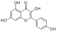
3,5,7-trihydroxy-2-(4-hydroxyphenyl)chromen-4-one
|
|
| 别名 |
3,4',5,7-Tetrahydroxyflavone; Pelargidenolon; Indigo Yellow; Kaempferol; Campherol
|
|
| HS Tariff Code |
2934.99.9001
|
|
| 存储方式 |
Powder -20°C 3 years 4°C 2 years In solvent -80°C 6 months -20°C 1 month |
|
| 运输条件 |
Room temperature (This product is stable at ambient temperature for a few days during ordinary shipping and time spent in Customs)
|
| 溶解度 (体外实验) |
|
|||
|---|---|---|---|---|
| 溶解度 (体内实验) |
配方 1 中的溶解度: ≥ 2 mg/mL (6.99 mM) (饱和度未知) in 10% DMSO + 40% PEG300 + 5% Tween80 + 45% Saline (这些助溶剂从左到右依次添加,逐一添加), 澄清溶液。
例如,若需制备1 mL的工作液,可将100 μL 20.0 mg/mL澄清DMSO储备液加入400 μL PEG300中,混匀;然后向上述溶液中加入50 μL Tween-80,混匀;加入450 μL生理盐水定容至1 mL。 *生理盐水的制备:将 0.9 g 氯化钠溶解在 100 mL ddH₂O中,得到澄清溶液。 配方 2 中的溶解度: ≥ 2 mg/mL (6.99 mM) (饱和度未知) in 10% DMSO + 90% (20% SBE-β-CD in Saline) (这些助溶剂从左到右依次添加,逐一添加), 澄清溶液。 例如,若需制备1 mL的工作液,可将 100 μL 20.0mg/mL澄清的DMSO储备液加入到900μL 20%SBE-β-CD生理盐水中,混匀。 *20% SBE-β-CD 生理盐水溶液的制备(4°C,1 周):将 2 g SBE-β-CD 溶解于 10 mL 生理盐水中,得到澄清溶液。 View More
配方 3 中的溶解度: ≥ 2 mg/mL (6.99 mM) (饱和度未知) in 10% DMSO + 90% Corn Oil (这些助溶剂从左到右依次添加,逐一添加), 澄清溶液。 配方 4 中的溶解度: 5 mg/mL (17.47 mM) in 0.5% CMC/saline water (这些助溶剂从左到右依次添加,逐一添加), 悬浊液; 超声助溶。 *生理盐水的制备:将 0.9 g 氯化钠溶解在 100 mL ddH₂O中,得到澄清溶液。 1、请先配制澄清的储备液(如:用DMSO配置50 或 100 mg/mL母液(储备液)); 2、取适量母液,按从左到右的顺序依次添加助溶剂,澄清后再加入下一助溶剂。以 下列配方为例说明 (注意此配方只用于说明,并不一定代表此产品 的实际溶解配方): 10% DMSO → 40% PEG300 → 5% Tween-80 → 45% ddH2O (或 saline); 假设最终工作液的体积为 1 mL, 浓度为5 mg/mL: 取 100 μL 50 mg/mL 的澄清 DMSO 储备液加到 400 μL PEG300 中,混合均匀/澄清;向上述体系中加入50 μL Tween-80,混合均匀/澄清;然后继续加入450 μL ddH2O (或 saline)定容至 1 mL; 3、溶剂前显示的百分比是指该溶剂在最终溶液/工作液中的体积所占比例; 4、 如产品在配制过程中出现沉淀/析出,可通过加热(≤50℃)或超声的方式助溶; 5、为保证最佳实验结果,工作液请现配现用! 6、如不确定怎么将母液配置成体内动物实验的工作液,请查看说明书或联系我们; 7、 以上所有助溶剂都可在 Invivochem.cn网站购买。 |
| 制备储备液 | 1 mg | 5 mg | 10 mg | |
| 1 mM | 3.4937 mL | 17.4685 mL | 34.9369 mL | |
| 5 mM | 0.6987 mL | 3.4937 mL | 6.9874 mL | |
| 10 mM | 0.3494 mL | 1.7468 mL | 3.4937 mL |
1、根据实验需要选择合适的溶剂配制储备液 (母液):对于大多数产品,InvivoChem推荐用DMSO配置母液 (比如:5、10、20mM或者10、20、50 mg/mL浓度),个别水溶性高的产品可直接溶于水。产品在DMSO 、水或其他溶剂中的具体溶解度详见上”溶解度 (体外)”部分;
2、如果您找不到您想要的溶解度信息,或者很难将产品溶解在溶液中,请联系我们;
3、建议使用下列计算器进行相关计算(摩尔浓度计算器、稀释计算器、分子量计算器、重组计算器等);
4、母液配好之后,将其分装到常规用量,并储存在-20°C或-80°C,尽量减少反复冻融循环。
计算结果:
工作液浓度: mg/mL;
DMSO母液配制方法: mg 药物溶于 μL DMSO溶液(母液浓度 mg/mL)。如该浓度超过该批次药物DMSO溶解度,请首先与我们联系。
体内配方配制方法:取 μL DMSO母液,加入 μL PEG300,混匀澄清后加入μL Tween 80,混匀澄清后加入 μL ddH2O,混匀澄清。
(1) 请确保溶液澄清之后,再加入下一种溶剂 (助溶剂) 。可利用涡旋、超声或水浴加热等方法助溶;
(2) 一定要按顺序加入溶剂 (助溶剂) 。
| NCT Number | Recruitment | interventions | Conditions | Sponsor/Collaborators | Start Date | Phases |
| NCT06060691 | Recruiting | Drug: Kaempferol | Female Sexual Dysfunction | Deraya University | September 20, 2023 | Phase 1 |
 CIMBA hydrochloride
 SRC-1 NR box peptide acetate
 ER ligand-12
 Diethylstilbestrol diphosphate
InvivoChem的所有产品仅用于作科学研究,不面向患者销售
Copyright 2020 InvivoChem LLC | All Rights Reserved 粤ICP备20063088号-1


 COA
COA


 463611831
