| 规格 | 价格 | 库存 | 数量 |
|---|---|---|---|
| 10 mM * 1 mL in DMSO |
|
||
| 1mg |
|
||
| 5mg |
|
||
| 10mg |
|
||
| 25mg |
|
||
| 50mg |
|
||
| 100mg |
|
||
| 250mg |
|
||
| Other Sizes |
|

| 靶点 |
TGF-β receptor type 1/ALK5 (IC50 = 38.2 nM)
Transforming Growth Factor-β type 1 receptor (TGFβRI) (IC50=0.8 nM); exhibits no significant inhibition on TGFβRII (IC50>10000 nM) [2] Transforming Growth Factor-β type 1 receptor (TGFβRI)[1] |
|---|---|
| 体外研究 (In Vitro) |
LY3200882 在体外以剂量依赖性方式有效抑制肿瘤和免疫细胞中 TGFβ 诱导的 SMAD 磷酸化[1]。 LY3200882 在三阴性乳腺癌原位 4T1-LP 模型中表现出有效的抗肿瘤作用,这种活性与肿瘤微环境中肿瘤浸润淋巴细胞的增加相关[1]。在体外免疫抑制实验中,LY3200882显示出能够拯救TGFβ1抑制或T调节细胞抑制的幼稚T细胞活性并恢复增殖[1]。 LY3200882 降低 NIH3T3 细胞活力,IC50 为 82.9 nM[2]。
作用机制研究表明LY3200882抑制多种促肿瘤活性。LY3200882在体外肿瘤和免疫细胞中有效抑制TGFβ介导的SMAD磷酸化。在体外免疫抑制实验中,LY3200882显示出挽救TGFβ1抑制或T调节性细胞抑制naïve T细胞活性和恢复增殖的能力。因此,LY3200882作为免疫调节剂具有良好的活性。此外,LY3200882在体外迁移试验中显示出抗转移活性,在体内实验转移性肿瘤模型(静脉注射三阴性乳腺癌EMT6-LM2模型)中也显示出抗转移活性[1]。 抑制TGFβRI激酶活性及下游Smad信号通路:LY3200882强效抑制重组人TGFβRI激酶活性,IC50为0.8 nM,而对TGFβRII及其他激酶抑制活性微弱(对TGFβRI的选择性比率>12500倍)。在A549细胞(IC50=3.2 nM)和MDA-MB-231细胞中,浓度依赖性阻断TGFβ1诱导的Smad2磷酸化(Western blot检测)[2] - 抑制TGFβ介导的细胞应答:在A549细胞中抑制TGFβ1诱导的上皮-间质转化(EMT),Western blot和免疫荧光显示间质标志物(波形蛋白vimentin、N-钙粘蛋白N-cadherin)表达降低,上皮标志物(E-钙粘蛋白E-cadherin)表达升高;在MDA-MB-231和A549细胞中抑制TGFβ1诱导的细胞迁移和侵袭(与单独TGFβ1组相比p<0.01)[2] - 癌细胞增殖抑制活性:通过MTT实验显示,对TGFβ应答型癌细胞系具有浓度依赖性生长抑制作用,A549(IC50=12.5 nM)、MDA-MB-231(IC50=15.8 nM)、HCT116(IC50=18.3 nM);以9.7 nM的IC50抑制A549细胞克隆形成,体现长期增殖抑制效应[2] - 阻断TGFβ诱导的靶基因表达:实时PCR定量显示,在A549细胞中减少TGFβ1诱导的靶基因(PAI-1、CTGF、纤连蛋白fibronectin)表达(与单独TGFβ1组相比p<0.001)[2] - 体外选择性抑制TGFβ信号:在浓度高达10 μM时,不影响骨形态发生蛋白(BMP)信号或其他激酶通路(如EGFR、VEGFR2、PI3K/Akt),证实高靶点选择性[1] |
| 体内研究 (In Vivo) |
LY3200882(60 mg/kg;口服灌胃;每天两次;21 天;雌性 BALB/C 小鼠)治疗可显着减缓 CT26 模型中肿瘤的形成[2]。在体内,LY3200882 显着且剂量依赖性地抑制皮下肿瘤中 TGFβ 介导的 SMAD 磷酸化[1]。在实验性转移肿瘤模型(三阴性乳腺癌静脉EMT6-LM2模型)中,LY3200882表现出体内抗转移功效[1]。
在临床前肿瘤模型中,LY3200882在三阴性乳腺癌原位4T1-LP模型中表现出较强的抗肿瘤活性,这种活性与肿瘤微环境中肿瘤浸润淋巴细胞增强有关。在原位4T1-LP模型中观察到持久的肿瘤消退,并在所有小鼠中再次挑战先天性肿瘤导致完全排斥。最后,LY3200882在同基因CT26模型中显示出具有检查点抑制(抗pd - l1)的组合抗肿瘤益处。[1] 抑制异种移植瘤生长:裸鼠皮下接种A549或MDA-MB-231细胞,待肿瘤体积达100–150 mm³时随机分组(每组n=6),实验组每日口服LY3200882 30 mg/kg,连续21天。每周测量两次肿瘤体积,处死时记录肿瘤重量。与溶媒对照组相比,LY3200882显著抑制肿瘤生长,A549模型抑制率65%,MDA-MB-231模型抑制率58%(p<0.01);肿瘤组织Western blot显示p-Smad2水平降低,证实体内TGFβRI信号抑制[1] - 增强抗肿瘤免疫应答:在MC38同源肿瘤模型中,LY3200882(30 mg/kg/天,口服)与抗PD-1抗体联合使用,显著增加肿瘤浸润CD8+ T细胞数量,减少调节性T细胞(Tregs),实现协同肿瘤生长抑制(与溶媒组相比抑制率78%,p<0.001)[1] |
| 酶活实验 |
ALK5抑制活性[2]
采用发光ADP检测法评价化合物与ALK5的结合能力。将10 mM原液在DMSO中连续稀释3倍,得到10点稀释曲线,最终化合物浓度范围为3.333 μM ~ 0.5 nM。测定在1X激酶反应缓冲液中进行,缓冲液中含有40 mM Tris pH 7.5, 20 mM MgCl2, 0.1% BSA, 1 mM DTT,最终测定体积为5 μL。在384孔的实验板中,每孔加入2.5 μL的ALK5蛋白,最终浓度为3 μg/mL,每孔中溶解100 nL每种浓度的测试化合物。2.5 μL TGF-βR1肽,终浓度3 μg/mL, ATP,终浓度1 mM。28℃孵育120 min后,加入5 μL ADP-Glo™试剂终止激酶反应,消耗剩余ATP。28℃孵育120 min后,加入10 μL激酶检测试剂,将ADP转化为ATP,记录发光。 体外p38α酶活性测定[2] 所有酶促反应在28°C下进行,反应时间为40 min。25 μL的反应混合物中含有50 mM HEPES, pH为7.5,0.0015% Brij-35, 25 ng激酶,10 μM ATP和fam标记的肽。化合物在100 μM, 3倍稀释,10倍浓度下进行检测。该检测由ChemPartner进行。它通过定量测定激酶反应后溶液中剩余ATP的量来测量激酶活性。荧光信号与存在的ATP量相关,与激酶活性的量呈负相关。在XLFit excel插件版本5.4.0.8中计算IC50值,使用公式:Y=Bottom +(Top-Bottom)/(1+(IC50/X) ^Hill Slope),其中X为化合物浓度,Y为抑制率(%)。 TGFβRI激酶活性测定:将重组人TGFβRI激酶结构域与ATP、特异性肽底物及系列稀释的LY3200882共同孵育,采用均相时间分辨荧光(HTRF) assay检测底物磷酸化水平。以溶媒处理组为100%活性,通过药物浓度与激酶活性百分比绘制曲线,计算IC50值[2] - 激酶选择性测定:采用放射活性激酶 assay,将10 μM LY3200882与468种人源激酶面板孵育,通过检测放射性ATP掺入底物的量确定每种激酶的抑制率,对比对TGFβRI与其他激酶的抑制作用评估选择性[2] - TGFβRII交叉反应性测定:采用与TGFβRI相同的HTRF assay,使用重组人TGFβRII激酶结构域,加入系列浓度LY3200882(0.1 nM–10 μM),测定IC50以评估交叉反应性[2] |
| 细胞实验 |
基于细胞的荧光素酶报告试验TGF-β 1型受体活性[2]
本实验的目的是在细胞基础上鉴定干扰SMAD 2,3依赖性基因表达的化合物,证明它们在细胞水平上抑制ALK5。以每孔4000个细胞的速度,在96孔板中,用DMEM培养基将已准备好的冷冻原液中的Luc-Smad 2/3-NIH3T3细胞进行平板培养。细胞贴壁过夜后,培养基改为2%胎牛血清。在DMSO中配制试验化合物,制成4mm的原液。将原液在DMSO中连续稀释4倍,得到8点稀释曲线,最终化合物浓度范围为20 μM ~ 1.22 nM,并加入待测化合物。24小时后,在每个孔中加入Glo Lysis Buffer和Bright-Glo Luciferase assay system,使孔体积翻倍。将等分液(180 μL)转移到白色实心底板上,在平板读取器上读取发光(读取1 s)。 Smad磷酸化Western blot实验:A549或MDA-MB-231细胞血清饥饿24小时,用LY3200882(0.1 nM–10 μM)预处理1小时,再用5 ng/ml TGFβ1刺激30分钟。收集细胞提取蛋白,用p-Smad2、总Smad2及内参GAPDH抗体进行Western blot,通过光密度分析定量p-Smad2与总Smad2的比值[2] - 细胞增殖MTT实验:将癌细胞(A549、MDA-MB-231、HCT116)接种于96孔板,用LY3200882(0.1 nM–10 μM)处理72小时,加入MTT试剂孵育4小时后检测吸光度,通过量效曲线计算IC50值[2] - 克隆形成实验:A549细胞低密度接种于6孔板,用LY3200882(0.1 nM–10 μM)处理14天,每3天更换培养基。克隆经固定、染色后计数,以溶媒对照组为基准计算克隆形成率[2] - 细胞迁移和侵袭实验:使用Transwell小室(侵袭实验加Matrigel),将经LY3200882(10 nM–1 μM)预处理1小时的MDA-MB-231或A549细胞接种于上室,下室加入5 ng/ml TGFβ1。迁移实验24小时后、侵袭实验48小时后,固定并染色下室膜上的细胞,计数统计[2] - EMT标志物表达实验:A549细胞用LY3200882(10 nM–1 μM)与5 ng/ml TGFβ1共同处理72小时,Western blot检测E-cadherin、N-cadherin、vimentin表达;免疫荧光染色观察E-cadherin定位[2] - 靶基因表达实时PCR实验:A549细胞用LY3200882(0.1 nM–10 μM)与5 ng/ml TGFβ1共同处理24小时,提取总RNA,实时PCR定量PAI-1、CTGF、fibronectin mRNA水平,以GAPDH为内参归一化[2] |
| 动物实验 |
动物/疾病模型: BALB/C雌性小鼠(5-8周龄)注射CT26细胞[2]
剂量: 60 mg/kg 给药途径: 灌胃(po);每日两次;持续21天 实验结果: 在CT26模型中观察到肿瘤生长显著延迟。 药代动力学程序[2] 本研究在BALB/C雄性小鼠中进行,以研究测试化合物的药代动力学特征。小鼠随机分为两组,每组3只。将测试化合物[15r(LY3200882类似物)]溶于PEG200-EtOH-solutol-生理盐水(4:1:1:14)中,配制成0.5 mg/mL的灌胃溶液和0.1 mg/mL的静脉注射溶液。样品采集方法如下:1)单次口服(PO)组:5 mg/kg,0.2 mL/10 g;2)单次尾静脉注射(IV)组:1 mg/kg,0.1 mL/10 g。分别于静脉或口服给药后 5、15、30 分钟、1、2、6、10 和 24 小时,从眼眶静脉丛采集血液样本至肝素化 EP 管中,并采用 LC-MS/MS (API 4500) 分析血液成分。 体内肿瘤异种移植模型[2] 采用成熟的肿瘤发生模型评估化合物15r(LY3200882 类似物)在 BALB/C 雌性小鼠模型中的抗肿瘤作用。所有小鼠均饲养于标准无特定病原体 (SPF) 条件下,动物实验严格遵守动物福利和伦理委员会 (AWEC) 批准的方案。将 1 × 10⁶ 个 CT26 细胞/只小鼠皮下注射至 5 至 8 周龄的 BALB/C 雌性小鼠体内。所有化合物均采用灌胃法给药。每周三次通过触诊检查小鼠肿瘤的生长情况,并使用公式 V = 0.5 × 长 × 宽² 计算肿瘤体积。实验和结果评估过程中,研究人员未采用盲法。小鼠由实验室的一名独立人员随机分为三组,每组 6 只。未采用统计学方法预先确定样本量。化合物的抗肿瘤效果通过肿瘤生长抑制率 (TGI) 或相对肿瘤增殖率 (T/C) 进行评估:TGI(%) = [1-(Vt1-Vt0)/(Vc1-Vc0)] × 100%,其中 Vc1 和 Vt1 分别为肿瘤取出时对照组和治疗组的平均肿瘤体积,Vc0 和 Vt0 分别为给药开始时相同组的平均肿瘤体积。 T/C (%) = TRTV/CRTV × 100%,其中 TRTV 为治疗组的相对肿瘤体积 (RTV),CRTV 为对照组的 RTV。(RTV = Vt/V0,Vt 为肿瘤取出时治疗组的平均体积,V0 为给药开始时各组的平均体积)。 异种移植瘤生长抑制试验:将 5×10^6 个 A549 或 MDA-MB-231 细胞皮下植入 6-8 周龄的雌性裸鼠体内。当肿瘤体积达到 100-150 mm³ 时,将小鼠随机分为载体组和 LY3200882 组(每组 n=6)。LY3200882 以 30 mg/kg 的剂量每日一次灌胃给药,持续 21 天。每周使用游标卡尺测量两次肿瘤体积,并在处死小鼠时记录肿瘤重量。收集肿瘤组织进行p-Smad2的Western blot分析[1] - 同源肿瘤免疫治疗实验:将1×10^6个MC38结肠癌细胞皮下植入C57BL/6小鼠体内。当肿瘤体积达到80–100 mm³时,将小鼠随机分为四组:载体组、LY3200882组(30 mg/kg/天,口服)、抗PD-1抗体组(10 mg/kg,腹腔注射,每周两次)以及联合治疗组。治疗持续14天,每周测量三次肿瘤体积。处死小鼠后,采用流式细胞术分析肿瘤浸润淋巴细胞[1] |
| 药代性质 (ADME/PK) |
口服生物利用度:在大鼠中,口服LY3200882(10 mg/kg)的口服生物利用度为52%,Cmax为1.8 μg/ml,AUC0–24h为12.6 μg·h/ml [1]
- 半衰期和清除率:在犬中,静脉注射LY3200882(5 mg/kg)显示末端半衰期(t1/2)为4.8小时,全身清除率为1.2 ml/min/kg。口服给药(10 mg/kg)后,Cmax 为 2.3 μg/ml,AUC0–24h 为 18.9 μg·h/ml [1] - 血浆蛋白结合率:通过平衡透析法测定,LY3200882 在人、大鼠和犬血浆中均表现出较高的血浆蛋白结合率(>95%)[2] |
| 毒性/毒理 (Toxicokinetics/TK) |
体外毒性:LY3200882在浓度高达10 μM时,对正常人支气管上皮细胞(BEAS-2B)未显示出显著的细胞毒性(细胞存活率>85% vs 对照组)[2]
- 体内毒性:小鼠连续21天口服LY3200882(30 mg/kg/天),未出现显著的体重减轻、血液学异常或主要器官(肝脏、肾脏、心脏、肺脏)的组织病理学改变[1] - 无药物相互作用:在浓度高达10 μM时,未抑制细胞色素P450酶(CYP1A2、CYP2C9、CYP2C19、CYP2D6、CYP3A4),表明其药物相互作用的可能性较低[2] |
| 参考文献 |
|
| 其他信息 |
LY-3200882 正在临床试验 NCT04158700(一项针对晚期癌症患者的 LY3200882 联合帕博利珠单抗研究)中进行研究。
TGFβ 抑制剂 LY3200882 是一种口服生物利用度高的药物,靶向转化生长因子-β (TGFβ),具有潜在的抗肿瘤活性。给药后,LY3200882 可特异性靶向并结合 TGFβ,从而阻止 TGFβ 与其受体 TGFβR 的结合以及 TGFβ 介导的信号转导。这可能导致 TGFβ 依赖性癌细胞增殖的减少。 TGFβ信号通路在肿瘤中常常失调,并在细胞生长、分化、凋亡、迁移、侵袭、血管生成以及多种免疫反应的调控中发挥关键作用。转化生长因子β (TGFβ) 信号通路是一种多效性细胞通路,在癌症中扮演着至关重要的角色。事实上,侵袭性肿瘤通常与高配体水平相关,因此在多种肿瘤类型中预后不良。癌细胞利用自分泌和旁分泌TGFβ信号来调节肿瘤细胞和肿瘤微环境,从而导致高度侵袭性和转移性表型,诱导并增加肿瘤血管生成,调节基质中的细胞外基质,并抑制免疫监视和抗肿瘤免疫。针对TGFβ通路的小分子抑制剂galunisertib(又名LY2157299一水合物)的临床研究提供了概念验证数据,支持TGFβ在癌症中的作用以及靶向TGFβ通路的实用性。本文描述了新一代TGF-β受体1型(TGFβRI)小分子抑制剂LY3200882的鉴定。该分子是一种高效、高选择性的TGFβRI抑制剂,其结构平台具有可规模化合成的合成路线。它是一种ATP竞争性抑制剂,可抑制TGFβRI的丝氨酸/苏氨酸激酶结构域。总之,我们开发了一种新型高效、高选择性的TGFβRI小分子抑制剂,可用于癌症治疗。[1] 抑制转化生长因子β(TGF-β)1型受体(ALK5)为治疗纤维化疾病和恶性肿瘤提供了一种可行的方法。在本研究中,我们设计并合成了一系列新的4-(吡啶-4-氧基)-3-(3,3-二氟环丁基)-吡唑衍生物,并对其作为TGF-β 1型受体抑制剂的生物学活性进行了评价。活性最强的化合物 15r 对 ALK5 酶和 NIH3T3 细胞活力的抑制 IC50 值分别为 44 nM 和 42.5 nM。化合物 15r 的口服血浆暴露量和生物利用度均优于 LY-3200882,并且在 CT26 异种移植小鼠模型中体内抑制了 65.7% 的肿瘤生长。[2] LY3200882 是一种新型高选择性 TGFβRI 小分子抑制剂,属于 4-(吡啶-4-氧基)-3-(3,3-二氟环丁基)-吡唑衍生物类。[2] 其作用机制涉及直接抑制 TGFβRI 激酶活性,从而阻断下游 Smad2/3 信号通路,该通路对肿瘤进展、EMT、转移和免疫抑制至关重要。[1][2] LY3200882 对 TGFβRI 的选择性优于其他激酶(包括 TGFβRII),从而最大限度地减少了脱靶效应。[2] 在临床前研究中, LY3200882在TGFβ反应性异种移植模型中显示出单药抗肿瘤活性,并且与免疫检查点抑制剂(抗PD-1)联合使用时具有协同疗效,凸显了其在联合癌症治疗中的潜力[1] LY3200882具有良好的药代动力学特性,包括良好的口服生物利用度、中等的半衰期和高血浆蛋白结合率,支持其作为口服治疗药物的开发[1][2] |
| 分子式 |
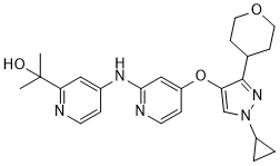
C24H29N5O3
|
|
|---|---|---|
| 分子量 |
435.518765211105
|
|
| 精确质量 |
435.227
|
|
| 元素分析 |
C, 66.19; H, 6.71; N, 16.08; O, 11.02
|
|
| CAS号 |
1898283-02-7
|
|
| 相关CAS号 |
|
|
| PubChem CID |
121249291
|
|
| 外观&性状 |
Off-white to light yellow solid powder
|
|
| LogP |
2.1
|
|
| tPSA |
94.3
|
|
| 氢键供体(HBD)数目 |
2
|
|
| 氢键受体(HBA)数目 |
7
|
|
| 可旋转键数目(RBC) |
7
|
|
| 重原子数目 |
32
|
|
| 分子复杂度/Complexity |
612
|
|
| 定义原子立体中心数目 |
0
|
|
| SMILES |
O1CCC(CC1)C1C(=CN(C2CC2)N=1)OC1C=CN=C(C=1)NC1C=CN=C(C=1)C(C)(C)O
|
|
| InChi Key |
PNPFMWIDAKQFPY-UHFFFAOYSA-N
|
|
| InChi Code |
InChI=1S/C24H29N5O3/c1-24(2,30)21-13-17(5-9-25-21)27-22-14-19(6-10-26-22)32-20-15-29(18-3-4-18)28-23(20)16-7-11-31-12-8-16/h5-6,9-10,13-16,18,30H,3-4,7-8,11-12H2,1-2H3,(H,25,26,27)
|
|
| 化学名 |
|
|
| 别名 |
|
|
| HS Tariff Code |
2934.99.9001
|
|
| 存储方式 |
Powder -20°C 3 years 4°C 2 years In solvent -80°C 6 months -20°C 1 month |
|
| 运输条件 |
Room temperature (This product is stable at ambient temperature for a few days during ordinary shipping and time spent in Customs)
|
| 溶解度 (体外实验) |
|
|||
|---|---|---|---|---|
| 溶解度 (体内实验) |
配方 1 中的溶解度: ≥ 2.5 mg/mL (5.74 mM) (饱和度未知) in 10% DMSO + 40% PEG300 + 5% Tween80 + 45% Saline (这些助溶剂从左到右依次添加,逐一添加), 澄清溶液。
例如,若需制备1 mL的工作液,可将100 μL 25.0 mg/mL澄清DMSO储备液加入到400 μL PEG300中,混匀;然后向上述溶液中加入50 μL Tween-80,混匀;加入450 μL生理盐水定容至1 mL。 *生理盐水的制备:将 0.9 g 氯化钠溶解在 100 mL ddH₂O中,得到澄清溶液。 配方 2 中的溶解度: ≥ 2.5 mg/mL (5.74 mM) (饱和度未知) in 10% DMSO + 90% (20% SBE-β-CD in Saline) (这些助溶剂从左到右依次添加,逐一添加), 澄清溶液。 例如,若需制备1 mL的工作液,可将 100 μL 25.0 mg/mL澄清DMSO储备液加入900 μL 20% SBE-β-CD生理盐水溶液中,混匀。 *20% SBE-β-CD 生理盐水溶液的制备(4°C,1 周):将 2 g SBE-β-CD 溶解于 10 mL 生理盐水中,得到澄清溶液。 View More
配方 3 中的溶解度: ≥ 2.5 mg/mL (5.74 mM) (饱和度未知) in 10% DMSO + 90% Corn Oil (这些助溶剂从左到右依次添加,逐一添加), 澄清溶液。 1、请先配制澄清的储备液(如:用DMSO配置50 或 100 mg/mL母液(储备液)); 2、取适量母液,按从左到右的顺序依次添加助溶剂,澄清后再加入下一助溶剂。以 下列配方为例说明 (注意此配方只用于说明,并不一定代表此产品 的实际溶解配方): 10% DMSO → 40% PEG300 → 5% Tween-80 → 45% ddH2O (或 saline); 假设最终工作液的体积为 1 mL, 浓度为5 mg/mL: 取 100 μL 50 mg/mL 的澄清 DMSO 储备液加到 400 μL PEG300 中,混合均匀/澄清;向上述体系中加入50 μL Tween-80,混合均匀/澄清;然后继续加入450 μL ddH2O (或 saline)定容至 1 mL; 3、溶剂前显示的百分比是指该溶剂在最终溶液/工作液中的体积所占比例; 4、 如产品在配制过程中出现沉淀/析出,可通过加热(≤50℃)或超声的方式助溶; 5、为保证最佳实验结果,工作液请现配现用! 6、如不确定怎么将母液配置成体内动物实验的工作液,请查看说明书或联系我们; 7、 以上所有助溶剂都可在 Invivochem.cn网站购买。 |
| 制备储备液 | 1 mg | 5 mg | 10 mg | |
| 1 mM | 2.2961 mL | 11.4805 mL | 22.9611 mL | |
| 5 mM | 0.4592 mL | 2.2961 mL | 4.5922 mL | |
| 10 mM | 0.2296 mL | 1.1481 mL | 2.2961 mL |
1、根据实验需要选择合适的溶剂配制储备液 (母液):对于大多数产品,InvivoChem推荐用DMSO配置母液 (比如:5、10、20mM或者10、20、50 mg/mL浓度),个别水溶性高的产品可直接溶于水。产品在DMSO 、水或其他溶剂中的具体溶解度详见上”溶解度 (体外)”部分;
2、如果您找不到您想要的溶解度信息,或者很难将产品溶解在溶液中,请联系我们;
3、建议使用下列计算器进行相关计算(摩尔浓度计算器、稀释计算器、分子量计算器、重组计算器等);
4、母液配好之后,将其分装到常规用量,并储存在-20°C或-80°C,尽量减少反复冻融循环。
计算结果:
工作液浓度: mg/mL;
DMSO母液配制方法: mg 药物溶于 μL DMSO溶液(母液浓度 mg/mL)。如该浓度超过该批次药物DMSO溶解度,请首先与我们联系。
体内配方配制方法:取 μL DMSO母液,加入 μL PEG300,混匀澄清后加入μL Tween 80,混匀澄清后加入 μL ddH2O,混匀澄清。
(1) 请确保溶液澄清之后,再加入下一种溶剂 (助溶剂) 。可利用涡旋、超声或水浴加热等方法助溶;
(2) 一定要按顺序加入溶剂 (助溶剂) 。
 KY1070
 Rinvatercept
 PF-07868489
 TGFβRI-IN-7
InvivoChem的所有产品仅用于作科学研究,不面向患者销售
Copyright 2020 InvivoChem LLC | All Rights Reserved 粤ICP备20063088号-1


 COA
COA
 463611831
