| 规格 | 价格 | 库存 | 数量 |
|---|---|---|---|
| 10mg |
|
||
| 25mg |
|
||
| 50mg |
|
||
| 100mg |
|
||
| 250mg | |||
| 500mg | |||
| Other Sizes |
| 靶点 |
ROS; mitochondria-targeted antioxidant
|
|---|---|
| 体外研究 (In Vitro) |
线粒体醌 (MitoQ) 是一种针对线粒体的抗氧化剂。在四个小时的冷藏 (CS) 期间,使用剂量反应测试来确定米托醌 (MitoQ) 和 DecylTPP 治疗的理想剂量。为了研究线粒体醌治疗对 CS 损伤的潜在保护作用,首先使用了 MitoSOX Red(一种靶向并检测线粒体超氧化物产生的荧光染料)。当CS应用于正常大鼠肾(NRK)细胞时,线粒体超氧化物导致细胞荧光比对照组增加约两倍。当涉及 CS 诱导的线粒体超氧化物产生时,线粒体中的醌可以显着抵抗它,而对照化学物质 DecylTPP 则没有这种作用。线粒体超氧化物治疗大大减少了线粒体超氧化物的产生,并且用 DecylTPP 治疗的肾脏的线粒体超氧化物水平与仅接受 CS 的肾脏相当[1]。
MitoQ对CS期间线粒体超氧化物生成的保护作用。[1] MitoQ和DecylTPP治疗的最佳剂量是从4-h CS期间的剂量反应实验中选择的。对于基于细胞的研究,从0.5、0.75、1.0、1.5和2μM的范围内选择1μM,而离体研究的最有效剂量是50、100和500μM的剂量反应研究中的100μM(数据未显示)。最初使用线粒体靶向荧光染料MitoSOX Red测试了MitoQ治疗对CS损伤的潜在保护作用,该染料可测量线粒体超氧化物的产生。如图2A所示,与未处理的细胞相比,暴露于CS的NRK细胞由于线粒体超氧化物而导致荧光增加了约2倍。MitoQ对CS诱导的线粒体超氧化物产生具有显著的保护作用;而对照化合物DecylTPP没有提供任何保护。使用基于荧光分光光度法的测定法在396和510 nm处检测MitoSOX Red荧光激发,进一步证实了这一点,其中396 nm是线粒体超氧化物的特异性指示剂,510 nm检测非特异性氧化剂的产生(Robinson等人,2006)(图2B)。此外,与对照组肾脏相比,单独暴露于CS的肾脏显示线粒体超氧化物生成增加(图2C)。MitoQ治疗显著降低了线粒体超氧化物的产生,而用DecylTPP治疗的肾脏的线粒体超氧化物水平与单独暴露于CS的肾脏相当(图2C)。 MitoQ减弱CS过程中硝基酪氨酸加合物的形成。[1] 免疫细胞化学和免疫组织化学用于评估CS期间的硝基酪氨酸蛋白加合物。如图3A所示,与未处理的细胞相比,单独暴露于CS的NRK细胞的硝基酪氨酸染色(红色荧光)显著增加。MitoQ将硝基酪氨酸的形成减少了2倍,而对照化合物DecylTPP没有减少CS介导的硝基酪氨酸的生成(图3A)。过氧化亚硝酸盐处理的细胞具有强烈的硝基酪氨酸形成(阳性对照),当硝基酪氨酸抗体被过量3-硝基酪氨酸预吸收时被阻断(阴性对照;ONOO-+阻断)。大鼠肾脏免疫组织化学数据与关于MitoQ和硝基酪氨酸作用的体外研究结果一致。图3B显示,与对照组肾脏相比,CS肾脏的远端和近端小管中硝基酪氨酸(棕色染色)的增加,肾小球中的增加程度较小。用MitoQ治疗的肾脏硝基酪氨酸形成较少。相比之下,DecylTPP治疗的肾脏与CS肾脏的硝基酪氨酸含量相似。使用用过量3-硝基酪氨酸预吸收的抗体(CS+阻断)也证实了硝基酪氨酸染色的特异性。 MitoQ防止CS期间线粒体呼吸复合物失活。[1] 在体外和离体肾模型中评估线粒体呼吸复合物活性,以研究4-h CS是否会改变线粒体呼吸功能。与未处理的细胞相比,NRK细胞CS后复合物I和II显著失活(图4)。MitoQ完全阻止了复合物I和II的失活,而DecylTPP对复合物活性没有显著影响。本研究未对复合物III和IV进行评估,因为我们之前已经证明,24小时CS对这两种复合物的活性都没有影响(Mitchell等人,2010);因此,没有必要对这些复杂的活动进行评估。与体外研究结果一致,大鼠肾脏的CS导致复合物I和II活性的部分失活,对复合物III和IV没有影响(图4)。MitoQ对CS肾脏的复合物I和II失活具有保护作用,而DecylTPP没有任何作用。 MitoQ减少NRK细胞CS+再武装过程中硝基酪氨酸的形成和细胞死亡。[1] 为了测试MitoQ是否可能在再灌注/移植后提供对氧化剂产生和细胞死亡的保护,细胞暴露于CS加RW。如图6A所示,NRK细胞的4-h CS加过夜(18小时)RW导致显著的硝基酪氨酸形成。在CS期间添加MitoQ显著减弱了CS期间的硝基酪氨酸染色。而对照化合物DecylTPP没有效果。LDH细胞毒性显示,暴露于CS的NRK细胞的细胞死亡显著增加。RW与未处理细胞的比较(图6B)。MitoQ在单独CS和CS期间将细胞死亡降低了约2倍。RW.DecylTPP在两个治疗组中都没有减少细胞死亡。 MitoQ清除分离的胰腺腺泡细胞中的ROS产生[2] 应用1 mM H2O2导致细胞(对照组)中ROS的稳定上升,这反映在CM-H2DCFDA荧光强度的增加上;用1预处理的细胞 μM MitoQ与对照组细胞相比,ROS产生显著减少,而1 μM dTPP,没有抗氧化活性的MitoQ亲脂性阳离子没有影响(图1(a))。MitoQ和dTPP本身均未诱导ROS的产生(图1(b))。 MitoQ不能保护分离的胰腺腺泡细胞免受AP沉淀剂引起的线粒体去极化[2] 未用1进行预处理 μM MitoQ和dTPP在分离的胰腺腺泡细胞中引起了ΔΨM的去极化,这与实验结束时应用原核CCCP以引发完全去极化形成鲜明对比。(图2(a))。然而,在10 μM的MitoQ和dTPP都诱导了ΔΨM本身的稳定降低,这对MitoQ来说更为深刻(图2(b)和2(c))。与单独用HEPES处理的对照细胞相比,CCK(10 nM)诱导了ΔΨm的去极化(图2(d)),这种作用不受1 μM MitoQ或dTPP(图2(e))。同样,TLCS(500μM)的灌注使ΔΨM去极化(图2(f)),而1不受影响 μM MitoQ或dTPP(图2(g))。 MitoQ导致胰腺腺泡细胞死亡并加重CCK诱导的坏死[2] 两者1 μM MitoQ和dTPP引起PI摄取增加,表明坏死;在2、6和10时观察到显著差异 h,用MitoQ或dTPP预处理的细胞与单独用HEPES处理的对照组细胞之间的差异(图3(a))。增加10 nM CCK诱导坏死的时间依赖性增加。然而,1 μM MitoQ对CCK诱导的细胞死亡没有任何保护作用。相反,与单独使用CCK相比,MitoQ和dTPP在2℃时均显著恶化了坏死 h、 而dTPP,而不是MitoQ,在以后的时间点加重了CCK诱导的细胞死亡(图3(b))。 |
| 体内研究 (In Vivo) |
米托醌 (MitoQ) 治疗可显着减少中性粒细胞浸润和胰腺水肿。 MitoQ 会剂量依赖性地增加血清淀粉酶;如果剂量较高,则大约翻倍。当以 10 mg/kg(剂量 1)给药时,MitoQ 治疗显着提高了血清 IL-6 水平,并使 Caerulein 诱导的肺 MPO 活性几乎增加了一倍 [2]。
MitoQ改善了CER-AP的整体胰腺组织病理学,但加重了全身损伤[2] 图4(a)显示了对照组和不同治疗组的代表性组织病理学幻灯片,图4(b)总结了单个成分的总体组织病理学评分和分解评分。腹腔注射生理盐水不会引起胰腺的任何明显的组织病理学变化,而蓝蛙素的过度刺激会引起AP的典型特征;胰腺导管边缘和实质明显水肿、空泡化、中性粒细胞浸润,局灶性腺泡细胞坏死明显12 h第一次注射蛙皮素后。与生理盐水对照组相比,CER-AP的特征还在于血清淀粉酶、胰蛋白酶和MPO活性以及肺MPO活性显著增加。 MitoQ两种剂量的治疗均显著减少了胰腺水肿和中性粒细胞浸润。然而,胰腺坏死没有得到预防,在较高剂量下有更大的坏死趋势,尽管这没有达到显著性。MitoQ剂量依赖性地增加血清淀粉酶,在较高剂量下约加倍(图5(a))。胰蛋白酶活性和MPO活性在任何剂量下都没有受到MitoQ的显著影响(图5(b)和5(c))。此外,MitoQ治疗使蓝蛙素诱导的肺MPO活性几乎翻了一番(图5(d)),在剂量1时,血清IL-6水平也明显升高(图5)。 非抗氧化剂类似物dTPP在两种剂量下均显著减少了水肿、中性粒细胞浸润和坏死,导致组织病理学评分总体降低。尽管dTPP降低了胰蛋白酶和MPO活性(图5(b)和5(c)),但血清淀粉酶没有受到显著影响(图5)。与MitoQ获得的结果类似,dTPP也显著增加了蓝蛙素诱导的肺MPO活性和血清IL-6水平(图5(d)和5(e))。 MitoQ无法抵御TLCS-AP[2] 假手术只引起胰腺腺泡细胞轻度水肿,没有明显的炎症和坏死迹象。输注3 通过胰管进入胰腺的mM TLCS在24小时时导致胰腺头部明显的组织病理学变化 h、 其特征是水肿、炎症、坏死显著增加,从而导致整体组织病理学评分(图6(a)(i-iv))。然而,胰腺的身体和尾部受到的影响要小得多(数据未显示)。与假手术组相比,TLCS-AP与血清淀粉酶、胰腺MPO活性和血清IL-6水平升高有关(图6(b)-6(d))。 较低剂量的MitoQ和dTPP均未诱导胰腺的组织病理学变化,水肿、炎症、坏死和总体组织病理学评分没有变化(图6(a)(i-iv))。同样,当TLCS-AP小鼠用MitoQ或dTPP治疗时,血清淀粉酶和胰腺MPO活性没有显著变化(图6(b)和6(c))。MitoQ或dTPP治疗使血清IL-6水平略有升高,但未达到统计学意义(图6(d))。在没有注射蓝蛙素或输注TLCS的情况下,单独对小鼠应用两种剂量的MitoQ或dTPP表明,MitoQ和dTPP本身都显著增加了肺MPO活性(数据未显示)。 MitoQ对PMA诱导的PMNs中ROS产生的双相效应[2] 图7(a)说明了1的效果 μM MitoQ或dTPP对PMA诱导的分离PMNs中ROS产生的影响。NAD(P)H氧化酶刺激剂PMA(50 ng/mL)在最初的几分钟内诱导PMNs周围细胞外溶液中ROS的急剧增加,在8 大约20分钟后,下降到平稳状态 mins.NAD(P)H氧化酶抑制剂DPI的应用降低了峰值相并完全抑制了ROS平台(图7(c))。MitoQ治疗对PMNs中ROS的产生产生了双相效应。因此,观察到PMA诱导的初始ROS峰值的浓度依赖性抑制,峰值时间延迟至10分钟(图7(a)-7(c))。应用1 与单独用PMA处理的细胞相比,μM dTPP对PMA诱导的PMNs中ROS产生的峰值没有显著影响。有趣的是,MitoQ在40℃时引起PMA诱导的ROS产生的浓度依赖性增强 mins(图7(c)),这是dTPP仅在较高浓度下共享的动作。 MitoQ治疗可减少CS诱导的肾损伤和细胞死亡。[1] 采用过碘酸希夫染色法检测大鼠肾脏CS期间的组织病理学变化。CS暴露后,出现了严重的广泛管状损伤,如扩张、刷状边界损失和细胞碎片/铸型形成(图5A)。MitoQ显著改善了肾脏组织学,而DecylTPP治疗没有逆转肾脏损伤。TUNEL染色,即细胞核的棕色染色,被用作细胞死亡的标志。如图5B所示,与对照组肾脏相比,CS导致细胞死亡(红色箭头)显著增加。与CS肾脏相比,MitoQ治疗将细胞死亡降低了约2倍,而DecylTPP对细胞死亡没有保护作用。 氧化应激引起的线粒体功能障碍可能在睾丸损伤和退化的发展中起关键作用,导致成年后生育能力受损。MitoQ作为线粒体靶向抗氧化剂已在许多疾病中使用了很长时间,但其对睾丸损伤的治疗作用“尚未有报道”。在这里,我们研究了MitoQ对雷公藤甲素(TP)诱导的氧化应激睾丸损伤的保护作用机制。在TP诱导的睾丸损伤模型中,小鼠口服MitoQ(分别为1.3、2.6和5.2mg/kg)14天。然后从形态学变化、精子发生评估、血睾屏障(BTB)完整性和细胞凋亡等方面对睾丸损伤进行综合评估。结果表明,MitoQ有效地增加了睾丸重量,保持了BTB的完整性,通过抑制氧化应激保护了睾丸组织的微观结构和精子形态。进一步的机制研究表明,MitoQ显著激活Keap1-Nrf2抗氧化防御系统,其特征是增加Nrf2及其靶基因HO-1和NQO1的表达。同时,MitoQ上调线粒体动力学蛋白Mfn2和Drp-1的表达,对线粒体具有保护作用。在此基础上,药代动力学研究结果表明,尽管绝对生物利用度较低,但口服后MitoQ可以进入睾丸组织,这为MitoQ治疗睾丸损伤提供了物质基础。更重要的是,MitoQ能够快速到达线粒体,并具有在塞尔托利细胞中靶向线粒体的突出特征。因此,这些结果为MitoQ在睾丸损伤疾病中的应用提供了信息[3]。 |
| 细胞实验 |
体外冷藏模型。[1]
将正常大鼠肾近端肾小管细胞保存在6孔100或150 mm或150 mm板中,置于37°C、含5%胎牛血清(FCS)的DMEM中,用5%CO2和95%空气充气的加湿培养箱中。细胞生长至60%融合,分为四个处理组:1)未处理(Untx),2)CS,3)CS+MitoQ,和4)CS+DecylTPP。未经处理的细胞在含有5%FCS的DMEM中保持在37°C(第1组)。CS是通过用冷PBS洗涤细胞两次并将其单独储存在UW/Viaspan溶液中(4°C下4小时)(第2组)、CS+MitoQ(1μM)(第3组)或CS+DecylTPP(1μM)(第4组)来启动的。在单独的实验中,通过将单独的UW溶液或含有MitoQ或DecylTPP的UW液替换为含有5%FCS的DMEM,将细胞暴露于CS加RW中过夜(37°C下18小时)。 ROS产生的测量[2] 如前所述,使用蔡司LSM510共聚焦显微镜,用探针5-氯甲基-2,7-二氯二氢荧光素二乙酰酯(CM-H2DCFDA)测量胰腺腺泡细胞中的实时ROS产生和氧化还原变化。将新鲜分离的小鼠胰腺腺泡细胞与1 μMMitoQ或dTPP,同时负载10 μM CM-H2DCFDA处理30分钟。然后用H2O2灌注细胞以诱导ROS。CM-H2DCFDA的荧光在488 nm,在505-550处收集发射 nm. 对于中性粒细胞中的ROS测量,使用POLARstar Omega平板读数器进行过氧化物酶增强鲁米诺化学发光测定。细胞以每孔500000的密度铺板,并用1 μMMitoQ或dTPP 10分钟,然后加入50 μM鲁米诺和75单位/mL辣根过氧化物酶。50℃诱导NAD(P)H氧化酶活化 ng/mL肉豆蔻酸佛波醇乙酸酯(PMA),同时使用1 μM二苯碘鎓(DPI)。440处的发光 ROS染料的nm记录为40 min.每只小鼠/每次运行的化学发光强度均归一化为阴性对照。 胰腺腺泡细胞中ΔΨm的测量[2] 在单独的实验中,如前所述,通过四甲基罗丹明甲酯测定法测定胰腺腺泡细胞的ΔΨm。简而言之,这些细胞装载了40 nM TMRM孵育30分钟,然后与1 μM或10 μM的MitoQ或dTPP。胆囊收缩素-8(CCK-8,10 nM)或胆汁酸牛磺胆酸3-硫酸盐(TLCS,500 μM)诱导ΔΨM去极化。灌注结束时,原卟啉羰基氰3-氯苯腙(CCCP,10 μM)诱导ΔΨM完全去极化。TMRM的荧光在543激发 nm,在560-650处收集发射 nm. 胰腺腺泡细胞体外细胞死亡试验[2] 胰腺腺泡细胞死亡是通过坏死细胞核吸收的荧光染料碘化丙啶(PI)的强度来检测的。对于CCK-8诱导的细胞死亡,使用了时间过程荧光板读数法。简而言之,从一只小鼠胰腺中分离细胞,离心,并重新悬浮于1 mL溶液。小心地将细胞移入单个孔中,以确保均匀性。细胞单独用CCK-8(10nM)处理,或在1 μM的MitoQ或dTPP。对于正常对照组,用等体积的细胞外溶液处理细胞。5之后 然后将PI(50μM)加入到通过自动搅拌混合的所有孔中。然后将微孔板放入POLARstar Omega板读数器(预热至37°C)中,通过激发543测定荧光 nm和发射620 nm,底部读数。该测定设置为以600秒的循环时间运行。所有荧光测量值均表示为与基础荧光(F/F 0比)的变化,其中F 0表示实验开始时记录的初始荧光,F表示特定时间点记录的荧光。 |
| 动物实验 |
Cold Storage Ex Vivo Model. [1]
Male Fischer 344 inbred rats weighing between 250 and 300 g were anesthetized with ethrane, followed by shaving and prepping with betadine. A 2-ml bolus of 0.9% (w/v) NaCl was administered intravenously, and an incision was made 1 cm superior to the symphysis pubis to the tip of the xiphoid process. Bulldog clamps were placed on the aorta and vena cava (proximal and distal to the renal vessels) to prevent blood flow to the kidneys. A 22-gauge surgical needle was used to puncture the rat's aorta to flush the renal grafts with saline (10 ml per kidney) using a small catheter. Once the kidneys started to turn light brown (perfusion), another vent was formed in the vena cava to allow blood flow from the kidneys. Once both kidneys were completely flushed, the right kidney was recovered and served as a control (group 1), and the left kidney was exposed to CS alone for 4 h at 4°C (group 2). In additional experiments, kidneys were flushed with saline through the aorta using a small catheter followed by flushing the right kidney with saline containing MitoQ (100 μM) and the left kidney with saline containing DecylTPP (100 μM) (10 ml per kidney). Tissues were recovered and stored in CS + MitoQ (100 μM; right kidney) or CS + DecylTPP (100 μM; left kidney) for 4 h at 4°C (groups 3 and 4, respectively). A thin middle section from all of the kidneys were cut and immediately fixed in 10% formalin before being embedded in paraffin for sectioning (4 μm) and histological evaluation. The remaining portion of the kidneys were quickly frozen in liquid nitrogen and stored in −80°C until needed for biochemistry analyses. Experimental AP Models [2] Seven intraperitoneal injections of a supramaximal dose (50 μg/kg) of caerulein, a CCK-8 analogue, were given on an hourly basis to induce hyperstimulation acute pancreatitis (CER-AP). Control mice received equal volumes of PBS injection. In the MitoQ treatment groups, MitoQ at 10 mg/kg (dose 1) or 25 mg/kg (dose 2) was given at the first and third injections of caerulein. Similarly, dTPP at 9.6 mg/kg (dose 1) or 24 mg/kg (dose 2) was given for the dTPP treatment group. MitoQ and dTPP were at the same molar concentration at doses 1 and 2. Mice were sacrificed at 12 h after the first caerulein injection to collect samples. Bile acid-induced AP was achieved by retrograde infusion of TLCS into the pancreatic duct (TLCS-AP). After induction of anesthesia, TLCS applied using a mini infusion pump at a speed of 5 μL/min for 10 minutes. Successful infusion of TLCS into pancreas was demonstrated by a diffuse light blue colour (methylene blue) appearing in the pancreatic head. Control mice received sham surgery without TLCS infusion. In the treatment groups, MitoQ (10 mg/kg) or dTPP (9.6 mg/kg) was given at 1 h and 3 h after TLCS infusion. Mice were sacrificed at 24 h after the TLCS infusion or sham surgery. In both animal models, analgesia was achieved by administration of 0.1 mg/kg buprenorphine hydrochloride. Pharmacokinetic and tissue distribution study in mice [3] Diet was prohibited for 12 h before the experiment while water was taken freely. All mice were randomly assigned to two groups for intravenous or oral administration of 2.6 mg/kg MitoQ, respectively. Blood sample was collected into heparinized tubes at 0 (pro-drug), 2 min, 5 min, 10 min, 15 min, 30 min, 45 min, 1 h, 2 h, 4 h, 6 h after tail injection (i.v.) administration. Meanwhile, blood sample was collected into heparinized tubes at 0 (pro-drug), 2 min, 5 min, 10 min, 15 min, 30 min, 45 min, 1 h, 1.5 h, 2 h, 3 h, 4 h, 6 h after orally administration. Blood samples of each time point were taken from 6 mice. Blood sample was immediately centrifuged at 3000 ×g for 10 min at 4 °C, and plasma was transferred into a new 1.5 mL Eppendorf tube and then stored at −80 °C until analysis. The pharmacokinetic parameters of MitoQ were calculated by DAS Software (version 3.0, China State Drug Administration) using non-compartmental methods. Absolute bioavailability was calculated by comparing the area under the curve (AUC) of oral administration with AUC of the same drug following intravenous administration at the same dose, as a previous report (Ma et al., 2014). The unit of the concentration (nmol/L) of MitoQ derived from the calibration curve was converted to ng/mL by multiplying relative molecular mass. Eighteen mice were randomly assigned to three groups (6 mice/group) to carry out tissue distribution study. Mice in three groups were sacrificed at 10 min, 1 h and 2 h respectively after intravenous injection 2.6 mg/kg pure MitoQ. Subsequently, testes were immediately removed, washed in normal saline and blotted dry with filter paper. An accurately weighed amount of the soft tissue samples (0.2 g) was individually homogenized with normal saline (0.5 ml) and stored at −80 °C until analysis. The unit of the concentration (nmol/L) of MitoQ derived from the calibration curve was converted to ng/mL by multiplying relative molecular mass. Dosage information/dosage regimen [3] Animals were randomly assigned to five groups (n = 10 per group). Control group: mice were given vehicle by gavage and intraperitoneal (i.p.) injection orderly once a day (ethanol/0.9% saline =1:9, vol); TP model group: mice were given an intraperitoneal injection with TP at a dose of 120 μg/kg after pre-treatment with above-mentioned vehicle; MitoQ group: mice were orally administrated with MitoQ at the dose of 1.3, 2.6, 5.2 mg/kg, respectively,for 1 h and then an i.p. injection with TP (120 μg/kg)every day. After the continuous treatment of MitoQ for 14 days, mice were euthanized, testicular tissues were removed and weighted to calculate the testis index (testis weight/body weight) and stored in liquid nitrogen for future studies. |
| 药代性质 (ADME/PK) |
Pharmacokinetic study of MitoQ in mice [3]
The validated LC–MS/MS method was successfully applied to the pharmacokinetic study of MitoQ after the single oral, tail injection administration of MitoQ at 2.6 mg/kg to mice. The main pharmacokinetic parameters calculated by the non-compartmental model are listed in Fig. 6 F. The mean plasma concentration-time curves of MitoQ are shown in Fig. 6 D and E. The time to peak concentration (Tmax) were observed at about 0.74 h aft er oral administration that indicated that MitoQ could be quickly absorbed into blood circulatory system. But peak plasma concentrations (Cmax) were found at a low level for only 9.77 ± 2.76 ng/ml. The half-lives (T1/2) of MitoQ were 1.066 ± 0.319 h and 0.74 ± 0.49 h after intravenous or oral administration, respectively. Low absolute bioavailability (17.95% ± 5.98%) of MitoQ was calculated after intravenous and oral administration. And the apparent volumes of distribution were 76.268 ± 13.534 L/kg and 324.52 ± 241.44 L/kg after intravenous or oral administration, respectively. The distribution of MitoQ in the testis tissues is listed in Fig. 6 G. The highest concentrations in testis tissues were 35.82 ± 8.233 ng/g and 194.55 ± 8.59 ng/g at 10 min following the orally and intravenous administration, respectively. However, MitoQ could not be detected in any tissue samples beyond 2 h. The data suggested that no accumulation was observed in tissues. The result also showed MitoQ could cross the blood-testis barrier and provided the material basis for testis injury. Mitoquinone (MitoQ10 mesylate) is a mitochondria-targeted antioxidant undergoing development for the treatment of neurodegenerative diseases. The aim of this study was to develop and validate an assay based on liquid chromatography/tandem mass spectrometry (LC/MS/MS) to determine mitoquinone and to detect and identify the metabolites of MitoQ10 in rat plasma after an oral dose. After a simple protein precipitation step, plasma samples were analyzed by reversed-phase liquid chromatography using gradient elution with acetonitrile/water/formic acid. Electrospray ionization in the positive ion mode with multiple reaction monitoring (MRM) was used to analyze mitoquinone employing the deuterated compound (d3-MitoQ10 mesylate) as internal standard. The calibration curve for mitoquinone was linear over the concentration range 0.5-250 ng/mL with a correlation coefficient>0.995. The method was sensitive (limit of quantitation 0.5 ng/mL) and had acceptable accuracy (relative error<8.7%) and precision (intra- and inter-day coefficient of variation<12.4%). Recoveries of mitoquinone at concentrations of 1.5, 20 and 200 ng/mL were in the range 87-114%. The method was successfully applied to a pharmacokinetic study in rat after a single oral dose in which four metabolites of MitoQ10 were tentatively identified as hydroxylated MitoQ10, desmethyl MitoQ10 and the glucuronide and sulfate conjugates of the quinol form of MitoQ10. [4] |
| 参考文献 |
|
| 其他信息 |
The majority of kidneys used for transplantation are obtained from deceased donors. These kidneys must undergo cold preservation/storage before transplantation to preserve tissue quality and allow time for recipient selection and transport. However, cold storage (CS) can result in tissue injury, kidney discardment, or long-term renal dysfunction after transplantation. We have previously determined mitochondrial superoxide and other downstream oxidants to be important signaling molecules that contribute to CS plus rewarming (RW) injury of rat renal proximal tubular cells. Thus, this study's purpose was to determine whether adding mitoquinone (MitoQ), a mitochondria-targeted antioxidant, to University of Wisconsin (UW) preservation solution could offer protection against CS injury. CS was initiated by placing renal cells or isolated rat kidneys in UW solution alone (4 h at 4°C) or UW solution containing MitoQ or its control compound, decyltriphenylphosphonium bromide (DecylTPP) (1 μM in vitro; 100 μM ex vivo). Oxidant production, mitochondrial function, cell viability, and alterations in renal morphology were assessed after CS exposure. CS induced a 2- to 3-fold increase in mitochondrial superoxide generation and tyrosine nitration, partial inactivation of mitochondrial complexes, and a significant increase in cell death and/or renal damage. MitoQ treatment decreased oxidant production ~2-fold, completely prevented mitochondrial dysfunction, and significantly improved cell viability and/or renal morphology, whereas DecylTPP treatment did not offer any protection. These findings implicate that MitoQ could potentially be of therapeutic use for reducing organ preservation damage and kidney discardment and/or possibly improving renal function after transplantation. [1]
In summary, this is the first report showing that mitochondrial superoxide increases significantly during early CS and contributes to mitochondrial and renal damage. We have identified the mitochondria-targeted antioxidant MitoQ to significantly protect against CS-mediated oxidative stress, mitochondrial dysfunction, cell death, and renal injury of renal proximal tubular cells and isolated rat kidneys. These findings suggest that infusion of MitoQ to kidneys before transplantation may be of therapeutic use to reduce CS damage, improve outcome for transplant recipients, and also increase the numbers of donated organs available for transplant, all of which could lead to a decline in health-care costs.[1] Although oxidative stress has been strongly implicated in the development of acute pancreatitis (AP), antioxidant therapy in patients has so far been discouraging. The aim of this study was to assess potential protective effects of a mitochondria-targeted antioxidant, MitoQ, in experimental AP using in vitro and in vivo approaches. MitoQ blocked H2O2-induced intracellular ROS responses in murine pancreatic acinar cells, an action not shared by the control analogue dTPP. MitoQ did not reduce mitochondrial depolarisation induced by either cholecystokinin (CCK) or bile acid TLCS, and at 10 µM caused depolarisation per se. Both MitoQ and dTPP increased basal and CCK-induced cell death in a plate-reader assay. In a TLCS-induced AP model MitoQ treatment was not protective. In AP induced by caerulein hyperstimulation (CER-AP), MitoQ exerted mixed effects. Thus, partial amelioration of histopathology scores was observed, actions shared by dTPP, but without reduction of the biochemical markers pancreatic trypsin or serum amylase. Interestingly, lung myeloperoxidase and interleukin-6 were concurrently increased by MitoQ in CER-AP. MitoQ caused biphasic effects on ROS production in isolated polymorphonuclear leukocytes, inhibiting an acute increase but elevating later levels. Our results suggest that MitoQ would be inappropriate for AP therapy, consistent with prior antioxidant evaluations in this disease.[2] In conclusion, the findings of this study further emphasize the unsuitability of antioxidant therapy in the treatment of AP, previously highlighted by a randomised, double-blind, and placebo-controlled clinical trial. There was no protection of experimental TLCS-AP by MitoQ and mixed effects observed in the milder CER-AP model, including elevations of inflammation markers. These results are in accordance with previous studies showing that suppression of ROS enhances pancreatic acinar cell necrosis by inhibiting a protective apoptotic mechanism, an action that would promote local pancreatic damage in AP.[2] In conclusion, this study demonstrates the novel beneficial effects of MitoQ on testicular injury induced by TP in vivo. MitoQ effectively restored microstructure of testicular tissue, recovered the integrity of BTB and maintained spermatogenesis in a mouse model of testicular damage induced by TP. The mechanisms underlying these effects may involve protecting testicular tissues from two aspects. On one hand, MitoQ played an antioxidant role by regulating the mitochondrial dynamics. On the other hand, MitoQ reduced oxidative stress through activating Nrf2/Keap1 signaling. The potency, efficacy and pharmacokinetic characteristics make it a prominent candidate for potentially clinical use in treating mitochondrial-related testicular injury and male infertility disease.[3] |
| 分子式 |
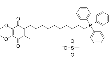
C38H47O7PS
|
|---|---|
| 分子量 |
678.8144
|
| 精确质量 |
678.278
|
| 元素分析 |
C, 67.24; H, 6.98; O, 16.50; P, 4.56; S, 4.72
|
| CAS号 |
845959-50-4
|
| 相关CAS号 |
845959-50-4 (mesylate);845959-52-6 (beta-CD complex);444890-41-9 (cation);336184-91-9 (bromide);
|
| PubChem CID |
11388331
|
| 外观&性状 |
Brown to orange ointment
|
| LogP |
7.706
|
| tPSA |
131.77
|
| 氢键供体(HBD)数目 |
0
|
| 氢键受体(HBA)数目 |
7
|
| 可旋转键数目(RBC) |
16
|
| 重原子数目 |
47
|
| 分子复杂度/Complexity |
965
|
| 定义原子立体中心数目 |
0
|
| SMILES |
O=C1C(C)=C(CCCCCCCCCC[P+](C2C=CC=CC=2)(C2C=CC=CC=2)C2C=CC=CC=2)C(=O)C(OC)=C1OC.O=S(C)([O-])=O
|
| InChi Key |
GVZFUVXPTPGOQT-UHFFFAOYSA-M
|
| InChi Code |
InChI=1S/C37H44O4P.CH4O3S/c1-29-33(35(39)37(41-3)36(40-2)34(29)38)27-19-8-6-4-5-7-9-20-28-42(30-21-13-10-14-22-30,31-23-15-11-16-24-31)32-25-17-12-18-26-32;1-5(2,3)4/h10-18,21-26H,4-9,19-20,27-28H2,1-3H3;1H3,(H,2,3,4)/q+1;/p-1
|
| 化学名 |
(10-(4,5-dimethoxy-2-methyl-3,6-dioxocyclohexa-1,4-dien-1-yl)decyl)triphenylphosphonium methanesulfonate
|
| 别名 |
Mito Q10; Mitoquinone methanesulfonate; MitoQ; MitoQ10 mesylate; MitoQ mesylate; MitoQ10; MitoQ-10; Mitoquinone mesylate; 845959-50-4; MitoQ; UNII-6E01CG547T; 6E01CG547T; Mitoquinone mesilate; Phosphonium, (10-(4,5-dimethoxy-2-methyl-3,6-dioxo-1,4-cyclohexadien-1-yl)decyl)triphenyl-, methanesulfonate (1:1);
|
| HS Tariff Code |
2934.99.9001
|
| 存储方式 |
Powder -20°C 3 years 4°C 2 years In solvent -80°C 6 months -20°C 1 month 注意: 请将本产品存放在密封且受保护的环境中,避免吸湿/受潮。 |
| 运输条件 |
Room temperature (This product is stable at ambient temperature for a few days during ordinary shipping and time spent in Customs)
|
| 溶解度 (体外实验) |
DMSO : ~100 mg/mL (~147.32 mM)
|
|---|---|
| 溶解度 (体内实验) |
配方 1 中的溶解度: ≥ 2.5 mg/mL (3.68 mM) (饱和度未知) in 10% DMSO + 40% PEG300 + 5% Tween80 + 45% Saline (这些助溶剂从左到右依次添加,逐一添加), 澄清溶液。
例如,若需制备1 mL的工作液,可将100 μL 25.0 mg/mL澄清DMSO储备液加入到400 μL PEG300中,混匀;然后向上述溶液中加入50 μL Tween-80,混匀;加入450 μL生理盐水定容至1 mL。 *生理盐水的制备:将 0.9 g 氯化钠溶解在 100 mL ddH₂O中,得到澄清溶液。 配方 2 中的溶解度: 2.5 mg/mL (3.68 mM) in 10% DMSO + 90% (20% SBE-β-CD in Saline) (这些助溶剂从左到右依次添加,逐一添加), 悬浊液; 超声助溶。 例如,若需制备1 mL的工作液,可将 100 μL 25.0 mg/mL澄清DMSO储备液加入900 μL 20% SBE-β-CD生理盐水溶液中,混匀。 *20% SBE-β-CD 生理盐水溶液的制备(4°C,1 周):将 2 g SBE-β-CD 溶解于 10 mL 生理盐水中,得到澄清溶液。 View More
配方 3 中的溶解度: 7.69 mg/mL (11.33 mM) in PBS (这些助溶剂从左到右依次添加,逐一添加), 澄清溶液; 超声助溶 (<60°C). 1、请先配制澄清的储备液(如:用DMSO配置50 或 100 mg/mL母液(储备液)); 2、取适量母液,按从左到右的顺序依次添加助溶剂,澄清后再加入下一助溶剂。以 下列配方为例说明 (注意此配方只用于说明,并不一定代表此产品 的实际溶解配方): 10% DMSO → 40% PEG300 → 5% Tween-80 → 45% ddH2O (或 saline); 假设最终工作液的体积为 1 mL, 浓度为5 mg/mL: 取 100 μL 50 mg/mL 的澄清 DMSO 储备液加到 400 μL PEG300 中,混合均匀/澄清;向上述体系中加入50 μL Tween-80,混合均匀/澄清;然后继续加入450 μL ddH2O (或 saline)定容至 1 mL; 3、溶剂前显示的百分比是指该溶剂在最终溶液/工作液中的体积所占比例; 4、 如产品在配制过程中出现沉淀/析出,可通过加热(≤50℃)或超声的方式助溶; 5、为保证最佳实验结果,工作液请现配现用! 6、如不确定怎么将母液配置成体内动物实验的工作液,请查看说明书或联系我们; 7、 以上所有助溶剂都可在 Invivochem.cn网站购买。 |
| 制备储备液 | 1 mg | 5 mg | 10 mg | |
| 1 mM | 1.4732 mL | 7.3658 mL | 14.7317 mL | |
| 5 mM | 0.2946 mL | 1.4732 mL | 2.9463 mL | |
| 10 mM | 0.1473 mL | 0.7366 mL | 1.4732 mL |
1、根据实验需要选择合适的溶剂配制储备液 (母液):对于大多数产品,InvivoChem推荐用DMSO配置母液 (比如:5、10、20mM或者10、20、50 mg/mL浓度),个别水溶性高的产品可直接溶于水。产品在DMSO 、水或其他溶剂中的具体溶解度详见上”溶解度 (体外)”部分;
2、如果您找不到您想要的溶解度信息,或者很难将产品溶解在溶液中,请联系我们;
3、建议使用下列计算器进行相关计算(摩尔浓度计算器、稀释计算器、分子量计算器、重组计算器等);
4、母液配好之后,将其分装到常规用量,并储存在-20°C或-80°C,尽量减少反复冻融循环。
计算结果:
工作液浓度: mg/mL;
DMSO母液配制方法: mg 药物溶于 μL DMSO溶液(母液浓度 mg/mL)。如该浓度超过该批次药物DMSO溶解度,请首先与我们联系。
体内配方配制方法:取 μL DMSO母液,加入 μL PEG300,混匀澄清后加入μL Tween 80,混匀澄清后加入 μL ddH2O,混匀澄清。
(1) 请确保溶液澄清之后,再加入下一种溶剂 (助溶剂) 。可利用涡旋、超声或水浴加热等方法助溶;
(2) 一定要按顺序加入溶剂 (助溶剂) 。
 Effect of MitoQ on mitochondrial superoxide generation during CS.
Effect of MitoQ on CS-induced renal injury and cell death.J Pharmacol Exp Ther.2011 Mar;336(3):682-92. |
|---|
 MitoQ attenuates nitrotyrosine formation during CS.
MitoQ decreases CS plus RW oxidant production and cell death.J Pharmacol Exp Ther.2011 Mar;336(3):682-92. |
 MitoQ prevents mitochondrial respiratory complex inactivation during CS.Individual mitochondrial respiratory complex activities were measured using isolated mitochondria from NRK cells (A and B) and isolated rat kidney mitochondria (C-F) exposed to CS, CS + MitoQ, or CS + DecylTPP (1 μM in vitro and 100 μM ex vivo). Values are expressed as percentage mean ± S.E.M. (n= 3 in vitro or 5 ex vivo) of respective controls (set to 100). **,P< 0.01 or ***,P< 0.001 compared with respective controls. †,P< 0.05 compared with CS.J Pharmacol Exp Ther.2011 Mar;336(3):682-92. |