| 规格 | 价格 | 库存 | 数量 |
|---|---|---|---|
| 1mg |
|
||
| 5mg |
|
||
| 10mg |
|
||
| 25mg |
|
||
| 50mg |
|
||
| 100mg |
|
||
| 250mg |
|
||
| Other Sizes |
|
| 靶点 |
DNA intercalator
|
|---|---|
| 体外研究 (In Vitro) |
Nemorubicin 对 HT-29、A2780、DU145、EM-2、Jurkat 和 CEM 细胞系表现出抗肿瘤活性,IC70 分别为 578 nM、468 nM、193 nM、191 nM、68 nM 和 131 ± 9 nM [1].
Nemorubicin 是一种由 CYP3A 激活的抗癌前药,能够生成更有效的代谢物 PNU-159682[1][2]. Nemorubicin 通过核苷酸切除修复发挥作用(NER)系统。与缺乏 XPG 的 L1210/0 细胞相比,保留 NER 的 L1210/DDP 细胞表现出更高水平的新霉素 (0-0.3 μM) 活性。对 Nemorubicin 具有抗性的细胞对紫外线损伤更加敏感[3]。 Nemorubicin 的 IC50 为 0.2 nM,比缺乏 P450 的 9L 细胞(IC50,23.9 nM)低 120 倍,表明它是对 9L/3A/4 细胞具有细胞毒性。此外,nemoribucicin 的 IC50 为 1.4 nM,可显着抑制 Adeno-3A4 感染的 U251 细胞。当 P450 还原酶过度表达时,新柔比星的细胞毒性更强[4]。 |
| 体内研究 (In Vivo) |
Nemorubicin 在大鼠、小鼠和狗的肝微粒体中被人肝细胞色素 P450 (CYP) 3A4 转变为 PNU-159682[2]。当对患有 9L/3A4 肿瘤的小鼠静脉内 (IV) 或瘤内 (it) 给药时,新柔比星 (60 µg/kg) 显着延缓肿瘤的生长,但对 scid 中 9L 肿瘤的肿瘤生长延迟没有明显影响老鼠。在患有 9L/3A4 肿瘤的小鼠中,新霉素(40 µg/kg,腹腔注射)没有显示出抗肿瘤活性,也没有宿主毒性[4]。
|
| 酶活实验 |
相关性研究。[1]
Nemorubicin /MMDX (20 μmol/L)与10个人肝脏微粒体部分孵育;孵育方案与上面描述的相同。在这些实验中获得的PNU-159682形成率与在相同微粒体样品中评估的几种已知的CYP形式选择性催化活性相关(数据由BD Gentest提供,硝苯地平氧化和红霉素n -去甲基化除外)。采用线性回归分析确定决定系数(r2)和P值。 化学和免疫化学抑制研究。[1] 在不存在(即对照)和存在已知的CYP形式选择性化学抑制剂的情况下,用混合HLMs评价了20 μmol/L Nemorubicin /MMDX形成PNU-159682的情况。以下抑制剂在先前确定的适当浓度下对HLMs产生CYP形式选择性抑制:7,8-苯黄酮(1 μmol/L, cyp1a2选择性),磺胺苯唑(20 μmol/L, cyp2c9选择性),奎尼丁(5 μmol/L, cyp2d6选择性),二乙基二硫代氨基甲酸酯(25 μmol/L;CYP2A6/ e1选择性)、troleandomycin (100 μmol/L, cyp3a选择性)和酮康唑(1 μmol/L, cyp3a选择性)。在可逆抑制剂(7,8-苯黄酮、奎尼丁、磺胺苯唑和酮康唑)的实验中,抑制剂与底物共孵育;孵育方案与上述相同。在以机制为基础的抑制剂,即二乙基二硫代氨基甲酸酯和troleandomycin的实验中,抑制剂与肝微粒体和NADPH (0.5 mmol)在37°C下预孵育15分钟,然后加入底物和额外的0.5 mmol NADPH。然后按照上述方法进行反应。 免疫化学抑制研究是用含有抑制单克隆抗体的小鼠腹水进行的,这种单克隆抗体已被证明对不同的人类CYP酶具有特异性。混合HLMs (0.25 mg微粒体蛋白/mL;在0.3 mol/L Tris (pH 7.4)中,与一定量含有抗CYP单抗(20-140 μg)的小鼠腹水在37℃下预孵育5分钟;然后在0.2 mL的总量中加入Nemorubicin /MMDX(终浓度,20 μmol/L)和NADPH(终浓度,0.5 mmol/L)引发反应,进行如上所述。这些试验中使用的每种单抗的最高浓度(即7 μg腹水蛋白/总CYP的pmol)先前已被证明可以在HLMs中达到饱和,以进行适当的CYP形式特异性反应。在没有单克隆抗体的情况下进行对照孵育。 Nemorubicin MMDX与cdna表达的人细胞色素P450酶[1] 孵育 MMDX与含有cdna表达的CYP酶的微粒体孵育按照HLMs的方法进行,但所用酶的量为50 pmol/mL,孵育60分钟后终止;底物浓度为20 μmol/L。所有的孵育都是一式两份。采用荧光高效液相色谱法分析各样品上清液PNU-159682含量。 Nemorubicin (3'-脱氨基-3'-[2''(S)-甲氧基-4''-吗啉基]阿霉素;MMDX)是一种目前处于肝细胞癌II/III期临床试验的研究药物。MMDX的生物活化产物,3'-脱氨基-3'',4'-脱水-[2''(S)-甲氧基-3''(R)-氧-4''吗啉基]阿霉素(PNU-159682),最近在该药物与补充NADPH的大鼠肝微粒体的孵育中被鉴定出来。本研究旨在获取MMDX在人体内转化为PNU-159682的信息,并探讨PNU-159684的抗肿瘤活性。 实验设计:使用人肝微粒体(HLM)和表达个体人细胞色素P450(CYP)的基因工程细胞系的微粒体来研究MMDX的生物转化。我们还分别使用一组体外培养的人肿瘤细胞系和荷瘤小鼠检测了PNU-159682的细胞毒性和抗肿瘤活性。 结果:HLMs将MMDX转化为主要代谢产物,其在液相色谱中的保留时间和串联质谱中的离子裂解时间与合成的PNU-159682相同。在来自10名供体的HLMs库中,PNU-159682的形成率与三种不同的CYP3A介导的活性显著相关。Troleandomycin和酮康唑均为CYP3A抑制剂,可显著减少HLMs形成PNU-159682;该反应也被CYP3A4/5单克隆抗体浓度依赖性地抑制。在检测的10个cDNA表达的CYP中,只有CYP3A4形成PNU-159682。此外,PNU-159682在体外的细胞毒性明显高于MMDX和阿霉素,并且在所测试的两种体内肿瘤模型中有效,即播散性小鼠L1210白血病和MX-1人乳腺癌异种移植物。 结论:CYP3A4是人类肝脏中的主要CYP,它将MMDX转化为一种更具细胞毒性的代谢产物PNU-159682,在体内保持抗肿瘤活性。[1] 研究人员最近证明,Nemorubicin (MMDX)是一种正在研究的抗肿瘤药物,可被人肝细胞色素P450(CYP)3A4转化为活性代谢产物PNU-159682。本研究的目的是:(1)研究实验室动物(小鼠、大鼠和狗)肝微粒体对MMDX的代谢,以确定这些物种是否也产生PNU-159682,并确定其形成的CYP形式;(2) 比较MMDX与人肝微粒体(HLMs)的动物代谢,以确定哪种动物最接近人类;(3) 探讨PNU-159682形成的差异是否是先前报道的MMDX宿主毒性与物种和性别相关差异的原因。MMDX的动物代谢被证明与HLM观察到的代谢在性质上相似,因为在所有受试物种中,MMDX主要通过单一的CYP3A形式转化为PNU-159682。然而,在动力学参数方面,物种间和物种内存在明显的定量差异。小鼠和雄性大鼠的V(max)和内在代谢清除率(CL(int))值最接近人类,表明这些物种是研究MMDX生物转化的最合适的动物模型。在这里测试的物种、性别和品系的动物中,MMDX CL(int)与之前报告的MMDX LD(50)值之间存在密切的负相关关系,表明MMDX体内毒性的差异很可能是由于PNU-159682形成程度的性别和物种相关差异造成的。 |
| 细胞实验 |
在药物治疗前 24 小时,将 9L 和 CHO 细胞铺在 96 孔板的一式三份孔中,每孔 3000 个细胞。四天内,将不同浓度的 IFA 或新霉素应用于细胞。用结晶紫 (A595) 对细胞染色后,计算相对细胞存活率。 Prism 4 用于根据数据点的半对数图计算 IC50 值 [4]。
体外细胞毒性[1]
采用Skehan等人描述的磺胺嘧啶B法评估阿霉素、Nemorubicin /MMDX和PNU-159682对贴壁肿瘤细胞系(HT-29、A2780和DU 145)的细胞毒性作用;通过使用ZM细胞计数器计算治疗期结束时的存活细胞数,评估药物对非贴壁肿瘤细胞系(CEM、Jurkat和EM-2)生长的影响。在处理前24小时给呈指数增长的细胞播种,药物处理1小时后,取出培养基,在无药培养基中培养72小时;对照细胞不接触药物。在每个实验中,测定分6次进行。然后用线性插值法从半对数浓度-响应曲线计算IC70值。数据以至少三个独立实验的平均值±SE表示。 |
| 动物实验 |
雄性ICR/Fox Chase SCID小鼠用于培养9L和9L/3A4细胞形成实体瘤。细胞在DMEM培养基中培养至75%汇合度后,用胰蛋白酶消化,PBS缓冲液洗涤,并调整细胞浓度至2 × 10⁷个细胞/mL(DMEM培养基不含胎牛血清)。将4 × 10⁶个细胞/0.2 mL细胞悬液皮下注射到4周龄SCID小鼠(18-20 g)的后侧腹部,移植9L或9L/3A4肿瘤细胞。从移植后第7天开始,每周两次使用游标卡尺测量肿瘤大小(长度和宽度)。当平均肿瘤大小达到 300 至 400 mm³ 时,将溶于 PBS 的奈莫霉素通过静脉注射 (IV) 或瘤内注射 (it) 给药(间隔 7 天,共注射 3 次,每次剂量为 60 µg/kg 体重)。瘤内注射使用 30 号针头和设定流速为 1 µL/s 的注射泵。瘤内注射治疗中,每个肿瘤注射 3 次,每只 25 g 小鼠每个肿瘤注射 50 µL。换句话说,对于一只 30 g 的小鼠,注射 120 µL 浓度为 15 µg/mL 的奈莫霉素溶液,每个注射点注射 20 µL × 每个肿瘤 3 个注射点 × 每只小鼠 2 个肿瘤。相同体积的 PBS 腹腔注射到未用药的对照组小鼠中。在某些实验中,腹腔注射(ip)奈莫霉素,剂量为40或60 µg/kg体重。在研究期间,每周测量两次体重和肿瘤大小。肿瘤体积的计算公式为V = π/6 (L × W)3/2。肿瘤消退百分比的计算公式为100 × (V1 - V2)/V1,其中V1代表给药当日的肿瘤体积,V2代表给药后肿瘤体积缩小最显著当日的肿瘤体积。肿瘤体积在药物治疗后翻倍所需的时间称为肿瘤倍增时间[4]。
播散性L1210白血病。 [1] 使用 8 周龄的近交系雌性 CD2F1 (BALB/c × DBA/2) 小鼠评估 PNU-159682 的治疗效果,并与 Nemorubicin /MMDX 进行比较。通过静脉注射 105 个 L1210 细胞诱导播散性肿瘤;1 天后,将动物随机分为实验组 (n = 10),分别接受单次静脉注射 MMDX、PNU-159682 或生理盐水(对照组)。通过比较治疗组和对照组的中位生存时间来评估治疗效果,并以寿命增加百分比表示,计算公式如下:寿命增加百分比 = (100 × 药物治疗组小鼠的中位生存时间 / 对照组小鼠的中位生存时间) − 100。组间统计学比较采用非参数Mann-Whitney检验。 皮下MX-1人乳腺癌异种移植瘤。[1] 使用4至6周龄的雌性CD-1无胸腺裸鼠评估PNU-159682对MX-1人乳腺癌异种移植瘤的活性。第0天,将MX-1肿瘤碎片皮下移植到动物(n = 14)的右侧腹部。8天后,将动物随机分为药物治疗组或对照组(每组n = 7只小鼠),并开始治疗。 PNU-159682 按照 q7dx3(每 7 天一次,共三次)方案静脉注射(4 μg/kg);对照组动物注射生理盐水。肿瘤体积通过游标卡尺测量,并使用以下公式估算:肿瘤体积 (mm³) = D × d² / 2;其中 D 和 d 分别为肿瘤的最长直径和最短直径。出于伦理考虑,对照组动物在第 21 天处死,此时该组的平均肿瘤体积约为 2,500 mm³;接受药物治疗的动物监测至第 50 天,然后处死。 |
| 药代性质 (ADME/PK) |
目的:奈莫霉素(3'-脱氨基-3'-[2''(S)-甲氧基-4''-吗啉基]阿霉素;MMDX)是一种在研药物,目前正在进行肝细胞癌的II/III期临床试验。MMDX的一种生物活化产物,3'-脱氨基-3'',4'-脱水-[2''(S)-甲氧基-3''(R)-氧基-4''-吗啉基]阿霉素(PNU-159682),最近在MMDX与补充NADPH的大鼠肝微粒体的孵育液中被发现。本研究旨在获得MMDX在人体内生物转化为PNU-159682的信息,并探讨PNU-159682的抗肿瘤活性。实验设计:本研究采用人肝微粒体(HLM)和表达特定人细胞色素P450(CYP)的基因工程细胞系微粒体,研究MMDX的生物转化。此外,我们还分别利用体外培养的人类肿瘤细胞系和荷瘤小鼠,检测了PNU-159682的细胞毒性和抗肿瘤活性。结果:HLM将MMDX转化为一种主要代谢物,该代谢物在液相色谱中的保留时间和串联质谱中的离子碎片与合成的PNU-159682完全一致。在来自10名供体的HLM库中,PNU-159682的生成速率与三种不同的CYP3A介导的活性显著相关。CYP3A抑制剂曲罗霉素和酮康唑均能显著降低HLM中PNU-159682的生成; CYP3A4/5单克隆抗体可浓度依赖性地抑制该反应。在所检测的10种cDNA表达的CYP酶中,只有CYP3A4能生成PNU-159682。此外,PNU-159682在体外表现出比MMDX和阿霉素更强的细胞毒性,并且在所测试的两种体内肿瘤模型(即播散性小鼠L1210白血病和MX-1人乳腺癌异种移植瘤)中均有效。结论:CYP3A4是人肝脏中的主要CYP酶,它能将MMDX转化为细胞毒性更强的代谢物PNU-159682,后者在体内仍保留抗肿瘤活性。 [5]
我们最近证实,研究性抗肿瘤药物奈莫霉素(MMDX)可通过人肝细胞色素P450(CYP)3A4转化为活性代谢物PNU-159682。本研究的目标是:(1)研究实验动物(雌雄小鼠、大鼠和犬)肝微粒体对MMDX的代谢,以确定PNU-159682是否也在这些物种中产生,并鉴定负责其生成的CYP酶;(2)比较动物和人肝微粒体(HLM)对MMDX的代谢,以确定哪种动物物种与人类最为接近;(3)探讨PNU-159682生成差异是否是造成先前报道的MMDX宿主毒性存在物种和性别差异的原因。 MMDX的动物代谢在性质上与人肝微粒体(HLM)中的代谢相似,因为在所有测试物种中,MMDX主要通过单一的CYP3A酶转化为PNU-159682。然而,动力学参数在不同物种间和同一物种内存在显著的定量差异。小鼠和雄性大鼠的V(max)和固有代谢清除率(CL(int))值最接近人类,表明这些物种是研究MMDX生物转化的最合适动物模型。MMDX的CL(int)与先前报道的相同物种、性别和品系动物的MMDX LD(50)值呈显著负相关,表明MMDX体内毒性的差异很可能是由于PNU-159682生成程度的性别和物种差异所致。[6] |
| 参考文献 |
|
| 其他信息 |
奈莫霉素属于吗啉类化合物,是一种蒽环类抗生素,同时也是一种伯α-羟基酮和叔α-羟基酮。它在功能上与阿霉素相关。
奈莫霉素是蒽环类抗生素阿霉素的吗啉类似物,具有抗肿瘤活性。奈莫霉素经P450 CYP3A酶代谢为一种高细胞毒性的衍生物。与大多数蒽环类抗生素不同,奈莫霉素是拓扑异构酶I抑制剂,其作用机制似乎是通过核苷酸切除修复(NER)系统实现的。此外,该药物与其他蒽环类药物无交叉耐药性。 我们近期证实,在研抗肿瘤药物奈莫柔比星 (MMDX) 可通过人肝细胞色素 P450 (CYP) 3A4 转化为活性代谢物 PNU-159682。本研究旨在:(1) 研究实验动物(雌雄小鼠、大鼠和犬)肝微粒体对 MMDX 的代谢,以确定 PNU-159682 是否也存在于这些物种中,并鉴定负责其生成的 CYP 酶;(2) 比较动物和人肝微粒体 (HLM) 对 MMDX 的代谢,以确定哪种动物物种与人类最为接近;(3) 探讨 PNU-159682 生成的差异是否是造成先前报道的 MMDX 宿主毒性存在物种和性别差异的原因。 MMDX 的动物代谢在性质上与人肝微粒体 (HLM) 中的代谢相似,因为在所有测试物种中,MMDX 主要通过单一的 CYP3A 酶转化为 PNU-159682。然而,动力学参数存在显著的种间和种内定量差异。小鼠和雄性大鼠的 V(max) 和固有代谢清除率 (CL(int)) 值最接近人类,表明这些物种是研究 MMDX 生物转化的最合适动物模型。MMDX 的 CL(int) 与先前报道的相同物种、性别和品系动物的 MMDX LD(50) 值之间存在密切的负相关性,表明 MMDX 体内毒性的差异很可能是由于 PNU-159682 生成程度的性别和物种差异造成的。来源:Biochem Pharmacol. 2008年9月15日;76(6):784-95。 抗体药物偶联物(ADC)通常由人源化抗体和小分子药物通过化学连接子连接而成。经过数十年的临床前和临床研究,一系列ADC已被广泛用于临床治疗特定类型的肿瘤,例如用于治疗复发性霍奇金淋巴瘤和系统性间变性大细胞淋巴瘤的布伦妥昔单抗(Adcetris®)、用于治疗急性髓系白血病的吉妥珠单抗(Mylotarg®)、用于治疗HER2阳性转移性乳腺癌的ado-曲妥珠单抗(Kadcyla®)、inotuzumab ozogamicin(Besponsa®)以及最近用于治疗B细胞恶性肿瘤的polatuzumab vedotin-piiq(Polivy®)。迄今为止,已有超过80种抗体偶联药物(ADC)在约600项临床试验中处于不同的临床阶段。本综述总结了ADC的关键要素,重点介绍了ADC的最新进展、从临床数据中汲取的重要经验教训以及未来的发展方向。[3] 背景:载有阿霉素和microRNA16a的靶向EDV纳米细胞在复发性胶质瘤和间皮瘤的I期临床试验中显示出良好的安全性。本研究计划对一项正在进行的首次人体开放标签I/IIa期临床试验(针对难治性转移性胰腺癌患者)进行安全性分析,以评估携带细胞毒性药物PNU-159682(旨在克服耐药性)的EGFR靶向EDV纳米细胞与携带免疫调节佐剂α-半乳糖基神经酰胺(旨在刺激抗肿瘤免疫反应)的EDV纳米细胞联合应用的安全性、生物学和临床活性。方法:9例晚期胰腺癌患者入组剂量递增阶段,以评估EDV联合方案的安全性。剂量从2×10⁹ EDV/剂逐渐递增至第7周的最大剂量7×10⁹ EDV/剂,随后以第1周期达到的最大剂量给药。每个周期后均采用iRECIST标准评估肿瘤反应,并采集血液样本进行细胞因子和外周血单个核细胞(PBMC)分析。结果:EDV联合方案耐受性良好,未发生剂量限制性毒性(DLT)和药物相关严重不良事件(SAE)。少数患者出现1级输注反应,经支持治疗后迅速缓解。9例患者中有8例在8周时达到部分缓解(PR)或疾病稳定(SD)(临床获益率89%),5例可评估患者中有4例在4个月时确认缓解(80%),其中2例患者的缓解持续时间超过6个月。探索性分析显示,几乎所有可评估患者(6/8)的 IFN-α 和 IFN-γ 水平升高。此外,我们观察到 CD8+ T 细胞(2/8)、iNKT 细胞、树突状细胞和 NK 细胞(3/8)水平升高,以及耗竭 CD8+ T 细胞(3/8)水平降低,提示先天性和适应性免疫反应均被激活。结论:携带细胞毒性药物和免疫佐剂的 EDV 安全且耐受性良好。早期信号表明其具有持久疗效,这可能与先天性和适应性免疫反应的产生以及对耐药肿瘤细胞的细胞毒性作用有关。IIa 期研究计划再招募 35 例患者,以进一步评估其安全性和抗肿瘤疗效。临床试验信息:ACTRN12619000385145。[4] |
| 分子式 |
C32H37NO13
|
|---|---|
| 分子量 |
643.63508
|
| 精确质量 |
643.226
|
| 元素分析 |
C, 59.71; H, 5.79; N, 2.18; O, 32.32
|
| CAS号 |
108852-90-0
|
| 相关CAS号 |
Nemorubicin;108852-90-0; 108943-08-4 (HCl)
|
| PubChem CID |
65907
|
| 外观&性状 |
Red to pink solid powder
|
| 密度 |
1.6±0.1 g/cm3
|
| 沸点 |
852.2±65.0 °C at 760 mmHg
|
| 闪点 |
469.2±34.3 °C
|
| 蒸汽压 |
0.0±0.3 mmHg at 25°C
|
| 折射率 |
1.681
|
| LogP |
4.7
|
| tPSA |
201.75
|
| 氢键供体(HBD)数目 |
5
|
| 氢键受体(HBA)数目 |
14
|
| 可旋转键数目(RBC) |
7
|
| 重原子数目 |
46
|
| 分子复杂度/Complexity |
1160
|
| 定义原子立体中心数目 |
7
|
| SMILES |
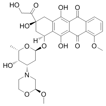
COC1=C2C(C(C3=C(O)C(C[C@](C(CO)=O)(O)C[C@]4([H])O[C@H]5C[C@H](N6CCO[C@H](OC)C6)[C@H](O)[C@H](C)O5)=C4C(O)=C3C2=O)=O)=CC=C1
|
| InChi Key |
CTMCWCONSULRHO-UHQPFXKFSA-N
|
| InChi Code |
InChI=1S/C32H37NO13/c1-14-27(36)17(33-7-8-44-22(12-33)43-3)9-21(45-14)46-19-11-32(41,20(35)13-34)10-16-24(19)31(40)26-25(29(16)38)28(37)15-5-4-6-18(42-2)23(15)30(26)39/h4-6,14,17,19,21-22,27,34,36,38,40-41H,7-13H2,1-3H3/t14-,17-,19-,21-,22-,27+,32-/m0/s1
|
| 化学名 |
(7S,9S)-6,9,11-trihydroxy-9-(2-hydroxyacetyl)-7-[(2R,4S,5S,6S)-5-hydroxy-4-[(2S)-2-methoxymorpholin-4-yl]-6-methyloxan-2-yl]oxy-4-methoxy-8,10-dihydro-7H-tetracene-5,12-dione
|
| 别名 |
Methoxymorpholinyldoxorubicin; PNU 152243; PNU-152243A; PNU152243A; PNU-152243A; PNU 152243A; Nemorubicin; Nemorubicin; 108852-90-0; Nemorubicin [INN]; Methoxymorpholino-doxorubicin; Methoxymorpholinyl doxorubicin; FCE-23762; Nemorubicin (GMP); 3'-DESAMINO-3'-(2-METHOXY-4-MORPHOLINYL)-DOXORUBICIN; methoxymorpholinyl-doxorubicin; 3′-deamino-3′-[2″(S)-methoxy-4″-morpholinyl]doxorubicin; MMDX
|
| HS Tariff Code |
2934.99.9001
|
| 存储方式 |
Powder -20°C 3 years 4°C 2 years In solvent -80°C 6 months -20°C 1 month |
| 运输条件 |
Room temperature (This product is stable at ambient temperature for a few days during ordinary shipping and time spent in Customs)
|
| 溶解度 (体外实验) |
DMSO: ~65 mg/mL (~101 mM)
|
|---|---|
| 溶解度 (体内实验) |
配方 1 中的溶解度: ≥ 3.25 mg/mL (5.05 mM) (饱和度未知) in 10% DMSO + 40% PEG300 +5% Tween-80 + 45% Saline (这些助溶剂从左到右依次添加,逐一添加), 澄清溶液。
例如,若需制备1 mL的工作液,可将100 μL 32.5 mg/mL澄清的DMSO储备液加入400 μL PEG300中,混匀;再将50 μL Tween-80+加入到上述溶液中,混匀;然后加入450 μL生理盐水定容至1 mL。 *生理盐水的制备:将 0.9 g 氯化钠溶解在 100 mL ddH₂O中,得到澄清溶液。 请根据您的实验动物和给药方式选择适当的溶解配方/方案: 1、请先配制澄清的储备液(如:用DMSO配置50 或 100 mg/mL母液(储备液)); 2、取适量母液,按从左到右的顺序依次添加助溶剂,澄清后再加入下一助溶剂。以 下列配方为例说明 (注意此配方只用于说明,并不一定代表此产品 的实际溶解配方): 10% DMSO → 40% PEG300 → 5% Tween-80 → 45% ddH2O (或 saline); 假设最终工作液的体积为 1 mL, 浓度为5 mg/mL: 取 100 μL 50 mg/mL 的澄清 DMSO 储备液加到 400 μL PEG300 中,混合均匀/澄清;向上述体系中加入50 μL Tween-80,混合均匀/澄清;然后继续加入450 μL ddH2O (或 saline)定容至 1 mL; 3、溶剂前显示的百分比是指该溶剂在最终溶液/工作液中的体积所占比例; 4、 如产品在配制过程中出现沉淀/析出,可通过加热(≤50℃)或超声的方式助溶; 5、为保证最佳实验结果,工作液请现配现用! 6、如不确定怎么将母液配置成体内动物实验的工作液,请查看说明书或联系我们; 7、 以上所有助溶剂都可在 Invivochem.cn网站购买。 |
| 制备储备液 | 1 mg | 5 mg | 10 mg | |
| 1 mM | 1.5537 mL | 7.7683 mL | 15.5366 mL | |
| 5 mM | 0.3107 mL | 1.5537 mL | 3.1073 mL | |
| 10 mM | 0.1554 mL | 0.7768 mL | 1.5537 mL |
1、根据实验需要选择合适的溶剂配制储备液 (母液):对于大多数产品,InvivoChem推荐用DMSO配置母液 (比如:5、10、20mM或者10、20、50 mg/mL浓度),个别水溶性高的产品可直接溶于水。产品在DMSO 、水或其他溶剂中的具体溶解度详见上”溶解度 (体外)”部分;
2、如果您找不到您想要的溶解度信息,或者很难将产品溶解在溶液中,请联系我们;
3、建议使用下列计算器进行相关计算(摩尔浓度计算器、稀释计算器、分子量计算器、重组计算器等);
4、母液配好之后,将其分装到常规用量,并储存在-20°C或-80°C,尽量减少反复冻融循环。
计算结果:
工作液浓度: mg/mL;
DMSO母液配制方法: mg 药物溶于 μL DMSO溶液(母液浓度 mg/mL)。如该浓度超过该批次药物DMSO溶解度,请首先与我们联系。
体内配方配制方法:取 μL DMSO母液,加入 μL PEG300,混匀澄清后加入μL Tween 80,混匀澄清后加入 μL ddH2O,混匀澄清。
(1) 请确保溶液澄清之后,再加入下一种溶剂 (助溶剂) 。可利用涡旋、超声或水浴加热等方法助溶;
(2) 一定要按顺序加入溶剂 (助溶剂) 。
|
|
 Pyridostatin trihydrochloride
 G-quadruplex ligand 4
 G4/hTERT-IN-4
 H2S probe 1
InvivoChem的所有产品仅用于作科学研究,不面向患者销售
Copyright 2020 InvivoChem LLC | All Rights Reserved 粤ICP备20063088号-1


 COA
COA


 463611831
