| 规格 | 价格 | 库存 | 数量 |
|---|---|---|---|
| 1mg |
|
||
| 5mg |
|
||
| 10mg |
|
||
| 25mg |
|
||
| 50mg |
|
||
| 100mg |
|
||
| 250mg |
|
||
| 500mg |
|
||
| Other Sizes |
|

| 靶点 |
Mitochondrial; neuroprotective
|
|---|---|
| 体外研究 (In Vitro) |
在不使用来自大脑、睫状体或胶质细胞的神经营养因子的情况下,原代胚胎大鼠脊髓 MN 在接种一小时后暴露于浓度范围为 0.1 至 10 µM 的 Olesoxime (TRO 19622) 中,可显着防止细胞损伤和死亡。这种保护在培养物中持续了三天。浓度为 10 µM 的 Olesoxime (TRO 19622) 通过神经营养因子混合物(包括大脑、睫状体和神经胶质细胞产生的因子)的作用维持 74±10% 的神经元存活。在此测试中,平均 EC50 为 3.2±0.2 µM。 Olesoxime (TRO 19622) 不仅可以保护 MN 细胞体,还可以促进神经突发育。在 1 µM 浓度下,olesoxime (TRO 19622) 仅略微提高细胞活力,但显着促进每个细胞的神经突发育达 54% [1]。人们发现了一类新型胆固醇肟,称为 Olesoxime (TRO 19622),因为它能够在缺乏神经营养因子的情况下提高纯运动神经元的存活率。 Olesoxime (TRO 19622) 选择性地靶向线粒体外膜中的蛋白质,重点关注线粒体并抑制氧化应激介导的通透性转变孔开放等过程[2]。
在细胞接种后1小时接触奥利索西(0.1-10 µM浓度范围),能显著保护原代培养的胚胎大鼠脊髓运动神经元(在缺乏脑源性、睫状神经营养因子和胶质细胞源性神经营养因子的条件下培养3天)免于死亡。当浓度为10 µM时,奥利索西维持了74±10%的神经元存活率(该比例与三种神经营养因子联合使用的效果相当)。本实验中测得的平均EC50值为3.2±0.2 µM。除保护神经元胞体外,奥利索西还促进神经突生长:在仅提高细胞存活率38%的1 µM浓度下,可使单个细胞的神经突总生长量增加54%。 化疗药物喜树碱能引起DNA链断裂并增加活性氧生成。皮质神经元与喜树碱共培养时,联合使用奥利索西可在16小时后产生剂量依赖性的细胞存活率提升,并降低活化的caspase-3和-7水平。这些效应与脑源性神经营养因子类似,但奥利索西的神经保护作用不依赖于ERK1/2或PI3K通路的激活。 微管靶向药物的体内给药常因周围神经病变发展而受限。体外实验显示,微管靶向药物会抑制大鼠和人类分化神经细胞的神经突生长,并引发末端结合蛋白(EB)1和EB2从微管解离至胞质。而同时暴露于奥利索西能维持EB蛋白分布和神经突生长。 通过啮齿类动物中枢神经系统细胞培养模型,在体外检测了奥利索西的髓鞘形成促进作用。奥利索西可剂量依赖性加速神经前体细胞向少突胶质前体细胞的分化,并在背根神经节神经元与少突胶质前体细胞的共培养体系中增强髓鞘形成[1]。 奥利索西(Olesoxime)是一种类胆固醇神经保护化合物,其作用靶点为线粒体电压依赖性阴离子通道(VDACs)。研究发现VDACs同样存在于质膜中,并在突触前成分中高度表达。本研究探讨了奥利索西与VDAC抑制剂对小鼠神经肌肉接头神经传递的影响。电生理分析显示:奥利索西能选择性抑制单次刺激和20Hz高频刺激诱发的神经递质释放,同时降低低频(0.5Hz)和20Hz刺激下FM1-43染料的流失速率(突触小泡胞吐作用指标)。提高细胞外Cl-浓度可增强奥利索西对胞吐的作用,且奥利索西能增加细胞内Cl-水平。奥利索西对诱发突触小泡胞吐及[Cl-]i的影响可被膜通透性与非通透性VDAC抑制剂阻断。免疫荧光标记证实突触膜上存在VDACs表达。鱼藤酮诱导线粒体功能障碍会干扰FM1-43的胞吐释放,而细胞通透性VDAC抑制剂(非奥利索西或非通透性VDAC抑制剂)可部分缓解鱼藤酮导致的FM1-43卸载异常和线粒体超氧化物生成。这表明奥利索西通过作用于质膜VDACs来限制神经传递——VDACs的激活可能通过增加神经末梢阴离子内流来抑制突触小泡胞吐[5]。 |
| 体内研究 (In Vivo) |
成年小鼠每天皮下注射 Olesoxime (TRO 19622)(3 或 30 mg/kg)超过两个月,耐受性良好,没有毒性或不良反应 [1]。当动物在损伤后口服治疗五天时,Olesoxime (TRO 19622) 以剂量依赖性方式增加运动神经元细胞体的存活率;在此剂量下,运动神经元存活率为 29 ±2% (n=18),与载体治疗的动物相比,存活率增加了 42% [3]。接受 3 mg/kg/d 或 30 mg/kg/d Olesoxime (TRO 19622) 预防性治疗的紫杉醇治疗大鼠分别具有 239±17.6 和 247±14.4 IENF/cm。对于两种剂量,下降幅度均显着小于用媒介物施用紫杉醇治疗的大鼠中观察到的 46%。
奥利索西(Olesoxime)是一种类胆固醇小分子化合物,最初在针对肌萎缩侧索硬化症及其他运动神经元退行性疾病的药物筛选中被发现。该药物不仅在体外和体内实验中表现出对运动神经元的神经保护与促再生作用,还在长春新碱和糖尿病引发的痛性周围神经病变大鼠模型中显示出镇痛效应。本研究采用紫杉醇诱导的痛性周围神经病变大鼠模型,旨在验证奥利索西能否逆转已形成的神经病理性疼痛,并探究其在紫杉醇暴露期间给药能否预防神经病理性疼痛综合征的发生及伴随的表皮感觉神经末梢分支退化。结果显示:奥利索西显著改善已形成的机械性异常性疼痛和机械性痛觉过敏,连续给药5天未出现耐受现象,且镇痛效果在末次给药后持续5-10天。在紫杉醇暴露期间联合给予奥利索西,可显著且持久地减轻机械性异常性疼痛/痛觉过敏的严重程度,并明显减少感觉神经末梢分支的退化。奥利索西通过靶向线粒体蛋白发挥作用,其效应支持紫杉醇诱发痛性周围神经病变的线粒体毒性假说。我们得出结论:奥利索西在临床上或可用于预防和治疗紫杉醇诱发的痛性周围神经病变。[4] 奥利索西在面神经轴切断术诱导的新生大鼠运动神经元退化模型中进行测试。神经切断术后7天,连续5天口服奥利索西(100 mg/kg)的大鼠比溶剂对照组表现出显著更高的运动神经元存活率。 为评估奥利索西能否促进周围神经再生,对坐骨神经压伤成年小鼠皮下注射奥利索西(0.3、3或30 mg/kg)。给药组在伤后2周开始出现剂量依赖性的神经再生加速,至第4周时所有剂量组与溶剂对照组均存在显著差异。第6周时,奥利索西治疗组小鼠神经肌肉功能恢复达假手术组的80%。组织学分析显示,溶剂对照组轴突横截面积总体小于对照组,而30 mg/kg奥利索西组轴突面积显著增大(7.6±0.1 vs 6.0±0.1 µm²;p<0.05)。第4周时,所有剂量组均显著减少"低髓鞘化"纤维数量。 在G93A高表达SOD1转基因ALS小鼠模型中,从出生后60天开始皮下注射Olesoxime/奥利索西(3或30 mg/kg)可改善运动功能、延迟疾病发作并使生存期延长10%。其中3 mg/kg剂量组体重下降起始时间延迟15天(p<0.01),两个剂量组网格测试功能衰退均延迟约11天(p<0.01)。 在链脲佐菌素(55 mg/kg)诱导的糖尿病性神经病变大鼠模型中,通过电生理检测和甩尾试验评估神经病变,采用热异常性疼痛及热/机械性痛觉过敏测试检测伤害感受。糖尿病诱导10天后开始口服Olesoxime/奥利索西(30和300 mg/kg/天)可显著缓解疼痛(p≤0.05),效果与3 mg/kg吗啡相当,同时显著降低复合肌肉动作电位潜伏期(运动神经传导指标)。单次口服奥利索西(10、30或100 mg/kg)可剂量依赖性逆转糖尿病性异常性疼痛,最高剂量组与溶剂对照组差异显著(p≤0.05)。连续给药5天后,所有剂量均显著改善触觉异常性疼痛,效果与加巴喷丁(50 mg/kg bid)相当。 在紫杉醇(第0、2、4、6天腹腔注射2 mg/kg)诱导的大鼠神经病理性疼痛模型中,预防性研究(-1至15天给药)和治疗性研究(第25天起连续5天给药)显示:口服Olesoxime/奥利索西(3或30 mg/kg/天)能显著缓解紫杉醇诱导的异常性疼痛/痛觉过敏至第40天(末次给药后25天),同时将表皮内神经纤维的损失率从紫杉醇组的46%降至22-25%。10或100 mg/kg/天剂量组在给药次日即显著改善痛觉过敏和异常性疼痛,虽然效果可逆,但末次给药后镇痛作用维持10天。 在长春新碱(第1、4、6天静脉注射200 µg/kg)诱导的大鼠神经病理性疼痛模型中,最高测试剂量(100 mg/kg口服)首次给药4小时后即显著减轻异常性疼痛(p<0.001)。连续给予10、30和100 mg/kg/天Olesoxime/奥利索西可显著缓解第11-14天的长春新碱诱导异常性疼痛[1]。 |
| 酶活实验 |
肌萎缩侧索硬化症(ALS)是一种致命的神经退行性疾病,其特征是皮层和脊髓运动神经元的逐渐死亡,目前尚无有效的治疗方法。使用能够在体外预防运动神经元细胞死亡的化合物的基于细胞的测定法,筛选了大约40000种低分子量化合物,以确定潜在的小分子治疗方法。我们报道了胆甾-4-烯-3-酮肟(TRO19622)作为治疗ALS的潜在候选药物的鉴定。在体外,TRO19622在缺乏营养支持的情况下以剂量依赖的方式促进运动神经元的存活[3]。
|
| 细胞实验 |
溶液与化学试剂 [5]
神经半膈肌标本固定于Sylgard包被的培养槽底部。肌肉组织以5 ml·min−1流速持续灌注生理溶液(129 mM NaCl、5 mM KCl、2 mM CaCl2、1 mM MgSO4、1 mM NaH2PO4、20 mM NaHCO3、11 mM葡萄糖及3 mM HEPES;pH=7.4),溶液经5% CO2/95% O2混合气体饱和。部分实验采用高Cl−浓度溶液(146 mM NaCl、5 mM KCl、2 mM CaCl2、1 mM MgCl2、13.5 mM胆碱氯化物及3 mM HEPES;pH=7.4)。 在20 Hz或5 Hz神经刺激前,使用0.4 μM 奥利索西预处理20分钟。Olesoxime/奥利索西溶解于DMSO,溶剂终浓度为0.001%。0.001-0.1%浓度的DMSO对小鼠膈肌神经肌肉传递无影响,故DMSO实验数据作为对照。VDAC抑制剂DIDS(50 μM,4,4'-二异硫氰酸二苯乙烯-2,2'-二磺酸;Tocris)和S-18(1 μM,S-18硫代磷酸随机寡核苷酸)在奥利索西给药前5分钟加入灌流液并维持至实验结束。鱼藤酮(10 μM,作用30分钟)用于诱导线粒体功能障碍。(−)维沙米可(2 μM)作为囊泡乙酰胆碱转运体抑制剂,用于阻断突触小泡乙酰胆碱再填充。 突触后电位记录 [5] 使用2.5 M KCl填充的标准玻璃微电极(尖端电阻5-10 MΩ)记录终板电位(EPPs)和微小终板电位(MEPPs)。信号检测采用Model 1600放大器和LA II数字I/O板,记录信号经0.03 Hz-10 kHz滤波后以50 kHz采样率数字化存储,供离线分析。通过定制程序处理数据,计算平均振幅、上升时间(峰值20%-80%时程)和衰减时间(峰值至50%时程)。MEPPs频率通过记录150-200个信号后统计得出,信噪比>7:1,检测阈值设为0.2 mV。神经刺激采用吸吮电极连接Model 2100隔离刺激器,施加0.1 ms矩形超强电脉冲(频率0.5 Hz或20 Hz)。为抑制肌肉收缩,记录前20分钟在灌流液中加入肌特异性Na+通道抑制剂μ-芋螺毒素-GIIIB(0.5 μM)。在0.5 Hz低频刺激下记录20分钟奥利索西处理的EPPs,随后对预处理肌肉的膈神经施加20 Hz高频刺激3分钟。 |
| 动物实验 |
在体内实验中,TRO19622 可挽救新生大鼠因轴突切断而死亡的运动神经元,并促进小鼠坐骨神经损伤后的神经再生。在家族性肌萎缩侧索硬化症 (ALS) 模型小鼠(SOD1G93A 转基因小鼠)中,TRO19622 治疗可改善运动功能,延缓临床疾病的发生,并延长生存期。TRO19622 可直接与线粒体通透性转换孔的两个组成部分结合:电压依赖性阴离子通道和 18 kDa 转运蛋白(或外周苯二氮卓受体),这提示了其发挥神经保护作用的潜在机制。 TRO19622 可能具有治疗 ALS 和其他运动神经元疾病及神经退行性疾病的潜力[3]。
在体内研究中,Olesoxime/TRO19622以羟丙基甲基纤维素或植物油悬浮液的形式通过灌胃给药,或通过皮下注射给药[3]。 Olesoxime 每日新鲜配制于玉米油中。Olesoxime 或赋形剂以 5.0 ml/kg 的体积通过灌胃给药。本研究中使用的奥来索肟剂量(3-100 mg/kg)是根据先前报道的神经保护和镇痛活性选择的。[4] 治疗方案[4] 为了确定奥来索肟是否对已建立的紫杉醇诱发疼痛具有镇痛作用,我们在疼痛高峰期前后,对动物每日口服奥来索肟后,检测了其撤退反应。行为学测试的基线反应在首次给予紫杉醇后的第23天和第24天进行(大约是疼痛高峰期平台期的开始),并建立了三个实验组,使每个实验组的机械性痛觉过敏和机械性痛觉超敏程度大致相等。随后,将各组(每组 n = 12)随机分配接受奥来索肟(10 mg/kg 或 100 mg/kg)或赋形剂,连续 5 天,从第 27 天开始。每天给药后 4 小时进行行为学测试。在最后一次给予奥来索肟/赋形剂后 1 天开始的洗脱期(洗脱期第 1 天;WD1),以及 WD3、WD5、WD10、WD14 和 WD18 也评估了行为学。行为学测试由一位对分组情况不知情的观察者进行。 预防范式 [4] 为了确定奥来索肟是否可以预防紫杉醇诱发的疼痛性周围神经病变的发生,比较了三个实验组(每组 n = 12)。各组分别接受赋形剂或奥来索肟(Olesoxime)治疗,剂量分别为每日3 mg/kg或30 mg/kg,连续17天,从首次注射紫杉醇前一天(D-1)开始,直至末次注射紫杉醇后9天(D15)。在末次注射紫杉醇后继续给药,是因为在出现具有统计学意义的疼痛过敏反应之前存在数天的延迟[8],因此疼痛病理的发生时间尚不确定。在需要同时给药的日期,奥来索肟于上午9:00给药,紫杉醇于下午1:00给药。从第16天开始,每3-5天重复进行行为学测试,直至第40天,由一位对分组情况不知情的观察者进行测试。 预防性治疗对紫杉醇诱导的表皮内神经纤维变性的影响[4] 本研究使用的紫杉醇模型已被证实与表皮内神经纤维(IENF)的显著丢失有关,即支配表皮的传入神经的感觉末梢受体树突[11, 22]。为了确定奥来索肟是否能预防这种变性,在三组大鼠中重复了上述预防性给药方案(奥来索肟剂量为3 mg/kg或30 mg/kg,或载体;每组n = 12)。在第29天和第30天进行行为学测试,以确认载体处理组中是否存在预期的紫杉醇诱发的疼痛过敏,以及3 mg/kg和30 mg/kg组中是否存在预期的镇痛效果。在第31天,从每组中随机选择8只大鼠处死,用于IENF的免疫细胞化学评估。另取4只相同年龄和体重的未接受任何处理的大鼠(即未接受紫杉醇或奥来索肟治疗)作为正常对照。 对紫杉醇诱发的自发性放电的影响[4] 紫杉醇诱发的疼痛性周围神经病变与A纤维和C纤维自发性放电的异常发生率相关。为了确定奥来索肟的急性镇痛作用是否与抑制这种放电有关,我们调查了连续两天分别接受赋形剂或100 mg/kg奥来索肟治疗的大鼠(每组n=6)中自发放电纤维的发生率(上述治疗方案研究发现,该治疗方案具有显著的抗异位痛和抗痛觉过敏作用)。所有大鼠均经确认存在紫杉醇诱发的疼痛(在第23-24天评估),随后在疼痛高峰期平台期(第27-44天)接受奥来索肟或赋形剂治疗。电生理实验在治疗第二天开始,即给药/赋形剂4小时后。将紫杉醇治疗组大鼠与四只未接受任何治疗的大鼠(既未接受紫杉醇也未接受奥来索肟治疗)进行比较。实验者对大鼠的分组情况不知情。纤维记录的手术准备大约需要1小时,数据采集在接下来的2-3小时内进行,此时口服奥来索肟后血浆浓度达到峰值。记录方法与之前描述的方法相同。简而言之,我们确定了每根微丝中可单独识别的纤维数量,并记录了具有自发放电的可单独识别纤维的发生率及其放电频率。我们测定了所有可单独识别纤维的传导速度。我们没有区分传导速度在A□和A□范围内的A纤维,因为基于此无法区分A纤维的功能类别。我们特意避免表征纤维对感受野刺激的反应。这样做需要反复施加有害刺激,这可能会使伤害感受器敏化。致敏的伤害感受器持续放电,这种放电无法与紫杉醇诱发的自发放电区分开来。 奥来索肟血浆水平[4] 行为学研究中动物的血液取自尾静脉,解剖学和电生理学研究中处死的动物的血液则通过心脏穿刺采集,血液收集于锂肝素抗凝管中,以3000 rpm离心10分钟,并将血浆置于干冰上冷冻。采用高效液相色谱-串联质谱法进行定量分析。该方法的检测限为0.01 μM。 |
| 药代性质 (ADME/PK) |
代谢和药代动力学[1]
奥来索肟以羟丙基甲基纤维素或植物油混悬液的形式经口灌胃给药于大鼠和小鼠,并以Cremophor EL/二甲基亚砜/乙醇/磷酸盐缓冲液(比例分别为5:5:10:80)的混合物进行皮下注射。为测定生物利用度,成年小鼠每日皮下注射奥来索肟,持续1周或6周,剂量分别为0.3、3和30 mg/kg。采用高效液相色谱-串联质谱法测定血浆和脑组织中奥来索肟的浓度。血浆和脑组织中奥来索肟的浓度呈剂量依赖性,1周后达到稳态,并在6周的治疗期间保持稳定。在3 mg/kg/天的剂量下,血浆和脑组织中奥莱索昔姆的浓度分别约为1.25 µM和0.5 µM。 大鼠药代动力学研究表明,奥莱索昔姆的消除半衰期约为24小时,导致药物蓄积,连续三次口服给药后,血浆浓度达到稳态。在糖尿病和长春新碱治疗的大鼠中,重复口服10 mg/kg/天的奥莱索昔姆,可使血浆稳态浓度达到2至4.5 µM。单次口服100 mg/kg奥莱索昔在两种模型中均导致血浆浓度介于14.2至37.5 µM之间。 在紫杉醇治疗的大鼠中,首次给予10 mg/kg奥莱索昔后,血浆浓度为0.82 µM,第五次给药后升高至1.39 µM。对于100 mg/kg剂量,第1天和第5天的血浆浓度分别为6.75 µM和8.91 µM。 一项I期随机、双盲、安慰剂对照、剂量递增临床试验评估了48名健康白种人志愿者连续11天口服四种剂量(50、150、250和500 mg,每日一次)奥莱索昔的药代动力学。在所有剂量下,奥莱索昔的吸收和消除均较慢:达峰时间 (Tmax) 约为 10 小时,给药后 19 天内仍可检测到奥莱索昔的浓度。各剂量组的平均半衰期 (t1/2) 相近,约为 120 小时。剂量分别以 3 倍、5 倍和 10 倍的比例递增(从 50 mg 分别增至 150 mg、250 mg 和 500 mg),但第 1 天的峰浓度 (Cmax) 分别以 2.2 倍、4.4 倍和 10.2 倍的比例递增,AUC0-τ 分别以 2.1 倍、4.6 倍和 10.8 倍的比例递增。在所有组均于第 11 天达到稳态时,Cmax 随剂量递增的比例分别为 2.1 倍、7.2 倍和 12.2 倍,AUC0-τ 分别以 2.0 倍、6.5 倍和 11.6 倍的比例递增。在第1天至第11天观察到的Cmax和Ctrough的平均累积比率约为4。不同志愿者和所有剂量组的血浆药代动力学特征相似:第11天Cmax和AUC0-τ的变异系数在21%至47%之间。 在一项针对ALS患者(n = 36)的Ib期临床试验中,评估了奥莱索昔(餐前给药)与利鲁唑联合用药1个月的药代动力学。在125 mg剂量的奥莱索昔下,男性和女性的Ctrough中位数分别为512和742 ng/ml;在250 mg剂量下分别为979和1685 ng/ml;在500 mg剂量下分别为2965和3310 ng/ml;这些数值在任何剂量下均未显示出性别差异。在500 mg剂量组中,第15天观察到谷浓度峰值(Ctrough)为5780 ng/ml。第15天和第30天的奥来索昔姆谷浓度值相似,表明在第15天已达到稳态。ALS患者的血浆谷浓度高于健康志愿者,这可能是由于与食物或利鲁唑合用所致。 在一项针对SMA患儿(n = 5)和成人(n = 3)患者的Ib期临床试验中,也评估了奥来索昔姆的药代动力学。单次口服奥来索昔姆(125 mg)后,将剂量调整为mg/kg,儿童和成人的Cmax和AUC值相当;Tmax、t1/2和总清除率也相同。每日一次给药后的结果相似。 采用多种方法研究了奥莱索肟在小鼠和大鼠中的脑渗透性。利用Q. Smith最初开发的原位大鼠脑灌注技术,评估了奥莱索肟相对于已知脑渗透化合物的脑渗透水平。共6只5至6周龄的大鼠接受了C4位标记的3H-奥莱索肟的灌注。在0.01至60的标度范围内,血脑屏障的渗透系数(Kin)为5.9 ± 3.0 µl/g/s。与参考化合物相比,奥莱索肟的渗透性介于秋水仙碱(一种渗透性依赖性化合物)和氟马西尼(一种通量依赖性化合物)之间。此外,还开发了一种提取和分析方法,用于检测和定量血浆和脑组织中的奥莱索肟。在多项药代动力学和疗效研究中,通过测定小鼠脑组织中奥莱索昔的含量,可以将脑组织浓度与同一研究中采集的血浆样本测得的AUC进行比较。在小鼠神经损伤模型中进行的慢性给药研究表明,奥莱索昔会随时间在脑组织中积累,而血浆浓度保持不变(表4)。结合这些不同的方法,可以得出结论:奥莱索昔能够进入大脑,并且与疗效相关的组织浓度可以与血浆浓度相关联。[2] |
| 毒性/毒理 (Toxicokinetics/TK) |
毒性[1]
成年小鼠每日皮下注射奥莱索肟(3 或 30 mg/kg),持续 2 个月以上,耐受性良好,未见毒性或不良反应。在接受比预期治疗剂量高 40 倍的剂量,持续 4 周的动物中,也未观察到毒性。截至本文发表时,尚无其他毒性数据。 通过培养皮层神经元动作电位的产生/传导,评估了奥莱索肟的早期中枢神经系统安全性。接种后约 10 天,使用多电极阵列记录皮层神经元的自发动作电位。在对照条件下(标准生理盐水),测量 10 分钟的放电频率(Hz/s),然后用奥莱索肟灌注 10 分钟。暴露于 10 µM 奥莱索肟后,皮层神经元的放电频率未发生任何改变。相比之下,灌注河豚毒素(100 nM TTX)后立即观察到完全的放电抑制(图 8A)。同样,将大鼠 E14 运动神经元急性暴露于 10 µM 奥莱索昔姆(3 分钟)后,20 Hz 刺激诱发的动作电位模式未发生改变(数据未显示)。[2] 副作用和禁忌症 [1] 在奥莱索昔姆(50、150、250 和 500 mg,口服,每日一次,持续 11 天)在健康志愿者中进行的 I 期临床试验中,未报告严重不良事件。共报告了 69 例治疗期间出现的不良事件 (TEAE),其中 18 例被认为可能与奥莱索昔姆相关,22 例被认为不太可能相关,27 例被判定与奥莱索昔姆无关。在可能相关的治疗期间出现的不良事件(TEAE)中,50 mg 剂量组发生 2 例,250 mg 剂量组发生 2 例,500 mg 剂量组发生 7 例,安慰剂组发生 7 例。大多数 TEAE 的严重程度为轻度(48 例)或中度(21 例)。最常报告的 TEAE 为腹泻(9 例)、头痛(7 例)、便秘(4 例)、咽炎(4 例)和背痛(4 例)。TEAE 与剂量无关。生命体征、心电图参数、实验室检查或体格检查均未见相关变化。 在奥列索昔(125、250 或 500 mg,每日一次,口服)联合利鲁唑(50 mg,每日两次,口服)治疗肌萎缩侧索硬化症(ALS)患者的 Ib 期临床试验中,所有剂量均耐受良好。共报告了 69 例治疗期间出现的不良事件 (TEAE),其中 2 例被认为可能与奥莱索昔姆相关,13 例可能相关,21 例不太可能相关,33 例与奥莱索昔姆无关;2 例被认为可能相关的 TEAE 均发生在安慰剂组。在可能相关的 TEAE 中,1 例发生在对照组,6 例发生在 125 mg 组,3 例发生在 250 mg 组,3 例发生在 500 mg 组。TEAE 的严重程度分为轻度 (n = 55)、中度 (n = 13) 和重度 (n = 2)。最常报告的 TEAE 为乏力(12 例,其中 9 例发生在奥莱索昔姆治疗后)、腹泻(6 例,其中 4 例发生在奥莱索昔姆治疗后)、肌肉痉挛(4 例,其中 3 例发生在奥莱索昔姆治疗后)和便秘(3 例,其中 1 例发生在奥莱索昔姆治疗后)。TEAE 的发生频率、严重程度和持续时间与剂量无关。在所有剂量组中,均未观察到生命体征、心电图参数、实验室检查或体格检查的相关变化。 在针对脊髓性肌萎缩症(SMA)患儿和成人患者的奥列索昔姆(125 mg 口服)Ib 期临床试验期间,以及在 1 个月的随访期内,均未报告任何安全性问题。 |
| 参考文献 |
|
| 其他信息 |
奥来索肟是一种胆固醇样小分子,在一系列体外和体内临床前模型中均展现出显著的神经保护作用。例如,它能够预防神经退行性变,增强神经功能,并加速神经损伤后的神经再生。
药物适应症 已在研究用于治疗神经系统疾病。 脊髓性肌萎缩症的治疗 作用机制 奥来索肟与一个生理相关靶点相互作用:线粒体通透性转换孔(mPTP)。线粒体是细胞死亡的核心介质,与大多数(如果不是全部)神经退行性疾病相关,无论其始动因素是什么:基因突变、兴奋性毒性、活性氧、缺血、化学毒性等。线粒体在所有细胞中都发挥着多种作用。在神经元中,尤其是在突触附近,线粒体是重要的钙缓冲细胞器,在这些区域,膜兴奋性会导致大量钙离子通过钙通道内流。线粒体还产生微管介导的轴浆运输以及维持离子和营养转运蛋白活性所需的ATP。如果神经元无法建立或维持其功能,线粒体会通过释放凋亡因子来清除它。奥来索肟通过与线粒体通透性转换孔(mPTP)的蛋白成分相互作用,阻止这些凋亡因子的释放,从而保护神经元。这种作用机制可能具有普遍的神经保护活性,并可用于其他治疗适应症。肌萎缩侧索硬化症(ALS)是一种致残且致命的运动神经元疾病,亟需有效的治疗方法。肌萎缩侧索硬化症 (ALS) 的细胞和动物模型正逐渐揭示运动神经元选择性脆弱性的生物学机制,其中线粒体和线粒体通透性孔 (mPTP) 发挥着重要作用。已知与 mPTP 相关的蛋白质在运动神经元中富集,而 mPTP 主要调节因子的基因缺失在 ALS 转基因小鼠中具有显著效果,可延缓疾病发作并延长生存期。因此,mPTP 是一个合理的、基于机制的 ALS 药物研发靶点。Trophos SA 公司发现了一种具有胆固醇样结构的小分子化合物——奥来索肟 (TRO-19622),该化合物在细胞培养和啮齿动物模型中均表现出对运动神经元的显著神经保护作用。奥来索肟似乎作用于线粒体,可能作用于 mPTP。奥来索肟的 I 期临床试验已成功完成。口服奥来索肟耐受性良好,且能达到预期的临床有效浓度。在美国,奥来索肟已被授予治疗肌萎缩侧索硬化症(ALS)的孤儿药资格;在欧盟,它也被授予治疗脊髓性肌萎缩症的孤儿药资格。目前正在欧洲进行II/III期临床试验。[1] 奥来索肟作为ALS潜在疗法的研发是线粒体疾病领域的一项重大进展。奥来索肟用于治疗ALS的理论依据相当充分,且基于其作用机制。靶向线粒体,特别是线粒体通透性转换孔(mPTP)的理论基础是细胞和动物模型的基础研究。奥来索肟的具体靶点被认为是TSPO和VDAC:TSPO被认为是mPTP的调节因子,而VDAC被认为是mPTP的一个非必需成分。另一项使用不同TSPO配体(Ro5-4864)的研究表明,该配体能够保护新生运动神经元免受轴突切断术诱导的细胞死亡,但在G93Ahigh-mSOD1小鼠中未观察到积极作用,这提示奥来索肟与VDAC的结合可能是成年运动神经元中对抗病理性mPTP开放的更具治疗意义的神经保护作用机制。奥来索肟的特性使其成为一种理想的神经治疗药物:它可以口服,能够穿过血脑屏障,且耐受性良好。然而,仍需更多关于该化合物对受损生物系统的作用和安全性的数据。需要开展更多基础生物学研究,以阐明奥来索肟在亚细胞和分子水平上的作用机制。首先,需要明确奥来索肟是一种作用于线粒体的药物。 VDAC有三种亚型,部分位于质膜、内质网以及线粒体外膜(OMM)。如果奥莱索肟确实靶向线粒体并调节线粒体通透性转换孔(mPTP),那么其对线粒体钙滞留的作用机制需要进一步阐明。此外,奥莱索肟也可能通过调节线粒体活性氧(ROS)的产生间接作用于mPTP。另外,还需要明确奥莱索肟保护的细胞类型。例如,体内奥莱索肟可能直接对运动神经元(MN)发挥保护作用,也可能通过作用于小胶质细胞、星形胶质细胞、雪旺氏细胞或骨骼肌细胞间接保护运动神经元。肌萎缩侧索硬化症(ALS)患者迫切需要有效的治疗方法。探索奥莱索肟作为一种新型小分子疗法带来了希望。 [1] 奥莱索肟 (TRO19622) 是一种新型线粒体靶向神经保护化合物,目前正在进行肌萎缩侧索硬化症 (ALS) 的关键性临床疗效研究,并正在开发用于治疗脊髓性肌萎缩症 (SMA)。它属于一类新型胆固醇肟类化合物,因其在缺乏神经营养因子的纯化运动神经元中促进细胞存活的活性而被发现。奥莱索肟靶向线粒体外膜蛋白,在线粒体中富集,并阻止由氧化应激等因素介导的线粒体通透性转换孔的开放。奥莱索肟在多种体外和体内模型中均显示出强大的神经保护作用。尤其是在运动神经元疾病(特别是 ALS)的动物实验模型中,奥莱索肟提供了显著的保护作用。奥莱索肟可口服,能够穿过血脑屏障,且耐受性良好。综合来看,其药理学特性表明奥列索肟有望成为治疗运动神经元疾病的候选药物。[2]肌萎缩侧索硬化症 (ALS) 是一种致命的神经退行性疾病,其特征是皮质和脊髓运动神经元进行性死亡,目前尚无有效治疗方法。我们利用一种基于细胞的体外实验筛选了约 40,000 种小分子化合物,以寻找潜在的小分子治疗药物。我们报道了胆甾-4-烯-3-酮肟 (TRO19622) 作为一种潜在的 ALS 治疗候选药物的发现。体外实验表明,TRO19622 能够在缺乏营养支持的情况下以剂量依赖的方式促进运动神经元的存活。体内实验表明,TRO19622 能够挽救新生大鼠因轴突切断而死亡的运动神经元,并促进小鼠坐骨神经损伤后的神经再生。在SOD1(G93A)转基因小鼠(一种家族性ALS模型)中,TRO19622治疗改善了运动功能,延缓了临床疾病的发生,并延长了生存期。TRO19622直接与线粒体通透性转换孔的两个组成部分结合:电压依赖性阴离子通道和18 kDa转运蛋白(或外周苯二氮卓受体),这提示了其神经保护作用的潜在机制。TRO19622可能具有治疗ALS及其他运动神经元疾病和神经退行性疾病的潜力。[3] |
| 分子式 |
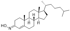
C27H45NO
|
|---|---|
| 分子量 |
399.6523
|
| 精确质量 |
399.35
|
| 元素分析 |
C, 81.14; H, 11.35; N, 3.50; O, 4.00
|
| CAS号 |
22033-87-0
|
| PubChem CID |
76971721
|
| 外观&性状 |
Typically exists as white to off-white solids at room temperature
|
| 密度 |
1.1
|
| 沸点 |
510ºC at 760mmHg
|
| 熔点 |
145-148ºC
|
| 闪点 |
341ºC
|
| 蒸汽压 |
1.56E-12mmHg at 25°C
|
| 折射率 |
1.583
|
| LogP |
7.858
|
| tPSA |
32.59
|
| 氢键供体(HBD)数目 |
1
|
| 氢键受体(HBA)数目 |
2
|
| 可旋转键数目(RBC) |
5
|
| 重原子数目 |
29
|
| 分子复杂度/Complexity |
663
|
| 定义原子立体中心数目 |
7
|
| SMILES |
C[C@@]12C(CC[C@]3([H])[C@]2([H])CC[C@@]4(C)[C@@]3([H])CC[C@@]4([C@]([H])(C)CCCC(C)C)[H])=CC(CC1)=NO
|
| InChi Key |
QNTASHOAVRSLMD-SIWSWZRQSA-N
|
| InChi Code |
InChI=1S/C27H45NO/c1-18(2)7-6-8-19(3)23-11-12-24-22-10-9-20-17-21(28-29)13-15-26(20,4)25(22)14-16-27(23,24)5/h17-19,22-25,29H,6-16H2,1-5H3/b28-21+/t19-,22+,23-,24+,25+,26+,27-/m1/s1
|
| 化学名 |
(8S,9S,10R,13R,14S,17R,E/Z)-10,13-dimethyl-17-((R)-6-methylheptan-2-yl)-1,2,6,7,8,9,10,11,12,13,14,15,16,17-tetradecahydro-3H-cyclopenta[a]phenanthren-3-one oxime
|
| 别名 |
E/Z-olesoxime; NSC 21311; NSC-21311; NSC21311; TRO-19622; TRO19622; TRO19622; RG6083; RG 6083; RG-6083;Olesoxime; Olesoxime, Z-; 22033-87-0; UNII-I2QN18P645; I2QN18P645; 66514-00-9; TRO 19622; (NE/Z)-N-[(8S,9S,10R,13R,14S,17R)-10,13-dimethyl-17-[(2R)-6-methylheptan-2-yl]-1,2,6,7,8,9,11,12,14,15,16,17-dodecahydrocyclopenta[a]phenanthren-3-ylidene]hydroxylamine;
|
| HS Tariff Code |
2934.99.9001
|
| 存储方式 |
Powder -20°C 3 years 4°C 2 years In solvent -80°C 6 months -20°C 1 month |
| 运输条件 |
Room temperature (This product is stable at ambient temperature for a few days during ordinary shipping and time spent in Customs)
|
| 溶解度 (体外实验) |
DMSO : ~50 mg/mL (~125.11 mM)
|
|---|---|
| 溶解度 (体内实验) |
配方 1 中的溶解度: ≥ 2.5 mg/mL (6.26 mM) (饱和度未知) in 10% DMSO + 40% PEG300 + 5% Tween80 + 45% Saline (这些助溶剂从左到右依次添加,逐一添加), 澄清溶液。
例如,若需制备1 mL的工作液,可将100 μL 25.0 mg/mL澄清DMSO储备液加入到400 μL PEG300中,混匀;然后向上述溶液中加入50 μL Tween-80,混匀;加入450 μL生理盐水定容至1 mL。 *生理盐水的制备:将 0.9 g 氯化钠溶解在 100 mL ddH₂O中,得到澄清溶液。 配方 2 中的溶解度: ≥ 2.5 mg/mL (6.26 mM) (饱和度未知) in 10% DMSO + 90% (20% SBE-β-CD in Saline) (这些助溶剂从左到右依次添加,逐一添加), 澄清溶液。 例如,若需制备1 mL的工作液,可将 100 μL 25.0 mg/mL澄清DMSO储备液加入900 μL 20% SBE-β-CD生理盐水溶液中,混匀。 *20% SBE-β-CD 生理盐水溶液的制备(4°C,1 周):将 2 g SBE-β-CD 溶解于 10 mL 生理盐水中,得到澄清溶液。 View More
配方 3 中的溶解度: ≥ 2.5 mg/mL (6.26 mM) (饱和度未知) in 10% DMSO + 90% Corn Oil (这些助溶剂从左到右依次添加,逐一添加), 澄清溶液。 1、请先配制澄清的储备液(如:用DMSO配置50 或 100 mg/mL母液(储备液)); 2、取适量母液,按从左到右的顺序依次添加助溶剂,澄清后再加入下一助溶剂。以 下列配方为例说明 (注意此配方只用于说明,并不一定代表此产品 的实际溶解配方): 10% DMSO → 40% PEG300 → 5% Tween-80 → 45% ddH2O (或 saline); 假设最终工作液的体积为 1 mL, 浓度为5 mg/mL: 取 100 μL 50 mg/mL 的澄清 DMSO 储备液加到 400 μL PEG300 中,混合均匀/澄清;向上述体系中加入50 μL Tween-80,混合均匀/澄清;然后继续加入450 μL ddH2O (或 saline)定容至 1 mL; 3、溶剂前显示的百分比是指该溶剂在最终溶液/工作液中的体积所占比例; 4、 如产品在配制过程中出现沉淀/析出,可通过加热(≤50℃)或超声的方式助溶; 5、为保证最佳实验结果,工作液请现配现用! 6、如不确定怎么将母液配置成体内动物实验的工作液,请查看说明书或联系我们; 7、 以上所有助溶剂都可在 Invivochem.cn网站购买。 |
| 制备储备液 | 1 mg | 5 mg | 10 mg | |
| 1 mM | 2.5022 mL | 12.5109 mL | 25.0219 mL | |
| 5 mM | 0.5004 mL | 2.5022 mL | 5.0044 mL | |
| 10 mM | 0.2502 mL | 1.2511 mL | 2.5022 mL |
1、根据实验需要选择合适的溶剂配制储备液 (母液):对于大多数产品,InvivoChem推荐用DMSO配置母液 (比如:5、10、20mM或者10、20、50 mg/mL浓度),个别水溶性高的产品可直接溶于水。产品在DMSO 、水或其他溶剂中的具体溶解度详见上”溶解度 (体外)”部分;
2、如果您找不到您想要的溶解度信息,或者很难将产品溶解在溶液中,请联系我们;
3、建议使用下列计算器进行相关计算(摩尔浓度计算器、稀释计算器、分子量计算器、重组计算器等);
4、母液配好之后,将其分装到常规用量,并储存在-20°C或-80°C,尽量减少反复冻融循环。
计算结果:
工作液浓度: mg/mL;
DMSO母液配制方法: mg 药物溶于 μL DMSO溶液(母液浓度 mg/mL)。如该浓度超过该批次药物DMSO溶解度,请首先与我们联系。
体内配方配制方法:取 μL DMSO母液,加入 μL PEG300,混匀澄清后加入μL Tween 80,混匀澄清后加入 μL ddH2O,混匀澄清。
(1) 请确保溶液澄清之后,再加入下一种溶剂 (助溶剂) 。可利用涡旋、超声或水浴加热等方法助溶;
(2) 一定要按顺序加入溶剂 (助溶剂) 。
A double-blind, randomized, multi-center study with 500 mg QD of TRO19622 versus placebo in patients with painful peripheral diabetic neuropathy
CTID: null
Phase: Phase 2 Status: Completed
Date:
InvivoChem的所有产品仅用于作科学研究,不面向患者销售
Copyright 2020 InvivoChem LLC | All Rights Reserved 粤ICP备20063088号-1


 COA
COA
 463611831
