| 规格 | 价格 | 库存 | 数量 |
|---|---|---|---|
| 1mg |
|
||
| 5mg |
|
||
| 10mg |
|
||
| 25mg |
|
||
| 50mg |
|
||
| 100mg |
|
||
| 250mg |
|
||
| Other Sizes |
|

| 靶点 |
FP ( Ki = 790 nM ); TP Receptor ( Ki = 2400 nM ); EP4 ( Ki = 1.3 nM ); EP3 ( Ki = 30 nM )
Prostaglandin E receptor EP4 subtype (Ki = 1.3 nM for EP4, as determined by competition-binding isotherms to displace radioligand binding to the respective prostanoid receptor). Ki values for other prostanoid receptors: 30 nM for EP3, 790 nM for FP, 2,400 nM for TP, and >10,000 nM for other prostanoid receptors. [1] ONO-AE3-208 selectively targets the EP4 receptor, one of the four E-type prostanoid (EP) receptors (EP1-EP4) that mediate the biological actions of prostaglandin E2 (PGE2). The compound acts as a high-affinity competitive antagonist of EP4, with a Ki value of 1.3 nM for the human EP4 receptor. It exhibits excellent selectivity: the Ki for the EP3 receptor is 30 nM (approximately 23-fold less potent), for the TP receptor is 2400 nM, and for the FP receptor is 790 nM. By blocking EP4, ONO-AE3-208 prevents PGE2 from activating downstream signaling pathways. EP4 is a G protein-coupled receptor (GPCR) that typically couples to Gs proteins, leading to activation of adenylate cyclase and increased intracellular cAMP levels. In the context of cancer, EP4 signaling promotes tumor growth, suppresses anti-tumor immunity, enhances tumor cell invasion and metastasis, and contributes to angiogenesis. ONO-AE3-208 antagonism of EP4 has been shown to reduce myeloid-derived suppressor cell (MDSC) infiltration, decrease Arg1 and Cox2 expression in MDSCs, enhance T-cell and NK-cell activity, and shift tumor-associated macrophages from M2-like to M1-like phenotypes. |
|---|---|
| 体外研究 (In Vitro) |
ONO-AE3-208以剂量依赖性方式抑制体外细胞侵袭和迁移,而不影响细胞增殖[2]。 ONO-AE3-208 在 EET 合成抑制剂 MS-PPOH 存在的情况下消除 CTGF。花生四烯酸 (AA) 会引起所附着的 Af-Art 的剂量依赖性扩张,这种效应会被 ONO-AE3-208 阻断[3]。
EP4拮抗剂ONO-AE3-208抑制前列腺癌细胞的侵袭和迁移而不影响细胞增殖[3]。 通过细胞增殖实验,观察ONO-AE3-208对PC3、LNCaP、LNCaP/mock、LNCaP/EP4+细胞增殖的影响。10µmol/L的ONO-AE3-208没有改变这些细胞的增殖率,尽管细胞表达了EP4。 ONO-AE3-208 能增强从野生型小鼠结肠分离的固有层单个核细胞(LPMNCs)的增殖(通过[³H]胸腺嘧啶摄入测定)。该增强作用与吲哚美辛处理相似。EP4激动剂AE1-734可抑制由吲哚美辛引起的增殖增加。在EP4缺陷小鼠的LPMNCs中,AE3-208和吲哚美辛均不影响增殖。[1] ONO-AE3-208 在LPS或LPS+ConA刺激下培养的LPMNCs中,能增加Th1细胞因子(IFN-γ和IL-2)的产生。[1] |
| 体内研究 (In Vivo) |
ONO-AE3-208 抑制小鼠 PC3 细胞的体内骨转移[2]。与ONO-AE3-208治疗组相比,对照组的光子肿瘤负荷以时间依赖性方式显着增加。前者的转移形成率明显高于后者。 ONO-AE3-208 治疗动物中转移形成的中位时间为 29 天,而对照组为 21 天[4]。
EP4抑制剂ONO-AE3-208可减轻链脲佐菌素糖尿病eNOS敲除小鼠的蛋白尿。 ONO-AE3-208可减少db/db小鼠的蛋白尿和系膜基质积聚。 ONO-AE3-208减轻亚全肾切除大鼠的肾损伤。[5] Connecting tubule glomerular feedback (CTGF) 实验Protocol[2] 1)实验#2和#3的时间控制:将CNT内的管腔NaCl从10 mmol/ l增加到80 mmol/ l,产生3条连续的浓度-响应曲线。 2) EP4拮抗剂ONO-AE3-208对CTGF的影响:CNT内管腔NaCl从10 mmol/L增加到80 mmol/L,形成3条连续的浓度-响应曲线。第二和第三条曲线分别添加EET合成抑制剂MS-PPOH(10−6 mol/L),第三条曲线添加EP4拮抗剂ONO-AE3-208(10−7 mol/L)。ONO-AE3-208的浓度是EP4受体Ki的77倍,EP2受体Ki的至少100倍。 3) EP4拮抗剂L161982对CTGF的影响:本实验与#2相似,但用EP4拮抗剂L161982(10−5 mol/L)代替ONO-AE3-208。L161982的浓度是EP4受体Ki的312倍,EP2受体Ki的6倍。 4)内皮破坏对CTGF的影响:CNT中NaCl从10 mmol/L增加到80 mmol/L,可诱导CTGF。将山羊抗血管性血液病因子抗人抗体(14.29 mg/ml稀释1:1000)加2%豚鼠补体灌注到Af-Art腔内10分钟,冲洗20分钟,再次诱导CTGF。为了确认内皮的完全功能去除,我们在Af-Art的管腔中添加了10 - 5 mol/L的乙酰胆碱,我们反复证明该浓度足以使Af-Art扩张。 5)外源性花生四烯酸(AA)对碳纳米管的影响:在NE预缩Af-Art后,在没有NaCl的情况下,将AA浓度从10−7到10−5 mol/L添加到碳纳米管的管腔中。在实验结束时,我们去除AA并将CNT管腔灌注液切换到80 mmol/L NaCl。 6) EP4拮抗剂对外源性AA诱导的CTGF的影响:本实验与#5相似,不同之处是将MS-PPOH添加到CNT的管腔中,并将ONO-AE3-208添加到管腔中。 在所有实验中,Af-Art直径都是在间隔3-5 μm的三个位点对NE的最大响应区域测量的,并表示为这三个测量值的平均值。用摄像机每隔5秒记录直径,用装有Metavue图像分析软件的计算机测量直径。 对接受3% DSS处理的野生型C57BL/6小鼠口服给予 ONO-AE3-208(10 mg/kg/天,通过饮水),可加重结肠炎,表现为体重减轻加剧、腹泻、便血评分和组织学损伤评分增加。此效应模拟了EP4缺陷小鼠的表型。[1] ONO-AE3-208 损害了接受3% DSS处理的野生型小鼠的黏膜屏障功能,表现为血清FITC-葡聚糖水平升高和黏膜下层FITC-葡聚糖浸润。[1] 在7% DSS诱导的结肠炎恢复期,给予 ONO-AE3-208 会抑制上皮再生(BrdU阳性上皮细胞减少),并增强黏膜下层CD4+ T细胞的活化和浸润。[1] |
| 酶活实验 |
通过竞争结合等温线实验测定 ONO-AE3-208 对前列腺素受体的Ki值。结果为:EP4为1.3 nM,EP3为30 nM,FP为790 nM,TP为2400 nM,其他前列腺素受体>10000 nM。[1]
表征 ONO-AE3-208 受体结合的主要非细胞方法是放射性配体受体结合实验。在该流程中,从表达特定 EP 受体亚型(如表达人 EP4 的 CHO 细胞)制备的膜制剂与放射性标记的高亲和力 EP 受体配体(如[³H]PGE2)在不同浓度的未标记 ONO-AE3-208 存在下共同孵育。孵育通常在 25°C 或 37°C 下进行 60-90 分钟以达到平衡。孵育后,通过使用预先在聚乙烯亚胺(PEI)中浸泡以降低非特异性结合的玻璃纤维滤膜(如 GF/B 或 GF/C 滤膜)进行快速真空过滤,将结合配体与游离配体分离。滤膜用冰冷的缓冲液多次洗涤以去除未结合的放射性。截留的放射性(代表与受体结合的[³H]PGE2 的量)通过液体闪烁计数测量。ONO-AE3-208 置换放射性标记配体的能力与放射性信号的降低成正比。半数抑制浓度(IC50)通过曲线拟合计算,抑制常数(Ki)使用 Cheng-Prusoff 公式推导。使用该方法,确定 ONO-AE3-208 对人 EP4 受体的 Ki 值为 1.3 nM,文献中报道的 pKi 值为 8.9。 |
| 细胞实验 |
细胞增殖试验[3]
采用细胞计数试剂盒-8 (CCK8)检测ONO-AE3-208对细胞增殖的影响。5 × 10~3细胞(PC3细胞)和1 × 10⁴细胞(LNCaP、LNCaP/mock和LNCaP/EP4+细胞)接种于96孔板。然后,每隔24 h,用10 μl CCK8试剂在37°温度下染色2 h,连续72 h,用自动板仪在450 nm处定量染色反应。每个实验重复三次,独立进行三次。 入侵检测[3] 采用BD BioCoat Matrigel侵袭室检测前列腺癌细胞的侵袭活性。用PBS洗涤细胞,在不含FBS的培养基中重悬,浓度为3 × 10⁴个细胞/ml。上腔涂布基质上加入细胞悬液500 μl,下腔中加入含1% FBS的培养基750 μl。在5% CO2培养箱中37°C孵育24 h后,用棉签擦拭滤膜上表面的细胞。滤网用70%乙醇固定,苏木精染色。在显微镜下随机选择6个视野对染色细胞进行计数。至少分析了来自三个不同实验的三个腔室。 愈合试验[3] 创面愈合实验如前所述进行。6孔培养皿中未融合的PC3、LNCaP、LNCaP/mock和LNCaP/EP4+细胞用塑料吸管尖划伤培养24 h。通过图像J (http://rsbweb.nih.gov/ij/)测量“创面”(划伤区域)的宽度,创面愈合比例按以下公式计算:100% - (24 h后的宽度/开始时的宽度)× 100%。每个实验重复三次,独立进行三次。 从野生型小鼠结肠分离固有层单个核细胞(LPMNCs),在含10% FBS的RPMI-1640培养基中培养。增殖实验:将细胞以10⁶个/ml密度接种于96孔板,24小时加入0.5 μCi [³H]胸腺嘧啶,72小时收集细胞,用液闪计数仪测定胸腺嘧啶掺入量。细胞因子测定:用LPS(10 μg/ml)单独或LPS+ConA(2 μg/ml)刺激LPMNCs 72小时,通过ELISA测定上清中IFN-γ和IL-2水平。[1] |
| 动物实验 |
动物模型 1:结肠炎诱导。[1]
将平均分子量为 5,000 的 DSS 以 3%(低剂量)或 7%(高剂量)的浓度添加到饮用水中,连续 7 天,喂给 8 周龄小鼠。在饮用水中添加 DSS 或下文提及的任何药物均不影响小鼠的饮水量。此外,还向饮用水中添加了吲哚美辛,剂量为 4 mg/kg/天,并在整个实验期间持续给动物服用。据报道,该剂量的吲哚美辛可在体内抑制大鼠和小鼠体内 PGE2 的产生。使用了一种 EP4 拮抗剂,ONO-AE3-208,4-{4-氰基-2-[2-(4-氟萘-1-基)丙酰氨基]苯基}丁酸 (AE3-208),以及一种 EP4 激动剂,ONO-AE1-734,甲基-7-[(1R, 2R, 3R)-3-羟基-2-[(E)-(3S)-3-羟基-4-(间甲氧基甲基苯基)-1-丁烯基]-5-氧代环戊基]-5-硫庚酸酯 (AE1-734)。通过竞争性结合等温线法测定ONO-AE3-208取代放射性配体与相应前列腺素受体结合的Ki值,EP4、EP3、FP和TP受体的Ki值分别为1.3、30、790和2400 nM,其他前列腺素受体的Ki值均大于10000 nM。AE1-734的Ki值,EP4、EP3和EP2受体的Ki值分别为0.7、56和620 nM,其余前列腺素受体的Ki值均大于10000 nM。ONO-AE3-208以10 mg/kg/天的剂量,通过饮用水口服给药。当以10 mg/kg的剂量口服该化合物时,给药后0.25小时达到血浆峰浓度677 ng/ml,生物利用度为18%。静脉注射实验测得该化合物的血浆半衰期为0.2小时。AE1-734从DSS处理前1天开始,每天两次皮下注射(每次0.1 mg/kg),直至实验结束。当以该剂量皮下注射AE1-734时,注射后10分钟达到血浆峰浓度100 ng/ml,生物利用度超过70%。血浆浓度下降的半衰期为30分钟。 为了评估黏膜完整性,采用了FITC-葡聚糖测定法。野生型C57BL/6小鼠分别在饮用水中添加ONO-AE3-208或载体。1天后,两组小鼠均在饮用水中添加3% DSS,并继续添加或不添加ONO-AE3-208。24小时后,每组小鼠口服200 μl FITC-葡聚糖(平均分子量4400)(溶于生理盐水,浓度为2 mg/ml)。给药4小时后测定血清FITC-葡聚糖浓度。EP4–/–小鼠及其野生型对照小鼠也采用相同方法处理,分别给予3% DSS和FITC-葡聚糖。将结肠组织速冻,并使用10 μm厚的冰冻切片进行荧光显微镜分析。 动物模型2:骨转移动物模型和生物发光成像[3] 为了建立骨转移模型,将1 × 10⁵个PC3/Luc细胞悬浮于100 µl PBS中,按照先前描述的方法接种到5周龄雄性裸鼠(NU/NU)的左心室(第0天)。在接种癌细胞前一天(第-1天),将小鼠分为两组(每组9只),然后对治疗组腹腔注射每日10 mg/kg的ONO-AE3-208,对对照组腹腔注射蒸馏水。后续转移情况的评估是通过在小鼠腹腔注射荧光素后7分钟,使用IVIS 100体内成像系统测量光子通量来进行的。每5-10天注射一次荧光素,持续60天,小鼠用1-3%异氟烷麻醉。 将34只6周龄裸鼠随机分为实验组和对照组,每组数量相等,分别腹腔注射ONO-AE3-208和双蒸水。然后,通过将荧光素稳定转染到前列腺癌PC3细胞中构建PC3/LUC细胞,并将这些细胞接种到小鼠左心室,以建立全身性骨转移动物模型。比较了两组小鼠的转移形成时间、光子肿瘤负荷以及建模后生存曲线的变化。[4] 动物模型3:链脲佐菌素(STZ)诱导的糖尿病eNOS−/−小鼠[5] 研究对象为8周龄的雄性C57BL/6和eNOS−/−(C57BL/6遗传背景)小鼠。小鼠禁食4小时后,连续5天,每天腹腔注射STZ(55 mg/kg,溶于0.1 M柠檬酸缓冲液,pH 4.5)或仅注射柠檬酸缓冲液。从首次注射STZ当天开始,连续3周,每天在饮用水中添加ONO-AE3-208(10 mg/kg),或仅饮用饮用水。在之前的报告中,小鼠口服10 mg/kg剂量的ONO-AE3-208后,0.25小时达到血浆峰浓度677 ng/ml,生物利用度为18%。将小鼠置于独立的代谢笼中饲养24小时后,采用ELISA法测定尿肾素含量(Exocell,费城,宾夕法尼亚州)和尿白蛋白排泄量。血糖采用OneTouch UltraMini血糖仪测定。为了确定广谱COX抑制剂的作用,雄性对照组、STZ糖尿病/6小鼠和eNOS−/−小鼠分别接受吲哚美辛(4 mg/kg/天,溶于饮用水中44,Cayman Chemical,Ann Arbor,MI)或仅饮用水处理,从首次腹腔注射STZ开始,持续两周(每组n = 10)。 动物模型4:db/db小鼠[5] 8周龄的BKS背景的雄性db/m和db/db小鼠被随机分配接受ONO-AE3-208(10 mg/kg/天,溶于饮用水中)或仅饮用水处理,持续八周。另一组db/db小鼠同时接受卡托普利治疗,剂量为20 mg/kg/天,溶于饮用水中18。血糖和尿白蛋白排泄量的测定方法如前所述。收缩压(SBP)采用CODA无创血压系统测定。血清肌酐采用高效液相色谱法(HPLC)测定。银染法中,将含0.5 µg肌酐的尿液样品溶解于样品缓冲液中,经SDS-PAGE电泳分离后,使用ProteoSilver Stain试剂盒进行染色。 动物模型5:部分肾切除大鼠[5] 8周龄雄性Sprague Dawley大鼠接受假手术或部分肾切除术,具体方法如前所述45。简而言之,在异氟烷麻醉下,对大鼠进行次全肾切除术,通过肾包膜下切除术切除右肾,并通过选择性结扎左肾动脉三支分支中的两支,使左肾三分之二发生梗死。假手术组则进行开腹手术,并在缝合伤口前对双肾进行操作。一周后,将大鼠随机分为两组,一组在饮用水中添加ONO-AE3-208(1 mg/kg/天或10 mg/kg/天),另一组仅饮用普通饮用水,并继续观察七周。采用尾套容积描记法测定收缩压(SBP),方法如前所述46。采用单次注射FITC菊粉清除率法,并通过尾静脉重复取样测定肾小球滤过率(GFR),方法如前所述。采用苯扎氯铵法测定24小时代谢笼养后的尿蛋白排泄量,并使用自动分析仪测定尿肌酐。 ONO-AE3-208以10 mg/kg/天的剂量通过饮用水灌胃给予C57BL/6小鼠。为诱导结肠炎,在饮用水中加入3% DSS,持续7天,并分别给予或不给予AE3-208。每日监测体重、粪便性状和隐血。第7天采集血液进行血细胞比容和白细胞计数,并取结肠进行组织学评分。[1] 为评估黏膜完整性,小鼠先用AE3-208或载体处理1天,然后给予3% DSS 24小时,最后灌胃给予FITC-葡聚糖(2 mg/ml,200 μl)。 4小时后测定血清FITC-葡聚糖水平。[1] 在恢复期实验中,小鼠先用7% DSS处理7天,随后用AE3-208处理3天,但不使用DSS。处死前2小时注射BrdU以标记增殖细胞。[1] |
| 药代性质 (ADME/PK) |
口服单次剂量为 10 mg/kg 时,ONO-AE3-208 在给药后 0.25 小时达到血浆峰浓度 677 ng/ml,生物利用度为 18%。静脉注射后的血浆半衰期为 0.2 小时。[1]
ONO-AE3-208 被表征为具有口服活性的化合物,具有适合体内使用的特性。该化合物在 DMSO 中具有良好的溶解性(81 mg/mL),在乙醇中具有中等溶解性(3 mg/mL),但不溶于水,因此需要适当的制剂用于口服或注射给药。对于口服给药,可在 CMC-Na(羧甲基纤维素钠)中制备浓度高达 5 mg/mL 的均匀混悬液。对于腹腔注射,有两种经过验证的制剂:(1)5% DMSO、40% PEG300、5% Tween80 和 50% ddH₂O 可实现 4.05 mg/mL(10.01 mM)的溶解度;(2)5% DMSO 和 95% 玉米油可实现 0.65 mg/mL(1.61 mM)的溶解度。在多项体内研究中,该化合物已通过口服和腹腔注射途径以 10 mg/kg/天的剂量有效给药。全面的 PK 参数如半衰期(t₁/₂)、分布容积(Vd)、清除率(CL)、生物利用度(F)和 Cmax 值在现有文献中尚未广泛发表。然而,该化合物在每日一次给药方案下对多种疾病模型的有效性表明其具有足够的药代动力学暴露量以产生生物活性。该化合物通常在-20°C 下储存,避光防潮,DMSO 储备液应分装并冷冻保存以防降解。 |
| 毒性/毒理 (Toxicokinetics/TK) |
ONO-AE3-208 的毒理学特征主要在动物疗效研究的背景下进行了评估,在治疗剂量下未报告显著的不良反应。在小鼠模型中,以 10 mg/kg/天的剂量给药 4 至 8 周通常耐受性良好。在治疗 8 周的 db/db 小鼠中,与溶剂对照组相比,ONO-AE3-208 未显著改变体重、肾脏重量、收缩压或血糖水平。在用 ONO-AE3-208(1 或 10 mg/kg/天)治疗 7 周的次全肾切除(SNx)大鼠中,与未治疗的 SNx 对照组相比,未观察到体重、左肾重量或血压的显著变化。同样,在未行肾切除的假手术大鼠中,ONO-AE3-208 对这些参数没有影响,表明在测试剂量下缺乏全身毒性。在胶质瘤模型中,接受 ONO-AE3-208 治疗的小鼠显示出完全的肿瘤排斥,没有明显的毒性或不良作用的迹象。在肾炎模型中,EP4 拮抗剂治疗不影响血压水平,这使其与可能升高血压的非选择性 COX 抑制剂有所区别。长期毒性研究(例如慢性致癌性、生殖毒性)尚未发表,因为该化合物仍主要处于临床前开发阶段。基于其作用机制——选择性 EP4 拮抗——潜在的安全性考虑包括对骨代谢的影响(EP4 参与骨形成)、肾功能(EP4 在肾血流动力学和盐处理中发挥作用)以及可能的胃肠道影响,尽管这些在已发表的研究中未见报道。总体而言,现有数据表明 ONO-AE3-208 在药理活性剂量下具有良好的安全性特征。
|
| 参考文献 |
|
| 其他信息 |
研究人员利用缺乏八种不同类型和亚型前列腺素受体的小鼠,研究了前列腺素在葡聚糖硫酸钠(DSS)诱导的结肠炎中的作用。在这些前列腺素受体缺陷小鼠中,只有EP4缺陷小鼠(而非DP、EP1、EP2、EP3、FP、IP或TP缺陷小鼠)在3% DSS处理后出现严重的结肠炎,而野生型小鼠仅出现轻微的结肠炎。在野生型小鼠中,给予EP4选择性拮抗剂(AE3-208)可模拟这种表型。EP4缺陷会损害黏膜屏障功能,并导致结肠上皮细胞丢失、隐窝损伤以及中性粒细胞和淋巴细胞聚集。相反,对野生型小鼠施用EP4选择性激动剂(AE1-734)可减轻通常由7% DSS诱导的严重结肠炎,而施用AE3-208则抑制结肠炎的恢复并显著诱导CD4+ T细胞增殖。体外实验表明,AE3-208增强了结肠固有层单核细胞的增殖和Th1细胞因子产生,而AE1-734则抑制了这些细胞的增殖和Th1细胞因子产生。DNA微阵列分析显示,EP4缺陷小鼠结肠中与免疫反应相关的基因表达升高,而与黏膜修复和重塑相关的基因表达降低。我们得出结论,EP4 通过维持黏膜完整性和下调免疫反应来维持肠道稳态。[1]连接小管-肾小球反馈 (CTGF) 是一种机制,其中连接小管 (CNT) 中的钠重吸收引起入球小动脉 (Af-Art) 扩张。CTGF 由类花生酸介导,包括前列腺素和环氧二十碳三烯酸;然而,它们的确切性质和来源仍然未知。我们假设在 CTGF 过程中,CNT 释放前列腺素 E2,后者与其 4 型受体 (EP4) 结合并扩张 Af-Art。我们显微解剖了具有完整附着 CNT 的兔 Af-Art,并对其进行灌注,然后用去甲肾上腺素预收缩。通过将 CNT 管腔内 NaCl 浓度从 10 mmol/L 增加到 80 mmol/L 来诱导 CTGF。我们在环氧二十碳三烯酸合成抑制剂MS-PPOH存在的情况下,分别在浴液中加入或不加入EP4受体阻断剂ONO-AE3-208来诱导CTGF。ONO-AE3-208完全抑制了CTGF的表达(对照组:9.4 ± 0.5 μM;MS-PPOH+ONO-AE3-208组:-0.6 ± 0.2 μM;P<0.001;n=6)。为了验证这些结果,我们使用了另一种特异性EP4阻断剂L161982(10⁻⁵ mol/L),结果也显示其同样抑制了CTGF的表达(对照组:8.5 ± 0.9 μM;MS-PPOH+L161982组:0.8 ± 0.4 μM;P<0.001;n=6)。为了证实介导 CTGF 的二十碳酸类物质是从 CNT 而非 Af-Art 释放的,我们首先使用抗体和补体破坏了 Af-Art 的内皮细胞。内皮细胞破坏并未影响 CTGF(7.9 ± 0.9 μm 对比 8.6 ± 0.6 μm;P=NS;n=7)。然后,我们在灌注液中保持 NaCl 浓度为零的情况下,向 CNT 管腔内添加花生四烯酸。花生四烯酸引起所连接的 Af-Art 呈剂量依赖性扩张(从 8.6 ± 1.2 μm 增至 15.3 ± 0.7 μm;P<0.001;n=6),并且这种作用可被 ONO-AE3-208 (10⁻⁷ mol/L) 阻断。我们得出结论,在CTGF期间,CNT释放前列腺素E2,后者作用于Af-Art上的EP4,诱导内皮非依赖性扩张。[2]
EP4是前列腺素E2受体之一,前列腺素E2是最常见的类前列腺素,与炎症性疾病和癌症相关。我们之前报道过,EP4的过表达是导致去势抵抗性前列腺癌进展的机制之一,而EP4拮抗剂ONO-AE3-208在体内通过调节雄激素受体的激活来抑制去势抵抗性进展。本研究旨在分析EP4与前列腺癌转移的关联以及ONO-AE3-208抑制转移的疗效。我们评估了前列腺癌细胞系LNCaP和PC3中EP4 mRNA的表达水平。构建了EP4过表达的LNCaP细胞系,并将其侵袭能力与对照LNCaP细胞(LNCaP/mock)进行比较。在不同浓度的ONO-AE3-208处理下,检测了这些细胞的体外增殖、侵袭和迁移能力。通过将表达荧光素酶的PC3细胞接种到裸鼠左心室,构建了体内骨转移小鼠模型。利用生物发光成像技术观察了给予或不给予ONO-AE3-208处理后的骨转移情况。结果显示,PC3细胞中EP4 mRNA的表达水平高于LNCaP细胞,且LNCaP细胞中EP4的过表达增强了其侵袭能力。ONO-AE3-208以剂量依赖的方式抑制了LNCaP细胞的体外侵袭和迁移,且不影响细胞增殖。ONO-AE3-208处理也抑制了PC3细胞的体内骨转移。 EP4表达水平与前列腺癌细胞的侵袭性相关,而EP4特异性拮抗剂ONO-AE3-208可抑制细胞侵袭、迁移和骨转移,表明其可能是一种治疗转移性前列腺癌的新型潜在疗法。[3] 目的:探讨EP4拮抗剂ONO-AE3-208对小鼠前列腺癌骨转移形成的影响。方法:将34只6周龄裸鼠随机分为实验组和对照组,每组数量相同,分别腹腔注射ONO-AE3-208和双蒸水。然后,将稳定转染荧光素的PC3/LUC细胞构建于前列腺癌PC3细胞中,并接种于小鼠左心室,建立全身性骨转移动物模型。本研究比较了两组小鼠在造模后转移灶形成时间、光子肿瘤负荷以及生存曲线的变化。结果:造模30天后,生物发光成像分析显示,与实验组相比,对照组的光子肿瘤负荷随时间显著增加(P < 0.01)。对照组的转移灶形成率也显著高于实验组(93.3% vs 33.3%,P < 0.001)。实验组动物骨转移形成的中位时间为 29 天(95% CI 26.547 - 35.262),而对照组为 21 天(95% CI 17.213 - 24.787)(P < 0.001)。结论:EP4 拮抗剂 ONO-AE3-208 可抑制小鼠前列腺癌骨转移的形成。[4] 连接小管-肾小球反馈 (CTGF) 是一种机制,其中连接小管 (CNT) 中的钠重吸收引起入球小动脉 (Af-Art) 扩张。CTGF 由类花生酸介导,包括前列腺素和环氧二十碳三烯酸;然而,它们的确切性质和来源仍不清楚。我们假设在CTGF期间,CNT释放前列腺素E2,后者与其4型受体(EP4)结合并扩张Af-Art。我们显微解剖了附着有完整CNT的兔Af-Art,进行灌注,并用去甲肾上腺素预收缩。通过将CNT管腔内NaCl浓度从10 mmol/L增加到80 mmol/L来诱导CTGF。我们在灌注液中加入或不加入EP4受体阻滞剂ONO-AE3-208,并在环氧二十碳三烯酸合成抑制剂MS-PPOH存在的情况下诱导CTGF。ONO-AE3-208完全抑制了CTGF(对照组:9.4 ± 0.5 μm;MS-PPOH+ONO-AE3-208组:-0.6 ± 0.2 μm;P<0.001;n=6)。为了验证这些结果,我们使用了一种不同的特异性EP4阻断剂L161982(10⁻⁵ mol/L),该阻断剂同样能消除CTGF(对照组:8.5 ± 0.9 μm;MS-PPOH+L161982组:0.8 ± 0.4 μm;P<0.001;n=6)。为了确认介导CTGF的二十碳酸类物质是从连接小管(CNT)而非动静脉内皮(Af-Art)释放的,我们首先使用抗体和补体破坏了Af-Art的内皮细胞。内皮细胞的破坏并未影响CTGF(7.9 ± 0.9 μm vs. 8.6 ± 0.6 μm;P=NS;n=7)。然后,我们在灌注液中保持NaCl浓度为零的情况下,向连接小管的管腔内添加了花生四烯酸。花生四烯酸可剂量依赖性地扩张附着的动静脉内膜(Af-Art)(从 8.6 ± 1.2 μm 扩张至 15.3 ± 0.7 μm;P<0.001;n=6),而这种作用可被 ONO-AE3-208 (10⁻⁷ mol/L) 阻断。我们得出结论,在 CTGF 期间,CNT 释放前列腺素 E2,后者作用于动静脉内膜上的 EP4,诱导内皮非依赖性扩张。[5] ONO-AE3-208 是一种 EP4 选择性拮抗剂,用于研究 EP4 在结肠炎中的作用。它通过损害黏膜屏障功能和增强 CD4+ T 细胞活化来加剧 DSS 诱导的结肠炎。[1] 该药物由日本小野药品工业株式会社提供。[1] |
| 分子式 |
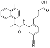
C24H21FN2O3
|
|---|---|
| 分子量 |
404.4335
|
| 精确质量 |
404.153
|
| 元素分析 |
C, 71.27; H, 5.23; F, 4.70; N, 6.93; O, 11.87
|
| CAS号 |
402473-54-5
|
| PubChem CID |
10111831
|
| 外观&性状 |
White to yellow solid powder
|
| 密度 |
1.3±0.1 g/cm3
|
| 沸点 |
662.4±55.0 °C at 760 mmHg
|
| 闪点 |
354.4±31.5 °C
|
| 蒸汽压 |
0.0±2.1 mmHg at 25°C
|
| 折射率 |
1.637
|
| LogP |
4.56
|
| tPSA |
90.19
|
| 氢键供体(HBD)数目 |
2
|
| 氢键受体(HBA)数目 |
5
|
| 可旋转键数目(RBC) |
7
|
| 重原子数目 |
30
|
| 分子复杂度/Complexity |
660
|
| 定义原子立体中心数目 |
0
|
| SMILES |
FC1=C([H])C([H])=C(C2=C([H])C([H])=C([H])C([H])=C21)C([H])(C([H])([H])[H])C(N([H])C1C([H])=C(C#N)C([H])=C([H])C=1C([H])([H])C([H])([H])C([H])([H])C(=O)O[H])=O
|
| InChi Key |
MTDIMKNAJUQTIO-UHFFFAOYSA-N
|
| InChi Code |
InChI=1S/C24H21FN2O3/c1-15(18-11-12-21(25)20-7-3-2-6-19(18)20)24(30)27-22-13-16(14-26)9-10-17(22)5-4-8-23(28)29/h2-3,6-7,9-13,15H,4-5,8H2,1H3,(H,27,30)(H,28,29)
|
| 化学名 |
4-[4-cyano-2-[2-(4-fluoronaphthalen-1-yl)propanoylamino]phenyl]butanoic acid
|
| 别名 |
AE 3-208; AE-3-208; AE3-208; ONO AE3 208; ONO-AE3-208; 4-[4-cyano-2-[2-(4-fluoronaphthalen-1-yl)propanoylamino]phenyl]butanoic Acid; 4-Cyano-2-[[2-(4-fluoro-1-naphthalenyl)-1-oxopropyl]amino]benzenebutanoic acid; DTXSID20435810; 4-(4-Cyano-2-(2-(4-fluoronaphthalen-1-yl)propanamido)phenyl)butanoic acid; 4-Cyano-2-[[2-(4-fluoro-1-naphthalenyl)-1-oxopropyl]amino]Benzenebutanoic acid; ONO-AE-3-208; ONO-AE 3-208
|
| HS Tariff Code |
2934.99.9001
|
| 存储方式 |
Powder -20°C 3 years 4°C 2 years In solvent -80°C 6 months -20°C 1 month |
| 运输条件 |
Room temperature (This product is stable at ambient temperature for a few days during ordinary shipping and time spent in Customs)
|
| 溶解度 (体外实验) |
DMSO: ~33.33 mg/mL (~82.4 mM)
|
|---|---|
| 溶解度 (体内实验) |
配方 1 中的溶解度: ≥ 2.08 mg/mL (5.14 mM) (饱和度未知) in 10% DMSO + 40% PEG300 + 5% Tween80 + 45% Saline (这些助溶剂从左到右依次添加,逐一添加), 澄清溶液。
例如,若需制备1 mL的工作液,可将100 μL 20.8 mg/mL澄清DMSO储备液加入400 μL PEG300中,混匀;然后向上述溶液中加入50 μL Tween-80,混匀;加入450 μL生理盐水定容至1 mL。 *生理盐水的制备:将 0.9 g 氯化钠溶解在 100 mL ddH₂O中,得到澄清溶液。 配方 2 中的溶解度: ≥ 2.08 mg/mL (5.14 mM) (饱和度未知) in 10% DMSO + 90% Corn Oil (这些助溶剂从左到右依次添加,逐一添加), 澄清溶液。 例如,若需制备1 mL的工作液,可将 100 μL 20.8 mg/mL 澄清 DMSO 储备液添加到 900 μL 玉米油中并混合均匀。 View More
配方 3 中的溶解度: 5%DMSO + 40%PEG300 + 5%Tween 80 + 50%ddH2O: 4.05mg/ml (10.01mM) 1、请先配制澄清的储备液(如:用DMSO配置50 或 100 mg/mL母液(储备液)); 2、取适量母液,按从左到右的顺序依次添加助溶剂,澄清后再加入下一助溶剂。以 下列配方为例说明 (注意此配方只用于说明,并不一定代表此产品 的实际溶解配方): 10% DMSO → 40% PEG300 → 5% Tween-80 → 45% ddH2O (或 saline); 假设最终工作液的体积为 1 mL, 浓度为5 mg/mL: 取 100 μL 50 mg/mL 的澄清 DMSO 储备液加到 400 μL PEG300 中,混合均匀/澄清;向上述体系中加入50 μL Tween-80,混合均匀/澄清;然后继续加入450 μL ddH2O (或 saline)定容至 1 mL; 3、溶剂前显示的百分比是指该溶剂在最终溶液/工作液中的体积所占比例; 4、 如产品在配制过程中出现沉淀/析出,可通过加热(≤50℃)或超声的方式助溶; 5、为保证最佳实验结果,工作液请现配现用! 6、如不确定怎么将母液配置成体内动物实验的工作液,请查看说明书或联系我们; 7、 以上所有助溶剂都可在 Invivochem.cn网站购买。 |
| 制备储备液 | 1 mg | 5 mg | 10 mg | |
| 1 mM | 2.4726 mL | 12.3631 mL | 24.7262 mL | |
| 5 mM | 0.4945 mL | 2.4726 mL | 4.9452 mL | |
| 10 mM | 0.2473 mL | 1.2363 mL | 2.4726 mL |
1、根据实验需要选择合适的溶剂配制储备液 (母液):对于大多数产品,InvivoChem推荐用DMSO配置母液 (比如:5、10、20mM或者10、20、50 mg/mL浓度),个别水溶性高的产品可直接溶于水。产品在DMSO 、水或其他溶剂中的具体溶解度详见上”溶解度 (体外)”部分;
2、如果您找不到您想要的溶解度信息,或者很难将产品溶解在溶液中,请联系我们;
3、建议使用下列计算器进行相关计算(摩尔浓度计算器、稀释计算器、分子量计算器、重组计算器等);
4、母液配好之后,将其分装到常规用量,并储存在-20°C或-80°C,尽量减少反复冻融循环。
计算结果:
工作液浓度: mg/mL;
DMSO母液配制方法: mg 药物溶于 μL DMSO溶液(母液浓度 mg/mL)。如该浓度超过该批次药物DMSO溶解度,请首先与我们联系。
体内配方配制方法:取 μL DMSO母液,加入 μL PEG300,混匀澄清后加入μL Tween 80,混匀澄清后加入 μL ddH2O,混匀澄清。
(1) 请确保溶液澄清之后,再加入下一种溶剂 (助溶剂) 。可利用涡旋、超声或水浴加热等方法助溶;
(2) 一定要按顺序加入溶剂 (助溶剂) 。
|
|
|
|
InvivoChem的所有产品仅用于作科学研究,不面向患者销售
Copyright 2020 InvivoChem LLC | All Rights Reserved 粤ICP备20063088号-1


 COA
COA


 463611831
