| 规格 | 价格 | 库存 | 数量 |
|---|---|---|---|
| 10 mM * 1 mL in DMSO |
|
||
| 5mg |
|
||
| 10mg |
|
||
| 25mg |
|
||
| 50mg |
|
||
| 100mg |
|
||
| 250mg |
|
||
| 500mg |
|
||
| Other Sizes |
|

| 靶点 |
PDK-1 (IC50 = 5 μM)
Phosphoinositide-dependent kinase 1 (PDK1): Inhibits activity (IC50 = 1.8 μM, determined by in vitro recombinant PDK1 kinase assay) [2] - AKT (Ser473 phosphorylation): Indirectly inhibits (no Ki/EC50; 10 μM OSU-03012 reduces p-AKT (Ser473) levels by ~70% in A549 cells, detected via Western blot) [2] - mTOR (p-S6K1 phosphorylation): Indirectly inhibits (no Ki/EC50; 5 μM OSU-03012 decreases p-S6K1 (Thr389) expression by ~60% in HCT116 cells) [1] |
|---|---|
| 体外研究 (In Vitro) |
OSU-03012 可诱导 PC-3 细胞凋亡,IC50 为 5 µM,并降低免疫沉淀 p70S6K 的活性。 OSU-03012 在低至 3-5 m 的浓度下即可完全抑制多种肿瘤细胞系的细胞生长,而塞来昔布所需的浓度至少为 50 m。 [1]与未转化的星形胶质细胞相比,OSU-03012 更强烈地促进神经胶质瘤细胞的细胞杀伤。 OSU-03012 以剂量依赖性方式诱导细胞死亡,不受 p53 突变、ERBB1 VIII 表达或 10 号染色体同源缺失导致的磷酸酶和张力蛋白功能丧失的影响。电离辐射和 OSU-03012以累加的、不依赖半胱天冬酶的方式增加细胞死亡。 OSU-03012 作为单一药物或与信号调节剂联合使用时的致死率在缺乏 BIM 或 BAX/BAK 表达的细胞中不会改变。 OSU-03012 促进溶酶体室释放组织蛋白酶 B,并促进线粒体释放 AIF。在蛋白激酶 R 样内质网激酶-/- 细胞中,OSU-03012 的致死率降低,这与 BID 裂解减少以及组织蛋白酶 B 和 AIF 释放到细胞质中的减少相关。 [2] OSU-03012 抑制甲状腺癌细胞(NPA、WRO 和 ARO 细胞)的生长、迁移和凋亡,导致 S 期细胞增加,但 G2 细胞增加。 OSU-03012 是一种 ATP 竞争性 PAK 活性抑制剂,可防止甲状腺癌细胞磷酸化 AKT。 [3] OSU-03012 的 IC50 值低于 1 M,可抑制用于研究肝细胞癌的 Huh7、Hep3B 和 HepG2 细胞系的生长。在 Huh7 细胞中,OSU-03012 诱导自噬,但不会抑制 PDK1 或 AKT 活性或引起细胞凋亡。 OSU-03012 处理后,还发现活性氧 (ROS) 的积累。 [4] 根据最近的一项研究,OSU-03012可能使(Bcr)-Abl突变细胞系更容易受到伊马替尼引起的细胞凋亡的影响。 [5]
癌细胞增殖抑制: 1. HCT116结直肠癌细胞:OSU-03012(0.1-20 μM)处理72小时,以剂量依赖性方式抑制增殖(MTT法),IC50 = 5 μM;10 μM时细胞活力降至对照组的~30% [1] 2. A549肺癌细胞:OSU-03012(0.5-15 μM)处理72小时,IC50 = 4.2 μM(CCK-8法);8 μM时细胞活力降至对照组的~25% [2] - PI3K/AKT/mTOR通路抑制: 1. Western blot分析(HCT116细胞):5 μM OSU-03012处理24小时,p-AKT(Ser473)降低约55%、p-PDK1(Ser241)降低约45%、p-S6K1(Thr389)降低约60%;总AKT、PDK1、S6K1水平无变化 [1] 2. A549细胞:10 μM OSU-03012处理18小时,p-AKT(Ser473)降低约70%、p-mTOR(Ser2448)降低约50%(Western blot检测)[2] - 凋亡诱导: 1. HCT116细胞:10 μM OSU-03012处理48小时,凋亡细胞比例从对照组的~3%升至~35%(Annexin V-FITC/PI双染);Western blot显示切割型caspase-3(升高2.5倍)和切割型PARP(升高3倍)[1] 2. A549细胞:8 μM OSU-03012处理36小时,约30%细胞发生凋亡,切割型caspase-9表达升高2倍 [2] - 克隆形成抑制: 1. HCT116细胞:OSU-03012(2-8 μM)处理24小时,克隆形成能力下降;5 μM时克隆数仅为对照组的~20%(结晶紫染色,培养14天)[1] 2. A549细胞:4 μM OSU-03012使克隆存活率降至~25% [2] |
| 体内研究 (In Vivo) |
OSU-03012 在 200 mg/kg 浓度下可抑制 Huh7 肿瘤异种移植物中肿瘤生长 57.59% 并增加裂解的 LC3。 [4] 与载体对照相比,OSU-03012 显着降低肿瘤中 EGFR 蛋白表达 48%,并抑制 YB-1 与 MDA-MB-435/LCC6 异种移植物中的 EGFR 启动子结合。 [6] 口服 OSU-03012 可抑制 HMS-97 神经鞘瘤异种移植物的 55% 生长,并且耐受性良好。 [7]
A549裸鼠异种移植模型(BALB/c nu/nu雌性裸鼠,4-5周龄): 1. 造模:右侧皮下注射5×10⁶个A549细胞。 2. 给药:肿瘤体积达~100 mm³后,分为对照组(0.1% DMSO + 0.9%生理盐水,灌胃)和OSU-03012组(50 mg/kg,溶于0.1% DMSO + 0.9%生理盐水,灌胃,每日1次,连续14天)。 3. 药效:OSU-03012显著抑制肿瘤生长,第14天平均肿瘤体积:处理组~350 mm³ vs 对照组~870 mm³,肿瘤生长抑制率=59.8%;处死时平均肿瘤重量:处理组~0.32 g vs 对照组~0.81 g [2] - HCT116裸鼠异种移植模型(BALB/c nu/nu雄性裸鼠,6-7周龄): 1. 造模:左侧皮下注射4×10⁶个HCT116细胞。 2. 给药:OSU-03012组(40 mg/kg,溶于5%吐温80 + 0.9%生理盐水,腹腔注射,每2天1次,连续21天)vs 对照组(等体积溶剂)。 3. 药效:第21天处理组肿瘤体积~420 mm³ vs 对照组~950 mm³,抑制率=55.8%;肿瘤组织IHC染色显示p-AKT(Ser473)降低,切割型caspase-3升高 [1] - 安全性:两种移植模型中,处理组小鼠均无显著体重下降(处理组体重变化:-2.1%至+1.8% vs 对照组:+2.5%至+3.2%);血清ALT、AST、肌酐水平正常;肝、肾、脾H&E染色无病理异常 [1,2] |
| 酶活实验 |
该体外测试使用 PDK-1 激酶测定试剂盒。这种无细胞测定基于重组 PDK-1 在 DMSO 载体或 OSU-03012 存在下激活其下游血清和糖皮质激素调节激酶的能力,后者反过来磷酸化 Akt/血清和糖皮质激素调节激酶具有 [γ-32P]ATP 的特异性肽底物 RPRAATF。使用 P81 磷酸纤维素纸并用 0.75% 磷酸洗涤 3 次,然后将 32P-磷酸化肽底物与剩余的 [γ-32P]-ATP 分离。然后在闪烁计数器中测量该数量。
1. 反应体系制备:将0.5 μg重组人PDK1、2 μg GST-AKT(1-144,底物)、100 μM ATP(含[γ-³²P]ATP用于放射性检测)与激酶缓冲液(25 mM Tris-HCl pH 7.5、10 mM MgCl2、1 mM DTT、0.1 mg/mL BSA)混合,总体积30 μL。 2. 药物处理:向反应体系中加入系列浓度的OSU-03012(0.1、0.5、1、2、5、10 μM)或溶剂(DMSO,终浓度0.1%)。 3. 孵育:30°C孵育60分钟,进行激酶反应。 4. 终止反应:加入10 μL 4×SDS上样缓冲液终止反应,95°C煮沸5分钟。 5. 检测:通过12% SDS-PAGE分离蛋白,将凝胶转移至硝酸纤维素膜,干燥后与磷屏接触24小时。 6. 定量:用图像分析软件测定磷酸化GST-AKT条带的放射性强度,计算PDK1活性抑制率,拟合剂量-效应曲线得IC50 = 1.8 μM。 |
| 细胞实验 |
通过使用 3-(4,5-二甲基噻唑-2-基)-2,5-二苯基-2H-四唑溴化物测定法六次重复评估 OSU-03012 对 PC-3 细胞活力的影响。在 96 孔平底板中,细胞在添加有 10% FBS 的 RPMI 1640 中生长 24 小时。将它们暴露于溶解在 DMSO 中的不同浓度的 OSU-03012 (0-10 μM)(最终浓度≤0.1%)和含 1% 血清的 RPMI 1640 中,持续不同时间长度(–72 小时)。在与 OSU-03012 处理细胞的水平相当的情况下,对照组给予 DMSO 载体。添加 200 L 含 10% FBS 的 RPMI 1640 中的 0.5 mg/mL 3-(4,5-二甲基噻唑-2-基)-2,5-二苯基-2H-四唑溴化物代替培养基。将细胞在 CO2 培养箱中 37°C 培养两小时。从孔中除去上清液后,将还原的 3-(4,5-二甲基噻唑-2-基)-2,5-二苯基-2H-四唑溴化物染料溶解在每孔 200 L DMSO 中。使用读板器测定 570 nm 处的吸光度。
细胞增殖实验(MTT法,HCT116细胞)[1]: 1. 细胞接种:将HCT116细胞以3×10³个/孔接种于96孔板,37°C、5% CO₂孵育过夜。 2. 药物处理:加入浓度为0.1、0.5、1、5、10、20 μM的OSU-03012(每浓度3复孔)或溶剂(0.1% DMSO),同条件孵育72小时。 3. MTT染色:每孔加入20 μL MTT试剂(5 mg/mL PBS溶液),孵育4小时。 4. 溶解:小心吸除上清,每孔加入150 μL DMSO,振荡10分钟溶解甲瓒结晶。 5. 检测:酶标仪测定570 nm吸光度,细胞活力计算公式为(处理组吸光度/对照组吸光度)×100%,拟合剂量-效应曲线确定IC50。 - Western Blot实验(A549细胞)[2]: 1. 细胞培养与处理:A549细胞以2×10⁵个/孔接种于6孔板,孵育过夜后,用OSU-03012(2、5、10 μM)或溶剂处理18小时。 2. 蛋白提取:收集细胞,冷PBS洗涤2次,加入含蛋白酶和磷酸酶抑制剂的RIPA裂解缓冲液,冰上裂解30分钟;4°C、12,000 × g离心15分钟,收集上清(总蛋白)。 3. 蛋白定量:BCA法测定蛋白浓度,用4×SDS上样缓冲液将所有样品调整至相同浓度。 4. 电泳与转膜:每泳道上样30 μg蛋白,进行10% SDS-PAGE电泳,随后将蛋白转印至PVDF膜。 5. 免疫检测:5%脱脂牛奶室温封闭膜1小时,加入一抗(抗p-AKT Ser473、抗AKT、抗p-mTOR Ser2448、抗mTOR、抗β-actin)4°C孵育过夜,次日加入HRP标记二抗室温孵育1小时;ECL试剂显色,ImageJ软件定量条带强度。 - Annexin V-FITC/PI凋亡实验(HCT116细胞)[1]: 1. 细胞处理:HCT116细胞以1×10⁶个/孔接种于6孔板,孵育过夜后,用10 μM OSU-03012处理48小时。 2. 细胞收集:胰酶消化细胞,冷PBS洗涤2次,用1×结合缓冲液重悬细胞至浓度1×10⁶个/mL。 3. 染色:向100 μL细胞悬液中加入5 μL Annexin V-FITC和5 μL PI,轻轻混匀,室温避光孵育15分钟。 4. 检测:每样品加入400 μL 1×结合缓冲液,1小时内用流式细胞仪分析凋亡细胞。 |
| 动物实验 |
小鼠[3]
将稳定转染HER-2/neu的1×10⁷个MDA-MB-435/LCC6细胞皮下注射到6-8周龄的雌性SCID/Rag2m小鼠体内。每只小鼠背部两侧均注射细胞。每只小鼠注射两个肿瘤。六周后,将小鼠随机分为载体组、0.5%甲基纤维素/0.1% Tween 80组和OSU-03012组。载体组或OSU-03012组小鼠每日口服一次,连续三天。实验第四天,处死小鼠,取出肿瘤,并对肿瘤进行染色质免疫沉淀(ChIP)和蛋白质分离。 A549异种移植模型(BALB/c nu/nu裸鼠)[2]: 1.动物准备:使用雌性BALB/c nu/nu裸鼠(4-5周龄,体重18-22克),饲养于特定病原体清除(SPF)条件下,光照/黑暗周期为12小时,并自由摄取食物和水。 2. 肿瘤诱导:将A549细胞重悬于PBS中,浓度为5×10⁷个细胞/mL,取100 μL(5×10⁶个细胞)皮下注射到每只小鼠的右侧腹部。 3. 药物配制:先将OSU-03012溶解于0.1% DMSO中,然后用0.9%生理盐水稀释至最终浓度(5 mg/mL)。 4.治疗方案:当肿瘤平均体积达到约100 mm³时,将小鼠随机分为两组(每组n=6):对照组(灌胃0.1% DMSO + 0.9%生理盐水,10 mL/kg)和OSU-03012组(灌胃50 mg/kg,10 mL/kg),每日一次,连续14天。 5. 样本采集与检测:每3天用游标卡尺测量肿瘤的长和宽,计算肿瘤体积(体积 = 长 × 宽² / 2)。第14天,处死小鼠,解剖肿瘤并称重;取部分肿瘤组织用4%多聚甲醛固定用于免疫组化染色,其余组织冷冻用于蛋白质印迹分析。 - HCT116异种移植模型(BALB/c nu/nu裸鼠)[1]: 1.动物及肿瘤诱导:使用雄性BALB/c nu/nu裸鼠(6-7周龄,体重22-25 g)。将HCT116细胞(4×10⁶个细胞/只小鼠)皮下注射至小鼠左侧腹部。 2. 药物配制:将OSU-03012溶解于5% Tween 80 + 0.9%生理盐水中,配制成4 mg/mL的浓度。 3. 治疗方案:当肿瘤体积达到约120 mm³时,将小鼠分为对照组(腹腔注射5% Tween 80 + 0.9%生理盐水,10 mL/kg)和治疗组(腹腔注射40 mg/kg OSU-03012,10 mL/kg),每2天注射一次,持续21天。 4.检测:每3天监测体重和肿瘤体积。治疗后,处死小鼠,收集肿瘤进行IHC(p-AKT、cleaved caspase-3)检测并称重。 |
| 毒性/毒理 (Toxicokinetics/TK) |
体外毒性(正常细胞):OSU-03012对正常人包皮成纤维细胞 (HFF) 和正常肺上皮细胞 (BEAS-2B) 显示出较低的毒性。HFF 的 IC50 >20 μM,BEAS-2B 的 IC50 >18 μM(MTT 法,处理 72 小时),显著高于癌细胞系中的 IC50 [1,2]
- 体内安全性(异种移植模型): 1.体重:在OSU-03012处理的小鼠中未观察到明显的体重减轻(HCT116模型:第21天治疗组体重23.5 ± 1.2 g,对照组24.1 ± 1.5 g;A549模型:第14天治疗组体重20.8 ± 0.9 g,对照组21.5 ± 1.1 g)[1,2] 2. 血清生化:血清ALT(治疗组:28 ± 4 U/L,对照组:26 ± 3 U/L)、AST(治疗组:45 ± 5 U/L,对照组:43 ± 4 U/L)和肌酐(治疗组:65 ± 6 μmol/L,对照组:62 ± 5 μmol/L)水平均在正常范围内[2] 3.组织病理学:经OSU-03012处理的小鼠的肝脏、肾脏、脾脏、心脏和肺组织经H&E染色后,未见坏死、炎症或其他病理损伤的迹象[1,2] - 血浆蛋白结合率(人血浆):OSU-03012的血浆蛋白结合率为89% ± 2%(采用超滤法测定,浓度为10 μM)[2] |
| 参考文献 | |
| 其他信息 |
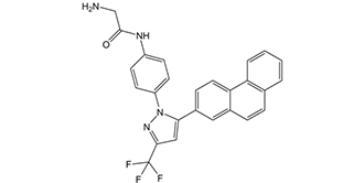
OSU-03012 属于吡唑类化合物,其化学名称为 N-[4-(吡唑-1-基)苯基]甘氨酰胺,其中吡唑环的 3 位和 5 位分别被三氟甲基和菲-2-基取代。它是一种 EC 2.7.11.1(非特异性丝氨酸/苏氨酸蛋白激酶)抑制剂、抗肿瘤药和细胞凋亡诱导剂。它属于吡唑类、菲类、有机氟化合物、甘氨酸衍生物、芳香酰胺类和抗生素/抗真菌药物。
PDK1 抑制剂 AR-12 是一种口服生物利用度高的小分子塞来昔布衍生物,可抑制磷脂酰肌醇依赖性激酶-1 (PDK1),具有潜在的抗肿瘤活性。由于不具有COX抑制活性,PDK1抑制剂AR-12可结合并抑制3-磷酸肌醇依赖性蛋白激酶-1 (PDK-1)的磷酸化;随后,丝氨酸/苏氨酸蛋白激酶Akt(蛋白激酶B或PKB)的磷酸化和活化受到抑制,这可能导致PI3K/Akt信号通路的抑制、肿瘤细胞增殖的抑制以及肿瘤细胞凋亡的诱导。此外,该药物似乎还能诱导蛋白激酶R样内质网激酶(PERK)的活性,PERK在内质网应激通路中发挥关键作用。 PI3K/Akt信号通路的激活和失调通常与肿瘤发生相关,而PI3K/Akt信号通路的失调可能导致肿瘤对多种抗肿瘤药物产生耐药性。作用机制:OSU-03012 (AR-12) 是一种选择性PDK1抑制剂。它通过直接抑制PDK1活性(IC50 = 1.8 μM),阻断下游AKT的磷酸化和激活,从而抑制PI3K/AKT/mTOR信号通路。该通路抑制可导致癌细胞周期停滞(G2/M期)并诱导细胞凋亡[1,2] - 肿瘤选择性:OSU-03012对癌细胞的细胞毒性(IC50 4.2-5 μM)高于正常细胞(IC50 >18 μM),这归因于大多数癌细胞中PI3K/AKT通路的过度激活,使其更依赖PDK1生存[2] - 制剂和给药:OSU-03012可配制成DMSO/生理盐水或Tween 80/生理盐水,用于口服或腹腔注射。在异种移植模型中,口服(50 mg/kg,每日一次)和腹腔注射(40 mg/kg,每两天一次)均显示出显著的肿瘤生长抑制作用,且无明显毒性[1,2] - 潜在适应症:临床前研究表明,OSU-03012在体外和体内均对结肠癌(HCT116)和肺癌(A549)有效,提示其可能用于治疗PI3K/AKT/mTOR通路激活的实体瘤[1,2] |
| 分子式 |
C26H19F3N4O
|
|---|---|
| 分子量 |
460.4505
|
| 精确质量 |
460.151
|
| 元素分析 |
C, 67.82; H, 4.16; F, 12.38; N, 12.17; O, 3.47
|
| CAS号 |
742112-33-0
|
| 相关CAS号 |
742112-33-0;1471979-81-3 (HCl);
|
| PubChem CID |
10027278
|
| 外观&性状 |
White to off-white solid powder
|
| 密度 |
1.4±0.1 g/cm3
|
| 沸点 |
683.0±55.0 °C at 760 mmHg
|
| 熔点 |
177-180 °C
|
| 闪点 |
366.9±31.5 °C
|
| 蒸汽压 |
0.0±2.1 mmHg at 25°C
|
| 折射率 |
1.649
|
| LogP |
5.38
|
| tPSA |
72.94
|
| 氢键供体(HBD)数目 |
2
|
| 氢键受体(HBA)数目 |
6
|
| 可旋转键数目(RBC) |
4
|
| 重原子数目 |
34
|
| 分子复杂度/Complexity |
711
|
| 定义原子立体中心数目 |
0
|
| SMILES |
FC(C1C([H])=C(C2C([H])=C([H])C3C4=C([H])C([H])=C([H])C([H])=C4C([H])=C([H])C=3C=2[H])N(C2C([H])=C([H])C(=C([H])C=2[H])N([H])C(C([H])([H])N([H])[H])=O)N=1)(F)F
|
| InChi Key |
YULUCECVQOCQFQ-UHFFFAOYSA-N
|
| InChi Code |
InChI=1S/C26H19F3N4O/c27-26(28,29)24-14-23(33(32-24)20-10-8-19(9-11-20)31-25(34)15-30)18-7-12-22-17(13-18)6-5-16-3-1-2-4-21(16)22/h1-14H,15,30H2,(H,31,34)
|
| 化学名 |
2-amino-N-{4-[5-(2-phenanthrenyl)-3-(trifluoromethyl)-1H-pyrazol-1-yl]-phenyl} acetamide
|
| 别名 |
AR12; AR 12; AR-12; OSU-03012; OSU03012; OSU 03012
|
| HS Tariff Code |
2934.99.9001
|
| 存储方式 |
Powder -20°C 3 years 4°C 2 years In solvent -80°C 6 months -20°C 1 month |
| 运输条件 |
Room temperature (This product is stable at ambient temperature for a few days during ordinary shipping and time spent in Customs)
|
| 溶解度 (体外实验) |
DMSO: ~11 mg/mL (~23.9 mM)
Water: <1 mg/mL Ethanol: <1 mg/mL |
|---|---|
| 溶解度 (体内实验) |
配方 1 中的溶解度: ≥ 2.5 mg/mL (5.43 mM) (饱和度未知) in 10% DMSO + 40% PEG300 + 5% Tween80 + 45% Saline (这些助溶剂从左到右依次添加,逐一添加), 澄清溶液。
例如,若需制备1 mL的工作液,可将100 μL 25.0 mg/mL澄清DMSO储备液加入到400 μL PEG300中,混匀;然后向上述溶液中加入50 μL Tween-80,混匀;加入450 μL生理盐水定容至1 mL。 *生理盐水的制备:将 0.9 g 氯化钠溶解在 100 mL ddH₂O中,得到澄清溶液。 配方 2 中的溶解度: 2.5 mg/mL (5.43 mM) in 10% DMSO + 90% (20% SBE-β-CD in Saline) (这些助溶剂从左到右依次添加,逐一添加), 悬浊液; 超声助溶。 例如,若需制备1 mL的工作液,可将 100 μL 25.0 mg/mL澄清DMSO储备液加入900 μL 20% SBE-β-CD生理盐水溶液中,混匀。 *20% SBE-β-CD 生理盐水溶液的制备(4°C,1 周):将 2 g SBE-β-CD 溶解于 10 mL 生理盐水中,得到澄清溶液。 View More
配方 3 中的溶解度: ≥ 2.5 mg/mL (5.43 mM) (饱和度未知) in 10% DMSO + 90% Corn Oil (这些助溶剂从左到右依次添加,逐一添加), 澄清溶液。 配方 4 中的溶解度: 0.5% methylcellulose+0.2% Tween 80: 30mg/mL 1、请先配制澄清的储备液(如:用DMSO配置50 或 100 mg/mL母液(储备液)); 2、取适量母液,按从左到右的顺序依次添加助溶剂,澄清后再加入下一助溶剂。以 下列配方为例说明 (注意此配方只用于说明,并不一定代表此产品 的实际溶解配方): 10% DMSO → 40% PEG300 → 5% Tween-80 → 45% ddH2O (或 saline); 假设最终工作液的体积为 1 mL, 浓度为5 mg/mL: 取 100 μL 50 mg/mL 的澄清 DMSO 储备液加到 400 μL PEG300 中,混合均匀/澄清;向上述体系中加入50 μL Tween-80,混合均匀/澄清;然后继续加入450 μL ddH2O (或 saline)定容至 1 mL; 3、溶剂前显示的百分比是指该溶剂在最终溶液/工作液中的体积所占比例; 4、 如产品在配制过程中出现沉淀/析出,可通过加热(≤50℃)或超声的方式助溶; 5、为保证最佳实验结果,工作液请现配现用! 6、如不确定怎么将母液配置成体内动物实验的工作液,请查看说明书或联系我们; 7、 以上所有助溶剂都可在 Invivochem.cn网站购买。 |
| 制备储备液 | 1 mg | 5 mg | 10 mg | |
| 1 mM | 2.1718 mL | 10.8589 mL | 21.7179 mL | |
| 5 mM | 0.4344 mL | 2.1718 mL | 4.3436 mL | |
| 10 mM | 0.2172 mL | 1.0859 mL | 2.1718 mL |
1、根据实验需要选择合适的溶剂配制储备液 (母液):对于大多数产品,InvivoChem推荐用DMSO配置母液 (比如:5、10、20mM或者10、20、50 mg/mL浓度),个别水溶性高的产品可直接溶于水。产品在DMSO 、水或其他溶剂中的具体溶解度详见上”溶解度 (体外)”部分;
2、如果您找不到您想要的溶解度信息,或者很难将产品溶解在溶液中,请联系我们;
3、建议使用下列计算器进行相关计算(摩尔浓度计算器、稀释计算器、分子量计算器、重组计算器等);
4、母液配好之后,将其分装到常规用量,并储存在-20°C或-80°C,尽量减少反复冻融循环。
计算结果:
工作液浓度: mg/mL;
DMSO母液配制方法: mg 药物溶于 μL DMSO溶液(母液浓度 mg/mL)。如该浓度超过该批次药物DMSO溶解度,请首先与我们联系。
体内配方配制方法:取 μL DMSO母液,加入 μL PEG300,混匀澄清后加入μL Tween 80,混匀澄清后加入 μL ddH2O,混匀澄清。
(1) 请确保溶液澄清之后,再加入下一种溶剂 (助溶剂) 。可利用涡旋、超声或水浴加热等方法助溶;
(2) 一定要按顺序加入溶剂 (助溶剂) 。
| NCT Number | Status | Interventions | Conditions | Sponsor/Collaborators | Start Date | Phases |
| NCT00978523 | Completed | Drug: AR-12 | Solid Tumors Lymphoma |
Arno Therapeutics | August 2009 | Phase 1 |
| NCT01171508 | Completed | Other: Sleep-diary | Anxiety Breast Cancer |
Melissa Voigt Hansen | February 2011 |
|
|
InvivoChem的所有产品仅用于作科学研究,不面向患者销售
Copyright 2020 InvivoChem LLC | All Rights Reserved 粤ICP备20063088号-1


 COA
COA

 463611831
