| 规格 | 价格 | 库存 | 数量 |
|---|---|---|---|
| 5mg |
|
||
| 10mg |
|
||
| 50mg |
|
||
| 100mg |
|
||
| 500mg |
|
||
| 1g |
|
||
| 2g |
|
||
| Other Sizes |
|

| 靶点 |
DNA synthesis; DNA alkylation
|
||
|---|---|---|---|
| 体外研究 (In Vitro) |
体外活性:奥沙利铂的主要作用机制是通过 DNA 加合物的形成介导的。奥沙利铂诱导原发性和继发性 DNA 损伤,导致细胞凋亡。 Oxaliplatin 对人黑色素瘤细胞系 C32 和 G361 有活性,IC50 分别为 0.98 mM 和 0.14 mM。奥沙利铂有效抑制膀胱癌细胞系 RT4 和 TCCSUP、卵巢癌细胞系 A2780、结肠癌细胞系 HT-29、胶质母细胞瘤细胞系 U-373MG 和 U-87MG、以及黑色素瘤细胞系 SK-MEL-2 和 HT-144 IC50 分别为 11 μM、15 μM、0.17 μM、0.97 μM、2.95 μM、17.6 μM、30.9 μM 和 7.85 μM。细胞测定:细胞毒性研究通过磺基罗丹明-B 微量培养比色测定进行。通常,细胞(RT4、TCCSUP、A2780、HT-29、U-373MG、U-87MG、SK-MEL-2 和 HT-144 细胞系)在第 0 天接种到 96 孔板中,并在第 1 天暴露于奥沙利铂1;磺胺罗丹明-B 测定在奥沙利铂暴露后 48 小时进行。除添加奥沙利铂时和最后测定期间外,板始终在 37°C、5% CO2 和 100% 相对湿度下孵育。用于测定的初始铺板细胞数量范围为 2-20 × 103 细胞/50 /nL/孔。用于铺板的细胞数量和药物暴露时间基于初步研究,使用以下标准:(a) 对照孔中的细胞在测定当天仍处于对数生长期; (b) 测定当天未经处理的对照的最大吸光度在 1.0 至 1.5 范围内; (c) 细胞在药物暴露期间经历>2倍倍增。每个浓度使用八个孔。使用与 IBM PC 兼容计算机连接的 Biotek Instruments 型号 EL309 微孔板读板机在 570 和/或 540 nm 处读取板。通过计算机程序DATALOG将数据传输并转换为LOTUS 1-2-3格式,并通过比较处理的药物与对照来计算存活分数。
|
||
| 体内研究 (In Vivo) |
每周向携带 HCCLM3 肝细胞肿瘤的裸鼠腹膜内注射 10 mg/kg 奥沙利铂,可显着减少肿瘤体积和凋亡指数。奥沙利铂(5mg/kg,第 1、5 和 9 天静脉注射)对 T 白血病淋巴瘤 L40 AKR 有活性,T/C 为 1.77。奥沙利铂对脑内移植的 L1210 白血病、MA 16-C 异种移植物、B16 黑色素瘤异种移植物、Lewis 肺异种移植物和 C26 结肠癌异种移植物也有效。奥沙利铂会诱导小鼠逆行神经元运输受损。
假治疗时,TTc转运导致胸椎上的荧光信号强度在注射后0至60分钟增加。从0到60分钟,荧光信号平均增加了722%+/-117%(平均值+/-SD)。奥沙利铂治疗的动物在基线时具有可比的转运(787%+/-140%),但在整个研究过程中转运迅速下降,连续一周降至363%+/-88%、269%+/-96%、191%+/-58%、121%+/-39%、75%+/-21%,并在7周时稳定在57%(+/-15%)左右。在大约3周时出现了统计学上显著的差异(p≤0.05,线性混合效应回归模型)。具有恒定截止阈值的定量免疫荧光组织学显示,与对照组相比,治疗动物在7周时脊髓中的TTc降低(5.2任意单位+/-0.52 vs 7.1 AU+/-1.38,p<0.0004,T检验)。NeuN染色显示,两组神经细胞质量没有显著差异(10.2+/-1.21 vs 10.5 AU+/-1.53,p>0.56,T检验)。 结论:我们首次证明,神经影像学体内分子成像可以显示奥沙利铂诱导的神经病变模型的影像学变化。逆行神经转运受损被认为是奥沙利铂诱导的神经病变病理生理学的重要组成部分。[6] |
||
| 细胞实验 |
磺基罗丹明-B 微量培养比色测定用于进行细胞毒性研究。磺基罗丹明-B 测试通常在细胞(RT4、TCCSUP、A2780、HT-29、U-373MG、U-87MG、SK-MEL-2 和 HT-144 细胞系)接种到 96-在第 0 天对孔板进行培养,并在第 1 天暴露于奥沙利铂。除了添加奥沙利铂时和最终测定期间外,板始终在 37°C、5% CO2 和 100% 相对湿度下孵育。该测定从 2–20 × 103 个细胞/50 nL/孔铺在载玻片上开始。检测当天,对照孔中的细胞必须仍处于对数生长期;未经处理的对照的最大吸光度必须介于 1.0 和 1.5 之间;并且在药物暴露期间,细胞必须经历两次以上的倍增。这些标准基于试点研究。一个浓度由八个孔组成。使用 IBM PC 兼容计算机作为接口,使用 Biotek Instruments 型号 EL309 酶标仪在 570-540 nm 处读取板。计算机程序 DATALOG 传输数据并将其转换为 LOTUS 1-2-3 格式。比较治疗药物和对照药物以确定存活分数。
|
||
| 动物实验 |
|
||
| 药代性质 (ADME/PK) |
吸收
活性奥沙利铂衍生物以游离铂的一部分形式存在于血浆超滤液中。单次静脉输注奥沙利铂 85 mg/m² 剂量 2 小时后,以超滤铂表示的药代动力学参数为 Cmax 0.814 mcg/mL。在 3 个疗程中评估的超滤铂暴露量(AUC0-48hr)的患者间和患者内变异性分别为 23% 和 6%。 消除途径 铂的主要消除途径是肾脏排泄。单次静脉输注 ELOXATIN 2 小时后 5 天,尿液排泄约占铂清除量的 54%,粪便排泄仅占约 2%。 分布容积 单次静脉输注奥沙利铂 85 mg/m² 剂量 2 小时后,分布容积为 440 L。2 小时输注结束后,约 15% 的给药铂进入体循环。剩余的 85% 迅速分布到组织或经尿液排出。 清除率 铂从血浆中的清除速率(10-17 L/h)与平均人肾小球滤过率 (GFR; 7.5 L/h) 相似或更高。超滤铂的肾清除率与 GFR 显著相关。 代谢/代谢物 奥沙利铂经历快速且广泛的非酶促生物转化。体外未发现细胞色素 P450 介导的代谢证据。在患者的血浆超滤液样本中已观察到多达 17 种含铂衍生物,其中包括几种细胞毒性物质(单氯二氢铂、二氯二氢铂、单水合二氢铂和双水合二氢铂)以及一些非细胞毒性的结合物。 生物半衰期 奥沙利铂给药后,超滤液中铂的浓度下降呈三相性,包括两个分布相:t1/2α;0.43 小时和 t1/2β;16.8 小时。随后是一个持续 391 小时的较长末端消除相 (t1/2γ)。 |
||
| 毒性/毒理 (Toxicokinetics/TK) |
肝毒性
服用奥沙利铂的患者中,相当一部分会出现血清转氨酶水平轻度且短暂的升高,但其与奥沙利铂的关系通常不明确。奥沙利铂化疗与肝脏组织学改变相关,其特征为肝窦扩张、充血和中央小叶坏死,提示肝窦阻塞综合征。这些改变通常为轻度至中度,在急性期无临床意义,但可进展为临床表现明显的肝窦阻塞综合征,或在长期治疗后发展为结节性再生性增生,并伴有脾肿大、血小板减少和食管静脉曲张。结节性再生性增生通常需要6至18个月才能发展,且多发生于多次奥沙利铂化疗之后。血清酶和胆红素升高程度轻微,主要的实验室检查结果是进行性且持续的血小板减少,反映了脾肿大和门静脉高压的发展。结节性再生性增生的首发临床表现可能是腹水、食管静脉曲张出血或肝性脑病。肝切除术、严重的胃肠道出血和败血症可能诱发肝功能失代偿和肝衰竭。有趣的是,一旦停止化疗,结节性再生性增生和门静脉高压往往会缓慢改善,但这些改变的长期后果尚不明确。 可能性评分:A(临床上明显的肝损伤的明确病因)。 蛋白结合:在患者中,铂类药物的血浆蛋白结合是不可逆的,且大于90%。主要的结合蛋白是白蛋白和γ球蛋白。铂还能与红细胞发生不可逆结合并积聚(约2倍),但似乎在红细胞内没有明显的活性。每两周给予85 mg/m²的铂后,在血浆超滤液中未观察到铂的积聚。 |
||
| 参考文献 | |||
| 其他信息 |
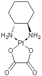
奥沙利铂是一种有机铂配合物,其中铂原子与1,2-二氨基环己烷和草酸根配体络合,草酸根配体被取代后生成活性奥沙利铂衍生物。这些衍生物形成DNA链间和链内交联,从而抑制DNA复制和转录。奥沙利铂是一种抗肿瘤药物,常与氟尿嘧啶和亚叶酸联合用于治疗转移性结直肠肿瘤。
另见:奥沙利铂(注释已移至)。 在已评估的新一代铂化合物中,近年来,以1,2-二氨基环己烷为载体配体的化合物(包括奥沙利铂)受到了广泛关注。分子生物学研究和美国国家癌症研究所的体外细胞毒性筛选表明,二氨基环己烷铂类药物(如奥沙利铂)属于一个独特的细胞毒性药物家族,与顺铂和卡铂不同,具有特定的细胞内靶点、作用机制和/或耐药机制。在I期临床试验中,奥沙利铂的剂量限制性毒性表现为短暂的急性感觉异常和累积性远端神经毒性,这些毒性在停药后数月内可逆。此外,在推荐剂量(130 mg/m²,每三周一次;或85 mg/m²,每两周一次,静脉输注两小时)下,奥沙利铂未显示任何听觉、肾脏和血液学方面的剂量限制性毒性。目前已在数百例晚期结直肠癌(ACRC)患者中开展了奥沙利铂抗肿瘤活性的II期临床试验。单药治疗在结直肠癌(ACRC)患者中的客观缓解率(ORR)总体而言,在既往接受过5-氟尿嘧啶(5-FU)治疗/难治的ACRC患者中为10%,在既往未接受过治疗的ACRC患者中为20%。临床前研究表明,奥沙利铂与胸苷酸合成酶抑制剂、顺铂/卡铂以及拓扑异构酶I抑制剂具有协同细胞毒性作用,且无血液学剂量限制性毒性,因此奥沙利铂成为联合用药的理想选择。在既往接受过5-FU治疗/难治的ACRC患者中,奥沙利铂联合5-FU和亚叶酸钙的II期临床试验显示,总缓解率在21%至58%之间,中位生存期为12至17个月。在既往未接受过治疗的ACRC患者中,奥沙利铂联合5-FU和亚叶酸钙的治疗方案显示,缓解率在34%至67%之间,中位生存期为15至19个月。两项共纳入 620 例既往未接受治疗的 ACRC 患者的随机对照试验,比较了 5-氟尿嘧啶 (5-FU) 联合亚叶酸钙与 5-FU 联合亚叶酸钙联合奥沙利铂的疗效。结果显示,第一项试验中奥沙利铂组的总缓解率为 34%,而 5-FU 联合亚叶酸钙组为 12%;第二项试验中,两组的总缓解率分别为 51.2% 和 22.6%。这些具有统计学意义的差异也体现在奥沙利铂组的疾病进展时间优势上(分别为 8.7 个月 vs. 6.1 个月和 8.7 个月 vs. 6.1 个月)。在接受奥沙利铂联合 5-FU 联合亚叶酸钙治疗的晚期结直肠癌患者中,已报道了少量但持续存在的组织学完全缓解病例,并且熟悉该联合方案的肿瘤科医生越来越多地开展二次转移灶切除术。基于临床前和临床报告显示奥沙利铂与多种抗癌药物(包括顺铂、伊立替康、拓扑替康和紫杉醇)之间存在叠加或协同作用,针对奥沙利铂单药治疗已显示出抗肿瘤活性的肿瘤类型(例如卵巢癌、非小细胞肺癌、乳腺癌和非霍奇金淋巴瘤),已开展或正在进行奥沙利铂与其他化合物联合用药的临床试验。其在卵巢癌中的单药和联合治疗数据证实其与顺铂/卡铂无交叉耐药性。尽管奥沙利铂在肿瘤内科中的作用尚未完全明确,但它似乎是一种重要的新型抗癌药物。[1]我们此前已证实顺铂和卡铂对人黑色素瘤细胞系具有体外活性。这两种药物是我们科室用于治疗晚期转移性黑色素瘤化疗方案的重要组成部分。本文报道了奥沙利铂与顺铂和卡铂相比,对人黑色素瘤细胞系的体外活性。结果发现,奥沙利铂对C32和G361细胞系均具有活性,其IC50值分别为49.48和9.07 μM(暴露1小时)、9.47和1.30 μM(暴露4小时)以及0.98和0.14 μM(暴露24小时)。在该体外系统中,奥沙利铂的细胞毒活性显著优于卡铂。随着暴露时间的延长,其活性逐渐接近顺铂。事实上,在24小时暴露后,奥沙利铂对G361细胞系的活性显著高于顺铂(p=0.0343)。奥沙利铂值得在临床上进行评估,既可作为单药治疗,也可与其他抗黑色素瘤药物联合治疗。[2] 本研究对新型非肾毒性铂类似物奥沙利铂进行了体外细胞毒性、蛋白结合、铂从全血到红细胞的分配、铂从红细胞到血浆的交换以及在血浆中的体外生物转化等方面的研究。细胞毒性研究针对一系列源自卵巢癌(A2780、A2780/cp)、膀胱癌(TCCSUP、RT4)、结肠癌(HT-29)、黑色素瘤(SKMEL-2、HTB144)和神经胶质瘤(U373MG 和 U87MG)的人类肿瘤细胞系进行。五种铂配合物的相对效力为:奥沙利铂 = 四铂 > 顺铂 > 异丙铂 > 卡铂。奥沙利铂对HT-29细胞有效,且与顺铂对A2780/cp细胞的交叉耐药性极低。两种膀胱癌细胞系、两种黑色素瘤细胞系以及两种胶质母细胞瘤细胞系中的一种均对奥沙利铂和四铂耐药。奥沙利铂-四铂和顺铂-卡铂两组药物的细胞毒性谱经Spearman秩相关检验显示具有统计学意义的相关性。奥沙利铂的蛋白结合率与顺铂和四铂相似;奥沙利铂(5、10或20微克/毫升)中85-88%的铂在给药后5小时内与血浆蛋白结合,平均半衰期为1.71±0.06小时。当奥沙利铂与全血(5、10 和 20 微克/毫升)孵育时,红细胞在 2 小时内吸收了总铂量的 37.1 ± 2.1%(最大吸收量),且这些铂无法交换到血浆中。因此,红细胞结合的铂不作为药物储存库。在血浆中,奥沙利铂在 0.5 小时时未发生变化,但在 1 小时时,血浆中 30% 的总铂出现在一个峰中,该峰的保留时间与四铂的主要生物转化产物 (反式-1,2-二氨基环己烷)二氯铂(II) 的保留时间相同。在 2 小时时,检测到了 (反式-1,2-二氨基环己烷)二氯铂(II) 和其他三个含铂峰,但未检测到未发生变化的奥沙利铂。所有铂在 4 小时时均在溶剂前沿附近的一个峰中洗脱出来。奥沙利铂和四铂细胞毒性的显著相似性可能是由于在组织培养基中形成了(反式-1,2-二氨基环己烷)二氯铂(II)所致。[3] 目的:铂类化疗药物奥沙利铂具有广泛的抗肿瘤活性。迄今为止,尚无关于奥沙利铂对肝细胞癌(HCC)细胞作用的详细数据。本文研究了奥沙利铂在体外和体内对HCCLM3和Hep3B细胞的抗增殖作用。研究方法:采用MTT法评估细胞活力,采用流式细胞术和透射电镜评估细胞凋亡。采用微阵列分析、定量逆转录-PCR和蛋白质印迹法评估HCCLM3细胞中凋亡相关蛋白的表达。本研究还利用异种移植瘤模型在体内研究了奥沙利铂的作用。结果显示,奥沙利铂抑制了HCCLM3和Hep3B细胞的生长。通过流式细胞术、透射电镜和末端脱氧核苷酸转移酶介导的dUTP缺口末端标记法(TUNEL)分析,我们发现细胞凋亡是奥沙利铂抑制肿瘤进展的主要机制。微阵列分析、定量逆转录PCR和蛋白质印迹分析进一步证实,在奥沙利铂诱导的细胞凋亡过程中,抗凋亡蛋白Bcl-2和Bcl-xL表达下调,而促凋亡蛋白Bax表达上调。结论:奥沙利铂对肝癌细胞的抗增殖作用是由于其诱导细胞凋亡所致。因此,奥沙利铂可能是一种治疗肝细胞癌的有效药物,其应用值得进一步深入研究。[4] 一种新型铂配合物——草酸铂(l-OHP)——在实验测试中,相同金属剂量下其疗效与顺铂相当,且在较低金属剂量下疗效优于卡铂;其对睾丸和卵巢等人类肿瘤的疗效与其他铂类似物相当,对黑色素瘤和乳腺癌的疗效更佳;且无肾毒性、心脏毒性或致突变性,血液毒性和神经毒性也极低。本文对其进行了描述,并与上述铂配合物进行了比较。草酸铂与5-氟尿嘧啶(5-Fu)联合使用,可使结直肠癌患者获得大量缓解,并已治愈部分无法手术的胃癌。与卡铂联合使用,在携带L1210突变的小鼠中取得了很高的治愈率,这是其他任何两种药物组合都无法达到的。[5] 结肠癌是世界上最常见的恶性肿瘤之一。奥沙利铂是一种第三代铂类化合物,广泛用于结肠癌的临床化疗。尽管近年来人们对奥沙利铂的抗肿瘤机制进行了研究,但与细胞对该化合物的反应相关的蛋白质组学变化仍知之甚少。在本研究中,我们对三种结肠癌细胞系(HT29、SW620和LoVo)进行了比较蛋白质组学分析,以研究奥沙利铂治疗后蛋白质表达水平的整体变化。二维凝胶电泳结合MALDI-TOF/TOF质谱分析显示,奥沙利铂处理后,三种细胞系(HT29、SW620和LoVo)中分别有57、48和53种差异表达蛋白。其中,21种蛋白在所有三种细胞系中均有表达。这些重叠蛋白参与多种细胞过程,例如细胞凋亡、信号转导、转录和翻译、细胞结构组织以及代谢。此外,免疫印迹实验证实了ezrin (EZRI)、热休克蛋白β-1 (HSPB1)、翻译调控肿瘤蛋白 (TCTP) 和细胞分裂控制蛋白2同源物 (CDC2) 的表达水平。这是首次对奥沙利铂处理的结肠癌细胞进行直接蛋白质组学分析。我们发现的几种有趣的蛋白值得进一步研究,因为它们可能在奥沙利铂的抗肿瘤作用中发挥重要作用。[8] |
| 分子式 |
C8H14N2O4PT
|
|
|---|---|---|
| 分子量 |
397.29
|
|
| 精确质量 |
397.06
|
|
| 元素分析 |
C, 24.19; H, 3.55; N, 7.05; O, 16.11; Pt, 49.10
|
|
| CAS号 |
61825-94-3
|
|
| 相关CAS号 |
|
|
| PubChem CID |
9887053
|
|
| 外观&性状 |
White solid powder
|
|
| 沸点 |
193.6ºC at 760 mmHg
|
|
| LogP |
0.614
|
|
| tPSA |
104.64
|
|
| 氢键供体(HBD)数目 |
4
|
|
| 氢键受体(HBA)数目 |
6
|
|
| 可旋转键数目(RBC) |
0
|
|
| 重原子数目 |
15
|
|
| 分子复杂度/Complexity |
191
|
|
| 定义原子立体中心数目 |
2
|
|
| SMILES |
[Pt+2].[O-]C(C(=O)[O-])=O.N([H])([H])[C@]1([H])C([H])([H])C([H])([H])C([H])([H])C([H])([H])[C@@]1([H])N([H])[H]
|
|
| InChi Key |
DRMCATBEKSVAPL-BNTLRKBRSA-N
|
|
| InChi Code |
InChI=1S/C6H12N2.C2H2O4.Pt/c7-5-3-1-2-4-6(5)8;3-1(4)2(5)6;/h5-8H,1-4H2;(H,3,4)(H,5,6);/q-2;;+2/t5-,6-;;/m1../s1
|
|
| 化学名 |
[(1R,2R)-2-azanidylcyclohexyl]azanide;oxalic acid;platinum(2+)
|
|
| 别名 |
L-OHP; diaminocyclohexane oxalatoplatinum; oxalatoplatin; oxalatoplatinum; DTXSID0036760; Oxalato(trans-l-1,2-cyclohexanediamine)platinum(II); cis-oxalato-trans-l-1,2-diaminocyclohexaneplatinum(II); US brand name: Eloxatin Foreign brand names: Dacotin; Dacplat; Eloxatine; Abbreviations: 1OHP; LOHP; Code names: JM83; RP54780; SR96669.
|
|
| HS Tariff Code |
2934.99.9001
|
|
| 存储方式 |
Powder -20°C 3 years 4°C 2 years In solvent -80°C 6 months -20°C 1 month 注意: 本产品在运输和储存过程中需避光。 |
|
| 运输条件 |
Room temperature (This product is stable at ambient temperature for a few days during ordinary shipping and time spent in Customs)
|
| 溶解度 (体外实验) |
|
|||
|---|---|---|---|---|
| 溶解度 (体内实验) |
Note: Oxaliplatin一般不用DMSO溶解,因为铂类药物在DMSO中易失活!另外, Oxaliplatin在溶液中不稳定,请现配现用!
配方 1 中的溶解度: 1.92 mg/mL (4.83 mM) in PBS (这些助溶剂从左到右依次添加,逐一添加), 澄清溶液; 超声助溶。 (<60°C). 配方 2 中的溶解度: 3.33 mg/mL (8.38 mM) in 5% w/v Glucose Solution (这些助溶剂从左到右依次添加,逐一添加), 澄清溶液; 超声助溶. 请根据您的实验动物和给药方式选择适当的溶解配方/方案: 1、请先配制澄清的储备液(如:用DMSO配置50 或 100 mg/mL母液(储备液)); 2、取适量母液,按从左到右的顺序依次添加助溶剂,澄清后再加入下一助溶剂。以 下列配方为例说明 (注意此配方只用于说明,并不一定代表此产品 的实际溶解配方): 10% DMSO → 40% PEG300 → 5% Tween-80 → 45% ddH2O (或 saline); 假设最终工作液的体积为 1 mL, 浓度为5 mg/mL: 取 100 μL 50 mg/mL 的澄清 DMSO 储备液加到 400 μL PEG300 中,混合均匀/澄清;向上述体系中加入50 μL Tween-80,混合均匀/澄清;然后继续加入450 μL ddH2O (或 saline)定容至 1 mL; 3、溶剂前显示的百分比是指该溶剂在最终溶液/工作液中的体积所占比例; 4、 如产品在配制过程中出现沉淀/析出,可通过加热(≤50℃)或超声的方式助溶; 5、为保证最佳实验结果,工作液请现配现用! 6、如不确定怎么将母液配置成体内动物实验的工作液,请查看说明书或联系我们; 7、 以上所有助溶剂都可在 Invivochem.cn网站购买。 |
| 制备储备液 | 1 mg | 5 mg | 10 mg | |
| 1 mM | 2.5171 mL | 12.5853 mL | 25.1705 mL | |
| 5 mM | 0.5034 mL | 2.5171 mL | 5.0341 mL | |
| 10 mM | 0.2517 mL | 1.2585 mL | 2.5171 mL |
1、根据实验需要选择合适的溶剂配制储备液 (母液):对于大多数产品,InvivoChem推荐用DMSO配置母液 (比如:5、10、20mM或者10、20、50 mg/mL浓度),个别水溶性高的产品可直接溶于水。产品在DMSO 、水或其他溶剂中的具体溶解度详见上”溶解度 (体外)”部分;
2、如果您找不到您想要的溶解度信息,或者很难将产品溶解在溶液中,请联系我们;
3、建议使用下列计算器进行相关计算(摩尔浓度计算器、稀释计算器、分子量计算器、重组计算器等);
4、母液配好之后,将其分装到常规用量,并储存在-20°C或-80°C,尽量减少反复冻融循环。
计算结果:
工作液浓度: mg/mL;
DMSO母液配制方法: mg 药物溶于 μL DMSO溶液(母液浓度 mg/mL)。如该浓度超过该批次药物DMSO溶解度,请首先与我们联系。
体内配方配制方法:取 μL DMSO母液,加入 μL PEG300,混匀澄清后加入μL Tween 80,混匀澄清后加入 μL ddH2O,混匀澄清。
(1) 请确保溶液澄清之后,再加入下一种溶剂 (助溶剂) 。可利用涡旋、超声或水浴加热等方法助溶;
(2) 一定要按顺序加入溶剂 (助溶剂) 。
Study of Pembrolizumab (MK-3475) Versus Chemotherapy in Chinese Participants With Stage IV Colorectal Cancer (MK-3475-C66)
CTID: NCT05239741
Phase: Phase 3 Status: Recruiting
Date: 2024-12-02
|
|---|
InvivoChem的所有产品仅用于作科学研究,不面向患者销售
Copyright 2020 InvivoChem LLC | All Rights Reserved 粤ICP备20063088号-1


 COA
COA
 Monohydrate(1)")
 463611831
