| 规格 | 价格 | 库存 | 数量 |
|---|---|---|---|
| 1mg |
|
||
| 5mg |
|
||
| 10mg |
|
||
| 25mg |
|
||
| 50mg |
|
||
| 100mg |
|
||
| 250mg |
|
||
| 500mg |
|
||
| Other Sizes |
|

| 靶点 |
HDAC1 ( Ki = 7 nM ); HDAC3/SMRT ( Ki = 8.2 nM ); HDAC6 ( Ki = 17 nM ); HDAC2 ( Ki = 19 nM ); HDAC10 ( Ki = 24 nM ); HDAC8 ( Ki = 280 nM ); MBLAC2 ( Ki < 10 nM )
|
|---|---|
| 体外研究 (In Vitro) |
体外活性:PCI-24781 对多种肿瘤细胞系表现出有效的抗肿瘤活性,GI50 范围为 0.15 μM 至 3.09 μM。 PCI-24781 对 HUVEC 内皮细胞也具有抗增殖作用,GI50 为 0.43 μM。 PCI-24781 治疗会导致 HCT116 或 DLD-1 细胞中乙酰化组蛋白和乙酰化微管蛋白呈剂量依赖性积累,诱导 p21 表达,并导致 PARP 裂解和 γH2AX 积累。 PCI-24781 对 HDAC 酶的抑制会导致与 HR 特别相关的基因(包括 RAD51)的转录显着减少。与 HR 的抑制一致,PCI-24781 处理会导致转染的 CHO 细胞中 I-SceI 诱导的染色体断裂的同源定向修复能力下降。 PCI-24781 诱导软组织肉瘤 (STS) 细胞 S 期耗竭、G2 细胞周期停滞和细胞凋亡。 PCI-24781 在 STS 细胞中诱导 Rad51 转录抑制,这可能是通过增强 E2F1 与 Rad51 近端启动子的结合介导的。 PCI-24781 在霍奇金淋巴瘤和非霍奇金淋巴瘤细胞系中通过 NF-κB 机制诱导 caspase 和活性氧依赖性细胞凋亡。激酶测定:使用连续胰蛋白酶偶联测定来测量 HDAC 活性。对于抑制剂表征,使用 96 孔测定板在 100 μL 反应体积中进行测量。对于每种同工酶,反应缓冲液中的 HDAC 蛋白 [50 mM HEPES、100 mM KCl、0.001% Tween 20、5% DMSO (pH 7.4),补充浓度为 0% (HDAC1)、0.01% (HDAC2) 的牛血清白蛋白、3、8 和 10) 或 0.05% (HDAC6)] 与不同浓度的 PCI-24781 混合,并孵育 15 分钟。添加胰蛋白酶至终浓度为 50 nM,添加乙酰基-Gly-Ala-(N-乙酰基-Lys)-AMC 至终浓度为 25 μM(HDAC1、3 和 6)、50 μM(HDAC2 和10) 或 100 μM (HDAC8) 来启动反应。阴性对照反应在不存在 PCI-24781 的情况下进行,重复八次。在荧光板读数器中监测反应。 30 分钟的滞后时间后,使用 355 nm 的激发波长和 460 nm 的检测波长在 30 分钟的时间范围内测量荧光。荧光随时间的增加被用作反应速率的量度。抑制常数 Ki(app) 使用程序 BatchKi 获得。细胞测定:将细胞培养至少两次倍增,并在 PCI-24781 暴露结束时使用阿拉玛蓝荧光细胞增殖测定监测生长。 PCI-24781 在 96 孔板的一式三份孔中以 9 个浓度进行测定,使用范围为 0.0015 μM 至 10 μM 的半对数间隔。每孔中的最终 DMSO 浓度为 0.15%。使用四参数逻辑方程通过非线性回归估计抑制细胞生长 50% (GI50) 和 95% 置信区间所需的浓度。
|
| 体内研究 (In Vivo) |
PCI-24781 以 200 mg/kg 的剂量每天一次(qod)给药,可显着抑制小鼠体内 HCT116 和 DLD-1 异种移植物的生长,分别达 69% 和 59%。在 HCT116 中以 20 mg/kg、40 mg/kg、80 mg/kg 或 160 mg/kg 的剂量每天一次给予 PCI-24781,连续 4 天,然后每周 3 天不治疗(每周 4 次)模型分别抑制肿瘤生长 48%、57%、82.2% 或 80.0%。
Abexinostat (PCI24781; CRA024781)的抗肿瘤活性和乙酰化诱导作用 [1] 为了评估CRA-024781的体内抗肿瘤活性,使用不同剂量和时间表对携带人结肠肿瘤异种移植物的小鼠静脉注射该化合物。在之前的HCT116异种移植物剂量计划研究中,静脉注射CRA-024781确定了两种具有良好治疗指标的方案:(a)每隔一天一次(q.o.d)或(b)连续4天每天一次,然后每周3天不治疗(q.d×每周4次;剂量计划数据未显示)。当根据第一种方案在HCT116或DLD-1异种移植物中以200mg/kg i.v.qo.d.的剂量评估CRA-024781时,观察到对肿瘤生长的统计学显著抑制(图4A和B)。HCT116和DLD-1模型的肿瘤生长抑制率分别为69%(P<0.000001)和59%(P<0.01)。尽管在DLD-1研究中观察到相对于溶媒对照组体重减轻了13%,但在HCT116研究中没有观察到体重减轻,这表明实验之间存在一些耐受性差异,200mg/kg接近每日一次给药的最大耐受剂量(数据未显示)。然后使用第二种方案(q.d×每周4次)对CRA-024781进行多剂量评估。在HCT116模型中,20、40、80和160 mg/kg的肿瘤生长抑制率分别为48%(P<0.05)、57%(P<0.01)、82.2%(P<0.0001)和80.0%(P<0.00001)。到研究结束时,这些剂量均未导致动物相对于载体的体重减轻(数据未显示)。在DLD-1模型中,CRA-024781每周4次,每天一次,没有显著抑制肿瘤生长,尽管在最高剂量160 mg/kg时观察到轻微的抑制趋势,显示出43%的抑制率(P=0.09),没有相关的体重减轻(数据未显示)。为了确定研究中获得的CRA-024781的血浆浓度是否足以在体内抑制HDAC酶,对外周血细胞进行了离体检查。如图5所示,给药后2小时和6小时,每个治疗组的微管蛋白乙酰化都有可测量的增加。总之,CRA-024781对HCT116和DLD-1人结直肠肿瘤异种移植物均表现出统计学上显著的抗肿瘤活性,尽管总体而言,在HCT116异种移植物模型中抗肿瘤活性似乎更为明显。 将Abexinostat(PCI24781;CRA024781)与化疗联合使用可在体内产生优异的抗STS作用。接下来,我们评估了PCI-24781单独和联合化疗在体内的影响。在四项武装研究中使用了肌肉内生长的SKLMS1和皮下生长的HT1080或作为实验性肺转移瘤,比较了低剂量阿霉素、顺铂、PCI-24781或PCI-24781联合化疗对SCID小鼠中人类STS局部和转移生长的影响。肿瘤形成后开始治疗(100mm3)。同样,在实验性肺转移模型中,只有在通过生物发光鉴定出已建立的转移后才开始治疗[3] 线性混合模型用于评估治疗组随时间的肿瘤生长(以对数转换的肿瘤体积表示),线性回归模型用于评估各治疗组的肿瘤重量。单独使用低剂量阿霉素治疗对SKLMS1异种移植物的生长没有显著影响(图3A)Abexinostat(PCI24781;CRA024781)单独诱导显著的肿瘤生长抑制(PCI-24781治疗的小鼠与对照组未治疗的肿瘤的肿瘤体积与时间的斜率;P=0.001)。最重要的是,与对照组、单独使用阿霉素或单独使用PCI-24781治疗肿瘤组相比,联合PCI-24781和低剂量阿霉素具有显著的抑制作用(P<0.0001)。研究结束时,对照组的平均肿瘤重量为1.62 g±0.47,阿霉素为1.34 g±0.43,PCI-24781为1.11 g±0.26,联合用药为0.64 g±0.24(图3B)。与对照组相比,单独使用PCI-24781显著降低了肿瘤重量(P=0.026)。此外,与其他治疗相比,PCI-24781联合化疗显著减轻了肿瘤重量(对照组、阿霉素和PCI-24781分别为P<0.0001、=0.0023和=0.047)。[3] 不同治疗组肿瘤的H&E染色显示,在阿贝诺司他(PCI24781;CRA024781)和联合治疗组中,肿瘤明显坏死(图3C);对活肿瘤切片进行免疫组织化学评估,以了解不同疗法对STS细胞增殖(PCNA)和凋亡(TUNEL)的影响。PCNA和TUNEL阳性染色核的平均值显示,对照组为88±2和20±2.4,阿霉素为85±1.2和16±1.4,PCI-24781为71±2.3和19±1.1,联合治疗为57±4.6和61±18.4,表明联合治疗具有最强的抗增殖和凋亡诱导作用(P<0.05)。还对切片进行了CD31染色,以检查对肿瘤相关血管系统的不同治疗效果。CD31计数没有显著差异;然而,在联合治疗组中,可以观察到大而通畅的血管数量显著减少[3]。 同样,在HT1080异种移植物中测试了Abexinostat (PCI24781; CRA024781)、顺铂及其组合的效果(图4)。与顺铂或PCI-24781治疗组和对照组小鼠相比,没有发现随时间推移的显著肿瘤生长差异。然而,联合治疗观察到肿瘤生长明显减少(与其他治疗组相比,P<0.001;图4A)。研究结束时,对照组的平均肿瘤重量为1.23 g±0.15,顺铂组为1.01 g±0.39,PCI-24781组为1.09 g±0.54,联合用药组为0.36 g±0.22(图4B)。单独使用PCI-24781或顺铂治疗没有明显减轻肿瘤重量,而联合治疗则观察到明显的肿瘤重量减轻(分别与对照组、顺铂组和PCI-24781组相比,P=0.0003、=0.0038和=0.0011)。免疫组织化学结果与上述SKLMS1治疗的肿瘤相似,显示PCI-24781和联合组坏死增加(图4C)。联合治疗组增殖减少和凋亡增强最为明显(对照组的平均PCNA和TUNEL阳性染色核分别为83±7.1和18±0.3,顺铂组为62±2.8和17±2.5,PCI-24781组为50±2.8和21±5.9,联合治疗组为38±14.1和33±8.6;P<0.05)。各组间CD31阳性率没有显著差异,而联合组大血管减少[3]。 最后,评估了各种疗法对STS肺转移的影响。采用实验性纤维肉瘤肺转移模型Abexinostat(PCI24781;CRA024781)与阿霉素或顺铂联合使用进行了测试(图4D)。随后对小鼠进行生物发光;代表性的连续图像如图4D所示,显示联合治疗小鼠的生物发光减少。H&E染色显示,在对照和化疗治疗的肿瘤中,大的转移性沉积物取代了许多肺实质;在PCI-24781组中观察到较小的病变,在联合治疗小鼠中观察到微小的显微镜病变。肺转移重量是通过从研究结束时的实际肺重量中减去估计的平均正常小鼠肺重量来计算的。对照组的平均肺转移重量为0.55 g±0.14,顺铂为0.62 g±0.26,阿霉素为0.48 g±0.33,PCI-24781组为0.33 g±0.20,PCI-24781-顺铂为0.15 g±0.19,PCI-2488-阿霉素为0.13 g±0.14。在PCI-24781治疗的小鼠中观察到转移负荷减少的趋势,但没有达到统计学意义,而与对照组或顺铂治疗组相比,PCI-24781联合任何一种化疗都能显著减轻肺转移的重量(P<0.05)。综上所述,这些数据表明,尽管PCI-24781在体外表现出显著的抗STS作用,但在体内作为单一疗法仅具有微弱的疗效。然而,将PCI-24781与低剂量常规化疗联合使用可在体内显著抑制STS肿瘤和转移生长,这是对潜在临床应用的观察[3]。 |
| 酶活实验 |
连续运行的胰蛋白酶偶联测定用于测量 HDAC 活性。使用 96 孔测定板在 100 μL 反应体积中进行测量,以进行抑制剂表征。 HDAC 蛋白与每种同工酶不同浓度的 PCI-24781 结合,并孵育 15 分钟。反应缓冲液为 50 mM HEPES、100 mM KCl、0.001% Tween 20、5% DMSO (pH 7.4),补充浓度为 0% (HDAC1)、0.01% (HDAC2、3、8 和 10) 的牛血清白蛋白),或 0.05% (HDAC6)。通过添加终浓度为 25 μM(HDAC1、3 和 6)、50 μM(HDAC2 和 10)或 100 μM(HDAC8)的乙酰基-Gly-Ala-(N-乙酰基-Lys)-AMC 开始反应。 )。添加胰蛋白酶至终浓度为 50 nM。一式八份,阴性对照反应在没有 PCI-24781 的情况下进行。荧光板读取器用于跟踪反应。在 30 分钟的滞后时间后,使用 355 nm 的激发波长和 460 nm 的检测波长在 30 分钟内测量荧光。使用荧光随时间的增加来计算反应速率。程序BatchKi用于获得抑制常数Ki(app)。
HDAC活动[1] 使用之前详细描述的连续胰蛋白酶偶联测定法测量HDAC活性(11)。为了表征抑制剂,使用96孔测定板在100μL的反应体积内进行了测量。对于每种同工酶,将反应缓冲液中的HDAC蛋白[50 mmol/L HEPES、100 mmol/L KCl、0.001%吐温20、5%DMSO(pH 7.4),补充有浓度为0%(HDAC1)、0.01%(HDAC2、3、8和10)或0.05%(HDAC6)的牛血清白蛋白]与不同浓度的抑制剂混合,并孵育15分钟。加入胰蛋白酶至终浓度为50 nmol/L,加入乙酰基Gly-Ala-(N-乙酰基-Lys)-AMC至终浓度25μmol/L(HDAC1、3和6)、50μmol/L(HADC2和10)或100μmol/L(HDAC8)以引发反应。阴性对照反应在没有抑制剂的情况下进行,重复8次。在荧光板读数器中监测反应。在30分钟的延迟时间后,使用355nm的激发波长和460nm的检测波长在30分钟内测量荧光。荧光随时间的增加被用作反应速率的度量。使用BatchKi程序获得抑制常数Ki(app)。 |
| 细胞实验 |
对 10 种肿瘤细胞系和 HUVEC 进行至少两次倍增培养后,使用阿拉玛蓝荧光细胞增殖测定法评估生长,以确定化合物暴露的终点。使用 0.0015 至 10 μmol/L 范围内的半对数间隔,在 96 孔板的一式三份孔中以九种浓度对该化合物进行测定。每孔的 DMSO 最终浓度为 0.15%。使用四参数逻辑方程通过非线性回归估计抑制细胞生长 50% 和 95% 置信区间所需的浓度[1]。
细胞增殖试验[1] 将10个肿瘤细胞系和HUVEC培养至少两倍,并在化合物暴露结束时使用如前所述的Alamar蓝荧光细胞增殖测定法监测生长。使用0.0015至10μmol/L的半对数间隔,在96孔板的三个孔中以9种浓度对化合物进行了测定。每个孔中的DMSO最终浓度为0.15%。使用四参数逻辑斯谛方程通过非线性回归估计抑制细胞生长50%(GI50%)和95%置信区间所需的浓度。 组蛋白和微管蛋白乙酰化、p21Cip1/WAF1积累、聚ADP核糖聚合酶切割和磷酸化组蛋白变体H2AX[2] 通过蛋白质印迹法检测了用Abexinostat (PCI24781; CRA024781)处理的细胞中乙酰化组蛋白、乙酰化微管蛋白、p21Cip1/WAF1、聚ADP核糖聚合酶(PARP)切割和磷酸化组蛋白变体H2AX(γH2AX)蛋白。肿瘤细胞和亚融合的HUVEC在浓度范围为0.01至10μmol/L的Abexinostat (PCI24781; CRA024781)存在下培养18小时。然后收集细胞并在含有蛋白酶抑制剂和磷酸酶抑制剂的裂解缓冲液中裂解。将裂解物溶解在SDS-PAGE还原样品缓冲液中,煮沸,然后在Novex Tris-甘氨酸凝胶中电泳。将凝胶印迹到硝化纤维上,用抗乙酰赖氨酸抗体检测乙酰化组蛋白、抗乙酰化微管蛋白抗体、抗p21Cip1/WAF1抗体、抗PARP抗体或抗γH2AX抗体。洗涤印迹,用适当的辣根过氧化物酶偶联的二抗孵育,并对印迹进行增强化学发光处理。 Annexin V染色法检测细胞凋亡。[2] 为了确定Abexinostat(PCI24781;CRA024781)和PARP抑制剂PJ34(EMD Biosciences)在HCT116细胞中的潜在协同作用,通过分析单独或联合使用特定剂量的药物96小时后膜联蛋白V-FITC(Biosource)的结合来评估细胞毒性。调整剂量,以保持两种药物在两种联合治疗中的比例恒定。使用FACSCalibur仪器(Becton Dickinson)定量细胞凋亡。CalcuSyn程序(Biosoft)用于根据参考文献29中所述计算的CI生成中值效应图。CI大于1表示拮抗作用,CI为1表示相加作用,CI小于1表示协同作用。为了确定HCT116细胞在24小时时的凋亡百分比,用0.2、0.5和1.0μM PCI-24781处理细胞,24小时后评估膜联蛋白V结合。 免疫荧光。[2] HCT116细胞在室载玻片上生长,用0.2μM的Abexinostat (PCI24781; CRA024781)处理24小时。然后将细胞暴露于10 Gy的辐射下,并在辐射后孵育1或16小时。然后将细胞固定在含有2%多聚甲醛、0.2%Triton X-100的PBS缓冲液中并透化。将细胞封闭在2%BSA的PBS中,用抗RAD51多克隆抗体探测,然后用AlexaFluor594偶联的二抗探测。染色后,使用带有DAPI的Vecta屏蔽安装介质 安装盖玻片,并使用荧光显微镜进行可视化。 TaqMan基因表达检测。[2] HCT116细胞用Abexinostat(PCI24781;CRA024781)处理不同时间,并使用RNeasy试剂盒 提取总RNA。使用核糖绿RNA定量试剂盒(分子探针)定量总RNA。根据制造商的说明,通过使用TaqMan主混合物 和50ng总RNA作为模板,建立一式三份的一步RT-PCR测定。重新测定每个反应孔中的RNA量,并用于正常化。 蛋白质印迹分析。[2] 用Abexinostat(PCI24781;CRA024781)处理细胞指定时间并裂解,使用BCA蛋白检测试剂盒 定量总蛋白。每条泳道中都装载了等量的蛋白质。用于阻断胱天蛋白酶切割的泛胱天蛋白酶抑制剂Q-VD-OPh购自MP Biomedicals。每种处理的总蛋白(30μg)在4-15%梯度SDS-PAGE凝胶 上溶解。将蛋白质转移到PVDF膜上,并使用与AlexaFluor680和IFdye800 偶联的适当一抗和二抗进行检测。使用Odyssey扫描仪 进行成像。 同源重组修复试验。[2] 如参考文献39所述,在DRAA8中国仓鼠卵巢细胞中测量同源重组修复活性。简而言之,DRAA8/CHO细胞包含一个无功能的GFP序列和一个整合到hprt基因座的内部GFP(iGFP)序列。突变的GFP序列包含一个18 bp的I-SceI识别位点,通过表达I-SceI-内切酶引入DSBs。通过使用核转染(Amaxa)转染I-SceI表达载体(3.5μg),并使用FACS通过测量GFP信号来定量使用下游iGFP重复序列诱导的DSB基因转化Abexinostat(PCI24781;CRA024781)转染后6小时以适当浓度加入,根据7-氨基放线菌素D(7-AAD)信号对活细胞进行门控,36小时后分析门控活细胞群中的GFP表达。 克隆生存试验。[2] 为了确定Abexinostat(PCI24781;CRA024781)处理对CHO-K1和NHEJ缺陷型Ku80突变XRS5细胞系集落形成的影响,将适当板密度的细胞铺在10cm的培养皿中并允许其附着。将细胞暴露于0.5、1.0或2.0μM浓度的药物中24小时,然后将细胞置于不含药物的新鲜生长培养基中,孵育7-10天。菌落用100%异丙醇固定,用1%结晶紫染色。对于每种情况,进行三次测定。在HCT116、NCI-H460和A549肺肿瘤细胞系中,为了确定PCI-24781治疗与辐射协同作用的适当时间,用1μM PCI-247811预处理培养物不同长度的时间,范围从2到24小时。将细胞暴露于0、2、4或6 Gy辐射(Gammacell 40,Atomic Energy of Canada,有限公司),并在7-12天后评估克隆生存率。根据每个剂量的镀覆效率,计算集落形成能力的百分比。 细胞增殖的测量。[3] 根据制造商的说明,细胞生长测定使用CellTiter96水性非放射性细胞增殖测定试剂盒。在HDACi(Abexinostat(PCI24781;CRA024781)/SAHA)治疗和阿霉素或顺铂单独或与HDACi联合治疗后48小时分析生长率;评估了几种给药顺序:用PCI-24781预处理24小时,然后加化疗24小时,反之,用化疗预处理,然后加PCI-24781,以及同时用这两种化合物治疗24小时。在490 nm波长下测量吸光度;处理过的细胞吸光度值以未处理细胞吸光度的百分比表示。 克隆试验。[3] STS细胞在培养皿中用DMSO(对照)和不同浓度的HDACi(Abexinostat(PCI24781;CRA024781)/SAHA)处理24小时。每孔重新接种100个细胞,然后在正常培养基中生长10天,然后用6%戊二醛、0.5%结晶紫溶液染色30分钟。从每个孔中倒出染色溶液,用去离子水洗涤细胞。计数保留染色溶液的单个菌落。 细胞周期分析。[3] STS细胞单层用Abexinostat(PCI24781;CRA024781)/SAHA处理不同时间。如所述进行碘化丙啶/荧光激活细胞分选(FACS)分析。 |
| 动物实验 |
将3×10⁶个肿瘤细胞(包括DLD-1和HCT116肿瘤细胞)皮下注射到雌性BALB/c裸鼠体内。当肿瘤体积平均达到-100 mm³时,开始使用阿贝西诺司他(CRA 024781)进行治疗[1]。为了评估阿贝西诺司他(CRA 024781)的抗肿瘤活性,将不同剂量和给药方案的药物静脉注射到人结肠肿瘤异种移植小鼠体内[1]。药代动力学分析[1]:阿贝西诺司他(PCI24781;CRA024781)溶于30% HP-环糊精水溶液中,并以10 mg/kg的剂量静脉注射到雌性BALB/c裸鼠(16-22 g)体内。在24小时内采集血样。使用锂肝素从每份血液样本中制备血浆,并采用液相色谱-串联质谱法测定阿贝西诺他(PCI24781;CRA024781)的含量。采用高效液相色谱-串联质谱法测定小鼠血浆样本中的CRA-024781含量。血浆样本采用乙腈(MeCN)进行蛋白质沉淀提取。使用Hypersil C-18色谱柱(50 × 2.1 mm)进行样品分离。在反相高效液相色谱中,以水/乙腈为流动相,流速为0.5 mL/min,采用线性梯度洗脱条件进行分离。样品经电喷雾电离后,通过多反应监测进行定量,记录分子离子到产物离子的转变:398 → 200 m/z。线性范围为 0.25–1,000 ng/mL,日间和日内变异系数 (%) 以及平均值与理论值的偏差 (%) 均在 ±15% 以内。定量下限为 0.25 ng/mL。药代动力学参数采用房室模型,使用 WinNonlin-Pro 4.1 版(Pharsight 公司,加利福尼亚州山景城)进行估算。药代动力学计算采用标称剂量和标称采集时间。
体内疗效研究 [1] 雌性 BALB/c nu/nu 小鼠在肿瘤植入前适应环境 3 至 5 天。所有小鼠均饲养于无菌半透明聚碳酸酯微隔离笼中,并保持无特定病原体 (SPF) 条件。将 HCT116 和 DLD-1 肿瘤细胞以每只小鼠 3 × 10⁶ 个细胞的密度皮下植入裸鼠体内。在开始治疗前,根据肿瘤体积对荷瘤小鼠进行随机分组。当平均肿瘤体积约为 100 mm³ 时,开始使用阿贝西诺他(Abexinostat,PCI24781;CRA024781)进行治疗。肿瘤体积的计算公式为:体积 = 0.5 × X² × Y,其中 X 为肿瘤宽度,Y 为肿瘤长度。所有研究中使用的溶剂均为 20% 的 HP-β-环糊精水溶液。给药方式为静脉注射,给药间隔为隔日一次(qod),或连续给药 4 天后停药 3 天,每周给药 4 次(qd × 4 周),具体给药方案见实验说明。肿瘤生长抑制率的计算公式为:100 × (1 − dT/dC),其中 dT 为治疗组自首次给药以来平均肿瘤体积的变化值,dC 为对照组自首次给药以来平均肿瘤体积的变化值。为了满足检验有效性的正态分布假设,对数转换后的数据进行单因素方差分析(ANOVA)进行统计分析,并使用 Dunnett 法校正 P 值以控制多重比较。对于单组比较,为了满足检验有效性的正态分布假设,对数转换后的数据进行 t 检验进行统计分析。 体内研究。[2] BALB/c 雌性裸鼠在接受 4.5 Gy(全身照射)24 小时后,将 3 × 10⁶ 个 HCT116 细胞与 Matrigel 以 1:1 的比例皮下植入右侧腹部。小鼠在植入后约 10 天或肿瘤体积达到至少 75–100 mm³ 时进入研究。共设置四种治疗方案,每组三只动物,每只动物接受 200 mg/kg 的剂量:(i)载体; (ii) 动物在研究结束前 4 小时接受单次口服给药 (1×);(iii) 动物在研究结束前 28 小时接受一次口服给药,并在 6 小时后接受第二次给药 (2×);(iv) 动物的给药方式与 2× 组相同,但额外在首次给药后 24 小时、研究结束前 4 小时,即第二天早上接受第三次给药 (3×)。动物被安乐死,提取肿瘤组织,并使用 BCA 蛋白测定试剂盒分离和定量总蛋白。每个肿瘤组织上样 30 μg 总蛋白,并进行蛋白质印迹分析以检测 RAD51、乙酰化微管蛋白和肌动蛋白的表达水平。 体内治疗研究。 [3] 台盼蓝染色证实了活的STS细胞(SKLMS1或HT1080细胞,1 × 10⁶/0.1 mL HBSS/只小鼠)被注射到6周龄雌性SCID小鼠(n = 40/实验)的侧腹部(SKLMS1肌注,HT1080皮下注射),每周测量两次生长情况;将HT1080GL细胞(稳定表达绿色荧光蛋白/荧光素酶)尾静脉注射,导致实验性肺转移,可通过生物发光进行追踪。当平均肿瘤体积达到100 mm³时,将小鼠随机分为四组(每组7-8只小鼠):(a)对照组(仅注射载体);(b)阿霉素/顺铂组(分别为1.2 mg/kg/每两周一次和2 mg/kg/每两周一次,腹腔注射); (c) Abexinostat (PCI24781; CRA024781)(50 mg/kg/d × 5 d/周,腹腔注射);以及 (d) Abexinostat (PCI24781; CRA024781)联合化疗(所有病例均在化疗前24小时开始使用PCI-24781)。PCI-24781的剂量和治疗方案均按照公司建议确定。肺转移治疗采用了类似的实验设计,当生物发光显示已形成肺转移时开始治疗;该研究包含六个组,以便评估Abexinostat (PCI24781; CRA024781)联合两种化疗药物的疗效。监测小鼠的肿瘤大小和体重,并在对照组肿瘤平均最大径达到1.5 cm或生物发光提示对照组肺部肿瘤负荷显著时处死小鼠。肿瘤被切除、称重后,冷冻或用福尔马林固定,并进行石蜡包埋,用于免疫组织化学研究。同样,肺组织也被切除、称重,并用福尔马林固定。 |
| 药代性质 (ADME/PK) |
体内药代动力学[1]
体外实验已证实Abexinostat (PCI24781; CRA024781)具有HDAC抑制和抗肿瘤特性,因此我们评估了该化合物的药代动力学,以评价其暴露量,从而为后续在疗效模型中进行测试奠定基础。我们向雌性小鼠静脉注射CRA-024781,并监测其血浆浓度随时间的变化(图3)。根据这些数据,计算得出清除率为297 mL/min/kg,中心室分布容积为3,750 mL/kg,稳态分布容积为9,230 mL/kg,主要血浆半衰期为6.7分钟(曲线下面积为73%),平均滞留时间为31分钟。这些数据表明,静脉注射CRA-024781后,其体内暴露量足以用于研究体内HDAC抑制剂的生物学效应。 CRA-024781的体内药代动力学特征表明,其药物暴露量足以在体内抑制HDAC。10 mg/kg的静脉注射剂量在15分钟内使血浆浓度高于测得的HCT116肿瘤细胞的细胞GI50%。然而,基于GI50%或机制生物标志物(如蛋白质乙酰化等)的出现来预测体内药物暴露量(曲线下面积)是否足以达到疗效,是难以预先确定的。因为这些数值是基于体外肿瘤细胞持续暴露于药物的情况,而体内生长的肿瘤则按照特定的给药方案(例如,每日一次,连续4天,持续16天)暴露于不同浓度的化合物中,这些浓度取决于药物在血浆中的药代动力学和蛋白结合率。此外,血浆中的药物浓度并不一定反映分布到肿瘤的药物浓度。事实上,相对较高的稳态分布容积(9230 mL/kg)表明CRA-024781可能很容易分布到组织中。然而,达到最大疗效所需的剂量(≥80 mg/kg)>10 mg/kg,这可能是因为每次给药后,HDAC 酶必须被抑制更长时间或更大程度才能达到抗肿瘤疗效。 |
| 毒性/毒理 (Toxicokinetics/TK) |
在之前的实验中,连续3天口服200 mg/kg剂量并未引起毒性反应(以体重减轻为指标)。选择200 mg/kg剂量给小鼠,也是因为该剂量与正在进行的临床试验中口服2.0 mg/kg剂量后预期的血浆浓度-时间曲线下面积相似。[2]
|
| 参考文献 |
|
| 其他信息 |
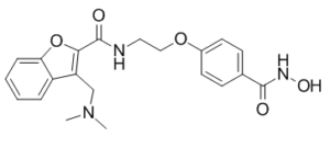
3-[(二甲氨基)甲基]-N-[2-[4-[(羟氨基)-氧甲基]苯氧基]乙基]-2-苯并呋喃甲酰胺属于苯并呋喃类化合物。
阿贝西诺司他已用于研究肉瘤、淋巴瘤、白血病、淋巴细胞性疾病和霍奇金淋巴瘤等多种疾病的治疗试验。它是一种新型的广谱羟肟酸类组蛋白去乙酰化酶 (HDAC) 抑制剂,具有潜在的抗肿瘤活性。 阿贝西诺司他是一种口服生物利用度高的羟肟酸类泛组蛋白去乙酰化酶 (HDAC) 抑制剂,具有潜在的抗肿瘤和放射增敏活性。给药后,阿贝西诺司他抑制 HDAC,导致高度乙酰化组蛋白的积累,进而诱导染色质重塑;选择性转录抑癌基因;阿贝西诺他通过抑制肿瘤抑制蛋白介导的肿瘤细胞分裂和诱导肿瘤细胞凋亡发挥作用。此外,阿贝西诺他还能降低DNA修复蛋白RAD51的表达,从而减少RAD51蛋白,阻止DNA双链断裂的修复,并增加肿瘤细胞对DNA损伤剂的敏感性。HDAC在多种肿瘤类型中表达上调,是一种负责染色质组蛋白去乙酰化的酶。 作用机制 阿贝西诺他是一种新型组蛋白去乙酰化酶(HDAC)抑制剂。HDAC抑制剂靶向HDAC酶,抑制癌细胞增殖并诱导癌细胞死亡(凋亡)。组蛋白去乙酰化是由一类相关的HDAC酶完成的。抑制这些酶会导致染色质结构和基因表达模式发生改变,从而抑制癌细胞增殖并诱导细胞凋亡。 CRA-024781 是一种新型广谱羟肟酸类组蛋白去乙酰化酶 (HDAC) 抑制剂,在体外和体内临床前研究中均显示出抗肿瘤活性,目前正在进行癌症 I 期临床试验。CRA-024781 对纯化的重组 HDAC1 的抑制常数 (Ki) 为 0.007 μmol/L,并且对其他 HDAC 同工酶 HDAC2、HDAC3/SMRT、HDAC6、HDAC8 和 HDAC10 的抑制作用也达到纳摩尔级。用 CRA-024781 处理体外培养的肿瘤细胞系可导致乙酰化组蛋白和乙酰化微管蛋白的积累,从而抑制肿瘤细胞生长并诱导细胞凋亡。对携带HCT116或DLD-1结肠肿瘤异种移植瘤的小鼠进行肠外给药CRA-024781,结果显示,在耐受性良好的剂量下(以体重衡量),肿瘤生长显著减少。肿瘤生长抑制伴随着外周血单核细胞中α-微管蛋白乙酰化水平的升高,以及肿瘤中多种基因表达的改变,其中包括一些参与细胞凋亡和细胞生长的基因。这些结果表明CRA-024781是一种新型的HDAC抑制剂,具有强大的抗肿瘤活性。[1] 组蛋白去乙酰化酶(HDAC)抑制剂,例如苯羟肟酸类药物PCI-24781,近年来已成为一类用于治疗癌症的药物。近期数据显示,HDAC抑制剂与电离辐射和其他DNA损伤剂具有协同作用,提示HDAC抑制剂可能部分通过抑制DNA修复发挥作用。本文提供的证据表明,HDAC酶在DNA双链断裂的同源重组修复中发挥着重要作用。PCI-24781与聚(ADP-核糖)聚合酶抑制剂PJ34(一种被认为能产生可通过同源重组(HR)修复的损伤的药物)的联合研究显示,二者对细胞凋亡具有协同作用。免疫荧光分析表明,HDAC抑制剂可完全抑制电离辐射诱导的亚核修复灶。机制研究揭示,PCI-24781对HDAC酶的抑制导致与HR特异性相关的基因(包括RAD51)的转录显著降低。体外和体内实验均显示,药物暴露24小时后,RAD51蛋白水平显著降低。与 HR 抑制一致,PCI-24781 处理导致转染 CHO 细胞中 I-SceI 诱导的染色体断裂的同源定向修复能力下降。此外,与野生型细胞相比,缺乏功能性非同源末端连接的 Ku 突变细胞的细胞杀伤作用增强。这些结果共同表明,HDAC 酶对于通过控制 HR 相关基因的表达和促进 HR 定向的亚核焦点的正确组装来维持功能性 HR 至关重要。[2] 目的:组蛋白去乙酰化酶抑制剂 (HDACi) 是一类很有前景的新型抗癌疗法;然而,人们对 HDACi 在软组织肉瘤 (STS)(一类起源于间叶组织的异质性恶性肿瘤)中的活性知之甚少。因此,我们研究了新型HDAC抑制剂PCI-24781单独使用或与传统化疗联合使用时,其潜在的抗软组织肉瘤(STS)作用及其潜在机制。实验设计:采用免疫印迹法评估PCI-24781对组蛋白和非组蛋白乙酰化以及潜在下游靶点表达的影响。利用细胞培养实验评估PCI-24781对STS细胞生长、细胞周期进程、细胞凋亡和化疗敏感性的影响。定量逆转录PCR、染色质免疫沉淀和报告基因实验有助于阐明PCI-24781诱导Rad51抑制的分子机制。本研究利用人软组织肉瘤(STS)异种移植模型,在体内测试了PCI-24781单药或联合化疗对肿瘤及转移生长的影响。结果显示,PCI-24781在体外表现出显著的抗STS增殖活性,可诱导S期细胞耗竭、G2/M期细胞周期阻滞并增加细胞凋亡。与化疗联合使用时,效果更佳。研究表明,PCI-24781可降低Rad51的表达,Rad51是DNA双链断裂同源重组修复的主要介质,这可能是PCI-24781增强化疗敏感性的机制之一。我们发现,PCI-24781通过Rad51近端启动子上的E2F结合位点转录抑制Rad51的表达。尽管单药PCI-24781对STS的生长和转移抑制作用有限,但与化疗联合使用时,抑制作用显著增强。[3] |
| 分子式 |
C21H23N3O5
|
|
|---|---|---|
| 分子量 |
397.42
|
|
| 精确质量 |
397.163
|
|
| 元素分析 |
C, 63.46; H, 5.83; N, 10.57; O, 20.13
|
|
| CAS号 |
783355-60-2
|
|
| 相关CAS号 |
|
|
| PubChem CID |
11749858
|
|
| 外观&性状 |
White to off-white solid powder
|
|
| 密度 |
1.3±0.1 g/cm3
|
|
| 折射率 |
1.624
|
|
| LogP |
1.68
|
|
| tPSA |
107.53
|
|
| 氢键供体(HBD)数目 |
3
|
|
| 氢键受体(HBA)数目 |
6
|
|
| 可旋转键数目(RBC) |
8
|
|
| 重原子数目 |
29
|
|
| 分子复杂度/Complexity |
550
|
|
| 定义原子立体中心数目 |
0
|
|
| SMILES |
O=C(NCCOC1=CC=C(C=C1)C(NO)=O)C2=C(C3=CC=CC=C3O2)CN(C)C
|
|
| InChi Key |
MAUCONCHVWBMHK-UHFFFAOYSA-N
|
|
| InChi Code |
InChI=1S/C21H23N3O5/c1-24(2)13-17-16-5-3-4-6-18(16)29-19(17)21(26)22-11-12-28-15-9-7-14(8-10-15)20(25)23-27/h3-10,27H,11-13H2,1-2H3,(H,22,26)(H,23,25)
|
|
| 化学名 |
3-[(dimethylamino)methyl]-N-[2-[4-(hydroxycarbamoyl)phenoxy]ethyl]-1-benzofuran-2-carboxamide
|
|
| 别名 |
CRA024781; PCI24781; CRA 024781; ABEXINOSTAT; 783355-60-2; PCI-24781; PCI 24781; CRA-024781; CRA-02478; CRA 024781; Abexinostat [USAN]; PCI-24781; CRA024781; PCI 24781; CRA-024781
|
|
| HS Tariff Code |
2934.99.9001
|
|
| 存储方式 |
Powder -20°C 3 years 4°C 2 years In solvent -80°C 6 months -20°C 1 month 注意: 该产品在溶液状态不稳定,请现配现用。 |
|
| 运输条件 |
Room temperature (This product is stable at ambient temperature for a few days during ordinary shipping and time spent in Customs)
|
| 溶解度 (体外实验) |
|
|||
|---|---|---|---|---|
| 溶解度 (体内实验) |
配方 1 中的溶解度: 6 mg/mL (15.10 mM) in 20% HP-β-CD in Saline (这些助溶剂从左到右依次添加,逐一添加), 悬浮液;超声助溶。
*生理盐水的制备:将 0.9 g 氯化钠溶解在 100 mL ddH₂O中,得到澄清溶液。 请根据您的实验动物和给药方式选择适当的溶解配方/方案: 1、请先配制澄清的储备液(如:用DMSO配置50 或 100 mg/mL母液(储备液)); 2、取适量母液,按从左到右的顺序依次添加助溶剂,澄清后再加入下一助溶剂。以 下列配方为例说明 (注意此配方只用于说明,并不一定代表此产品 的实际溶解配方): 10% DMSO → 40% PEG300 → 5% Tween-80 → 45% ddH2O (或 saline); 假设最终工作液的体积为 1 mL, 浓度为5 mg/mL: 取 100 μL 50 mg/mL 的澄清 DMSO 储备液加到 400 μL PEG300 中,混合均匀/澄清;向上述体系中加入50 μL Tween-80,混合均匀/澄清;然后继续加入450 μL ddH2O (或 saline)定容至 1 mL; 3、溶剂前显示的百分比是指该溶剂在最终溶液/工作液中的体积所占比例; 4、 如产品在配制过程中出现沉淀/析出,可通过加热(≤50℃)或超声的方式助溶; 5、为保证最佳实验结果,工作液请现配现用! 6、如不确定怎么将母液配置成体内动物实验的工作液,请查看说明书或联系我们; 7、 以上所有助溶剂都可在 Invivochem.cn网站购买。 |
| 制备储备液 | 1 mg | 5 mg | 10 mg | |
| 1 mM | 2.5162 mL | 12.5811 mL | 25.1623 mL | |
| 5 mM | 0.5032 mL | 2.5162 mL | 5.0325 mL | |
| 10 mM | 0.2516 mL | 1.2581 mL | 2.5162 mL |
1、根据实验需要选择合适的溶剂配制储备液 (母液):对于大多数产品,InvivoChem推荐用DMSO配置母液 (比如:5、10、20mM或者10、20、50 mg/mL浓度),个别水溶性高的产品可直接溶于水。产品在DMSO 、水或其他溶剂中的具体溶解度详见上”溶解度 (体外)”部分;
2、如果您找不到您想要的溶解度信息,或者很难将产品溶解在溶液中,请联系我们;
3、建议使用下列计算器进行相关计算(摩尔浓度计算器、稀释计算器、分子量计算器、重组计算器等);
4、母液配好之后,将其分装到常规用量,并储存在-20°C或-80°C,尽量减少反复冻融循环。
计算结果:
工作液浓度: mg/mL;
DMSO母液配制方法: mg 药物溶于 μL DMSO溶液(母液浓度 mg/mL)。如该浓度超过该批次药物DMSO溶解度,请首先与我们联系。
体内配方配制方法:取 μL DMSO母液,加入 μL PEG300,混匀澄清后加入μL Tween 80,混匀澄清后加入 μL ddH2O,混匀澄清。
(1) 请确保溶液澄清之后,再加入下一种溶剂 (助溶剂) 。可利用涡旋、超声或水浴加热等方法助溶;
(2) 一定要按顺序加入溶剂 (助溶剂) 。
| NCT Number | Recruitment | interventions | Conditions | Sponsor/Collaborators | Start Date | Phases |
| NCT03939182 | Active Recruiting |
Drug: Abexinostat Drug: Ibrutinib |
Diffuse Large B-cell Lymphoma Mantle Cell Lymphoma |
Memorial Sloan Kettering Cancer Center |
May 29, 2019 | Phase 1 |
| NCT03600441 | Active Recruiting |
Drug: Abexinostat | Follicular Lymphoma | Xynomic Pharmaceuticals, Inc. | August 27, 2018 | Phase 2 |
| NCT03590054 | Active Recruiting |
Drug: Abexinostat Biological: Pembrolizumab |
Locally Advanced Melanoma Metastatic Melanoma |
Rahul Aggarwal | August 20, 2018 | Phase 1 |
| NCT03934567 | Recruiting | Drug: Abexinostat | Lymphoma, Follicular | Xynomic Pharmaceuticals, Inc. | April 22, 2020 | Phase 2 |
| NCT03936153 | Recruiting | Drug: abexinostat | Diffuse Large B-cell Lymphoma (DLBCL) |
Xynomic Pharmaceuticals, Inc. | January 20, 2020 | Phase 2 |
|
|---|
|
|
InvivoChem的所有产品仅用于作科学研究,不面向患者销售
Copyright 2020 InvivoChem LLC | All Rights Reserved 粤ICP备20063088号-1


 COA
COA
")
")
")
 463611831
