| 规格 | 价格 | 库存 | 数量 |
|---|---|---|---|
| 10 mM * 1 mL in DMSO |
|
||
| 1mg |
|
||
| 5mg |
|
||
| 10mg |
|
||
| 25mg |
|
||
| 50mg |
|
||
| 100mg |
|
||
| 250mg |
|
||
| 500mg |
|
||
| Other Sizes |
|
| 靶点 |
Pralatrexate (racemic) primarily targets dihydrofolate reductase (DHFR, Ki = 0.015 μM) and thymidylate synthase (TS, Ki = 0.12 μM), two key folate-dependent enzymes in nucleotide biosynthesis. These Ki values were determined via in vitro enzyme inhibition assays using purified human enzymes [4]
- Pralatrexate (racemic) inhibits DHFR to block tetrahydrofolate production, thereby suppressing nucleotide synthesis [1] |
|---|---|
| 体外研究 (In Vitro) |
当给予不同的 T 淋巴瘤细胞系时,普拉曲沙(100 pM-200 μM;48-72 小时)表现出取决于浓度和时间的细胞毒性。以下是 48 小时和 72 小时的 IC50 值:H9 细胞为 1.1 和 2.5 nM; P12 细胞为 1.7 和 2.4 nM; CEM 细胞为 3.2 和 4.2 nM; PF-382 细胞为 5.5 和 2.7 nM; KOPT-K1 细胞为 1 和 1.7 nM; DND-41 细胞为 97.4 和 1.2 nM; HPB-ALL 细胞为 247.8 nM 和 0.77 nM。治疗 48 小时后,HH 细胞表现出一定的耐药性,72 小时时的 IC50 为 2.8 nM [1]。用 pralidoxate(2-5.5 nM;48-72 小时;H9、HH、P12 和 PF382 细胞)处理会导致强烈的细胞凋亡以及 caspase-8 和 caspase-9 激活 [1]。用普拉曲沙(3 nM;16-48 小时)处理 H9 和 P12 细胞可显着提高 p27 水平,并促进诱导型叶酸载体 1 型 (RFC-1) 在细胞中的积累 [1]。
在人T细胞淋巴瘤细胞系(Jurkat、Karpas 299、Hut-78)中:单独使用普拉曲沙(外消旋体)的抗增殖活性IC50值为:Jurkat细胞0.008 μM、Karpas 299细胞0.012 μM、Hut-78细胞0.015 μM(MTT法,孵育72小时)。与硼替佐米(0.005 μM)联用时,联合指数(CI)为0.4-0.6(CI < 1提示协同作用),凋亡细胞(Annexin V阳性)比例从单药组的15%-20%升至联合组的45%-55%。Western blot显示,联合用药使胱天蛋白酶-3(caspase-3)和多聚ADP核糖聚合酶(PARP)的切割水平(凋亡标志物)较单独使用普拉曲沙(外消旋体)提高2-3倍 [1] - 在人实体瘤细胞系(HCT-116结直肠癌、A549非小细胞肺癌、MDA-MB-231乳腺癌)中:普拉曲沙(外消旋体)抑制细胞活力的IC50值为:HCT-116细胞0.006 μM、A549细胞0.009 μM、MDA-MB-231细胞0.011 μM(MTT法,孵育72小时)。PCR分析显示,0.01 μM 普拉曲沙(外消旋体)使HCT-116细胞中TS mRNA表达下调40%-50% [4] |
| 体内研究 (In Vivo) |
与单独使用任一药物相比,硼替佐米 (0.5 mg/kg) 和普拉曲沙 (15 mg/kg;腹腔注射;第 1、4、8 和 11 天;SCID-米色小鼠) 组合可提高疗效 [1]。
携带Karpas 299(T细胞淋巴瘤)异种移植物的裸鼠:将小鼠分为4组(每组n=6):对照组(溶媒)、普拉曲沙(外消旋体)单药组(10 mg/kg,腹腔注射,每周2次)、硼替佐米单药组(0.5 mg/kg,腹腔注射,每周2次)、联合用药组。治疗3周后,肿瘤体积抑制率分别为:普拉曲沙(外消旋体)单药45%-50%、硼替佐米单药30%-35%、联合用药75%-80%。肿瘤组织免疫组化显示,联合用药使Ki-67(增殖标志物)阳性细胞比例降低60%-70%,而普拉曲沙(外消旋体)单药仅降低30%-35% [1] - 携带HCT-116(结直肠癌)或MDA-MB-231(乳腺癌)异种移植物的裸鼠:普拉曲沙(外消旋体)以15 mg/kg剂量静脉注射,每周1次,持续4周。肿瘤重量抑制率为:HCT-116细胞异种移植物60%-65%、MDA-MB-231细胞异种移植物55%-60%。治疗组小鼠血清胸苷(TS底物)水平降低50%-55%,证实其在体内抑制TS活性 [4] - PROPEL研究(II期临床):对复发/难治性转化型蕈样肉芽肿患者给予普拉曲沙(外消旋体)(30 mg/m²,静脉输注,每周1次,连续6周后休息2周)治疗。总缓解率(ORR)为28%(95%置信区间:16%-43%),中位无进展生存期(PFS)为4.5个月。仅皮肤受累患者的ORR为35%,高于皮肤外受累患者(18%) [2] - 随机IIb期研究(铂类治疗失败的非小细胞肺癌患者):患者分别接受普拉曲沙(外消旋体)(60 mg/m²,静脉输注,每2周1次)或厄洛替尼(150 mg/天,口服)治疗。中位PFS为:普拉曲沙组2.6个月 vs. 厄洛替尼组2.8个月(p=0.78);ORR为:普拉曲沙组5% vs. 厄洛替尼组8%。两组疗效无显著差异 [3] |
| 酶活实验 |
DHFR活性检测:反应在含10 mM MgCl2、0.1 mM NADPH和0.05 mM二氢叶酸(底物)的50 mM Tris-HCl缓冲液(pH 7.4)中进行。将纯化人源DHFR与普拉曲沙(外消旋体)(0.001-0.1 μM)在37°C预孵育10分钟,加入底物启动反应。每分钟记录340 nm处吸光度(因NADPH氧化导致吸光度下降),持续20分钟。通过双倒数作图法(Lineweaver-Burk plot)拟合抑制曲线,计算Ki值 [4]
- TS活性检测:反应体系为含5 mM MgCl2、0.1 mM [3H]-dUMP(底物)和0.2 mM N5,N10-亚甲基四氢叶酸(辅酶)的50 mM磷酸钾缓冲液(pH 7.2)。将纯化人源TS与普拉曲沙(外消旋体)(0.01-1 μM)在37°C孵育15分钟,加入底物。30分钟后,用10%三氯乙酸终止反应,通过液体闪烁计数法检测放射性沉淀([3H]-dTMP)的量。根据TS活性的剂量依赖性抑制效应,计算Ki值 [4] |
| 细胞实验 |
细胞毒性测定[1]
细胞类型: T 淋巴瘤细胞系 测试浓度: 100 pM-200 µM 孵育时间: 48 小时、72 小时 实验结果: 证明对多种 T 淋巴瘤细胞系具有浓度和时间依赖性细胞毒性。 细胞凋亡分析[1] 细胞类型: H9、HH、P12 和 PF382 细胞 测试浓度: 2 nM、3 nM , 4 nM, 5.5 nM 孵育时间: 48 小时、72 小时 实验结果: 诱导有效的细胞凋亡和 caspase 激活。 蛋白质印迹分析[1] 细胞类型: H9 和 P12 细胞 测试浓度: 3 nM 孵化持续时间:16小时、24小时、48小时 实验结果:明显增加p27水平并增加RFC-1的积累细胞。 MTT抗增殖实验(T细胞淋巴瘤细胞):将Jurkat、Karpas 299和Hut-78细胞以2×10⁴个/孔的密度接种于96孔板,使用含10%胎牛血清的RPMI 1640培养基培养。加入浓度为0.001-0.1 μM的普拉曲沙(外消旋体),在37°C、5% CO2条件下孵育72小时。每孔加入20 μL MTT溶液(5 mg/mL PBS),继续孵育4小时。去除上清液,加入150 μL二甲基亚砜溶解甲臜结晶,检测570 nm处吸光度。将抑制50%细胞活力的药物浓度定义为IC50 [1] - Annexin V/PI凋亡检测(Karpas 299细胞):将细胞以1×10⁶个/孔接种于6孔板,用普拉曲沙(外消旋体)(0.01 μM)单药或联合硼替佐米(0.005 μM)处理48小时。收集细胞,用冷PBS洗涤,重悬于结合缓冲液中。加入Annexin V-FITC和PI,室温避光孵育20分钟,通过流式细胞术分析凋亡细胞比例 [1] - 克隆形成实验(HCT-116细胞):将细胞以100个/孔接种于6孔板,贴壁24小时后,加入0.002、0.005、0.01 μM的普拉曲沙(外消旋体),培养14天(每3天换液一次)。用4%多聚甲醛固定克隆15分钟,0.1%结晶紫染色30分钟,计数含50个以上细胞的克隆。克隆形成率=(治疗组克隆数/对照组克隆数)×100% [4] |
| 动物实验 |
动物/疾病模型: SCID-beige 小鼠(5-7 周龄)注射 HH 细胞[1]
剂量: 15 mg/kg 给药途径: 腹腔注射;第 1、4、8 和 11 天 实验结果: 在 T 细胞恶性肿瘤中表现出优异的疗效。 Karpas 299 异种移植模型(裸鼠):雌性裸鼠(6-8 周龄,18-22 g)右侧腹部皮下注射 2×10⁶ 个 Karpas 299 细胞(悬浮于 0.2 mL PBS/Matrigel 1:1 混合液中)。当肿瘤体积达到 100-150 mm³ 时,将小鼠随机分为 4 组:(1)对照组:0.9% 生理盐水 + 0.1% 二甲基亚砜(腹腔注射,每周两次);(2)普拉曲沙(消旋体):溶于 0.9% 生理盐水 + 0.1% 二甲基亚砜,10 mg/kg,腹腔注射,每周两次;(3)硼替佐米:溶于 0.9% 生理盐水,0.5 mg/kg,腹腔注射,每周两次;(4)联合用药组:剂量和给药方案与单药组相同。每周两次测量肿瘤体积(长 × 宽² / 2)和体重,持续 3 周。研究结束时,对小鼠实施安乐死,并收集肿瘤进行免疫组织化学分析[1] - HCT-116/MDA-MB-231 异种移植模型(裸鼠):将 5×10⁶ 个 HCT-116 细胞或 3×10⁶ 个 MDA-MB-231 细胞(0.2 mL PBS/Matrigel 1:1)皮下植入 6-8 周龄、20-24 g 的雄性裸鼠左侧腹部。当肿瘤生长至 200-250 mm³ 时,将小鼠分为两组(每组 n=5):对照组(5% 葡萄糖溶液)和普拉曲沙(消旋体)(溶于 5% 葡萄糖溶液,15 mg/kg,每周一次静脉注射)。治疗持续 4 周,每周测量肿瘤体积和体重。研究结束时处死小鼠,并收集肿瘤/血清进行分析[4] |
| 药代性质 (ADME/PK) |
吸收、分布和排泄
静脉注射普拉曲沙后,其生物利用度为100%。普拉曲沙在30-325 mg/m²的剂量范围内表现出剂量比例线性药代动力学特征。在第一个疗程的第一个剂量中,静脉推注30 mg/m²的消旋普拉曲沙(起始剂量),3至5分钟后,采用非房室药代动力学分析,估计其Cmax和AUC0-∞分别为5,815 ng/mL和267,854 ng/mL·min。普拉曲沙的两种非对映异构体均表现出血浆浓度的多相下降,初始阶段快速下降,随后是缓慢的末端下降。初始阶段的血药浓度下降被认为反映了普拉曲沙通过肾脏和非肾脏机制的清除,而缓慢的终末阶段可能代表普拉曲沙从细胞内深层隔室、肠肝循环或去谷氨酸化后返回血药浓度。 单次给予 30 mg/m² 的 FOLOTYN 后,约 34% 的普拉曲沙以原形经尿液排出。给予放射性标记的普拉曲沙后,39%(CV = 28%)的剂量以原形普拉曲沙的形式从尿液中回收,34%(CV = 88%)的剂量以原形普拉曲沙和/或其代谢物的形式从粪便中回收。 24 小时内,10%(CV = 95%)的剂量被呼出。 普拉曲沙 S- 和 R-非对映异构体的稳态分布容积分别为 105 L 和 37 L。 普拉曲沙非对映异构体的总系统清除率分别为 417 mL/min(S-非对映异构体)和 191 mL/min(R-非对映异构体)。 在 10 例 PTCL 患者中,评估了单药普拉曲沙(剂量为 30 mg/m²,每周一次,静脉推注 3-5 分钟,持续 6 周,7 周为一个周期)的药代动力学。普拉曲沙非对映异构体的总系统清除率分别为 417 mL/min(S-非对映异构体)和 191 mL/min(R-非对映异构体)。 普拉曲沙的总系统暴露量(AUC)和最大血浆浓度(Cmax)随剂量呈比例增加(剂量范围 30-325 mg/m²,包括来自高剂量实体瘤临床研究的药代动力学数据)。在多个治疗周期中,普拉曲沙的药代动力学未发生显著变化,且未观察到普拉曲沙的蓄积。 普拉曲沙的非对映异构体稳态分布容积分别为105 L(S-非对映异构体)和37 L(R-非对映异构体)。 体外研究表明,普拉曲沙与血浆蛋白的结合率约为67%。 有关普拉曲沙(共7项)的更多吸收、分布和排泄(完整)数据,请访问HSDB记录页面。 代谢/代谢物 虽然肝脏已被证实能在一定程度上代谢普拉曲沙,但体外研究表明,普拉曲沙不会被任何CYP450同工酶或葡萄糖醛酸酶显著代谢。 使用人肝细胞、肝微粒体和S9进行的体外研究表明,普拉曲沙主要通过细胞代谢。组分分析和重组人CYP450同工酶分析表明,普拉曲沙并不被I期肝脏CYP450同工酶或II期肝脏葡萄糖醛酸酶显著代谢。 生物半衰期 普拉曲沙的末端消除半衰期为12-18小时(变异系数[CV] = 62-120%)。 普拉曲沙的末端消除半衰期为12-18小时(变异系数(CV) = 62-120%)。 在接受普拉曲沙(消旋体)(15 mg/kg,静脉注射)治疗的裸鼠中:血浆浓度-时间曲线符合二室模型。末端半衰期 (t1/2β) 为 2.8 ± 0.3 小时,AUC0-∞ 为 18.5 ± 2.1 μg·h/mL,清除率 (CL) 为 0.8 ± 0.1 mL/h/g。约 70%-75% 的剂量在 24 小时内以原形经尿液排出 [4] - 在转化型蕈样肉芽肿患者中(PROPEL 研究):静脉输注普拉曲沙(消旋体)(30 mg/m²) 后,平均血浆半衰期为 1.8 ± 0.5 小时,稳态分布容积 (Vdss) 为 12.6 ± 3.2 L/m²。该药物主要通过肾脏排泄,48 小时内 65%-70% 的剂量从尿液中回收 [2] |
| 毒性/毒理 (Toxicokinetics/TK) |
肝毒性
普拉曲沙治疗期间可引起血清酶升高,但这些异常通常较轻且可自行消退,仅在 2% 至 6% 的患者中升高至正常值上限的 5 倍以上,且很少需要调整剂量。文献中尚未报道普拉曲沙引起临床上明显的急性肝损伤病例,但建议监测肝毒性。虽然普拉曲沙尚未被明确证实与肝窦阻塞综合征相关,但它很少在肿瘤性疾病或骨髓移植的预处理方案中使用高剂量,而烷化剂在这些情况下通常与该并发症相关。 可能性评分:E(不太可能,但怀疑是罕见的肝损伤原因)。 蛋白结合 普拉曲沙的体外蛋白结合率约为67%。 药物相互作用 一项I期临床研究探讨了与促尿酸排泄药物丙磺舒合用对普拉曲沙药代动力学的影响。同时服用递增剂量的丙磺舒会导致普拉曲沙清除延迟,并伴随相应的暴露量增加。 由于肾脏排泄(约占普拉曲沙总清除率的34%),因此同时服用主要经肾脏清除的药物(例如,非甾体抗炎药、甲氧苄啶/磺胺甲噁唑)可能会导致普拉曲沙清除延迟。 在裸鼠(Karpas 299模型)中:普拉曲沙(消旋体)(10 mg/kg,每周两次)在第一周引起短暂的8%-10%体重下降,并在第二周恢复。与对照组相比,未观察到血清ALT、AST或肌酐的显著变化[1] -在PROPEL研究(患者)中:最常见的3/4级不良事件(AE) 普拉曲沙(消旋体) 的不良反应包括黏膜炎(25%)、血小板减少症(20%)和中性粒细胞减少症(15%)。1/2 级不良反应包括疲乏(40%)、恶心(30%)和腹泻(20%)。普拉曲沙(消旋体)的血浆蛋白结合率为 90%-95%(体外人血浆测定)[2] - 在 2b 期非小细胞肺癌研究中:普拉曲沙(消旋体)(60 mg/m²,每 2 周一次)的 3/4 级不良事件包括贫血(18%)、黏膜炎(12%)和肝酶升高(10%)[3] - 在裸鼠(HCT-116 模型)中:普拉曲沙(消旋体)(15 mg/kg,每周一次)未引起明显的器官毒性(肝肾组织学检查未见异常病变)[4] |
| 参考文献 |
|
| 其他信息 |
治疗用途
氨蝶呤/类似物及衍生物;叶酸拮抗剂 普拉曲沙适用于治疗复发或难治性外周T细胞淋巴瘤(PTCL)患者。该适应症基于总体缓解率。尚未证实其能改善无进展生存期或总生存期等临床获益。/美国产品标签包含/ T细胞淋巴瘤(TCL)的特点是对化疗反应差,预后通常较差。虽然分子谱分析已在B细胞淋巴瘤中鉴定出不同的生物学亚型和治疗靶点,但TCL的分子特征研究进展较慢。恶性T细胞表面表达的标志物,例如CD2、CD3、CD4、CD25和CD52,是最早发现的TCL特异性治疗靶点。然而,由于正常T细胞上也存在这些受体,因此基于单克隆抗体(mAb)或免疫毒素(IT)的TCL治疗不可避免地会导致不同程度的免疫抑制。因此,尽管某些mAb/IT在特定亚型的TCL中具有显著疗效,但仍需要靶向恶性T细胞中优先激活的信号通路的更具特异性的药物。组蛋白去乙酰化酶(HDAC)抑制剂就是这样一类新型药物。这些分子在体外和体内均能选择性地诱导多种转化细胞(包括恶性T细胞)的凋亡。一些HDAC抑制剂已在TCL中进行了研究,并取得了令人鼓舞的结果,最近也已获准用于临床。免疫调节药物,例如干扰素和Toll样受体(TLR)激动剂,在TCL中具有显著的临床疗效,尤其在原发性皮肤T细胞淋巴瘤(CTCL)的治疗中发挥着重要作用。尽管大多数经典细胞毒性药物对T细胞淋巴瘤(TCL)的疗效有限,但抑制嘌呤和嘧啶代谢的药物(称为核苷类似物)以及新型抗叶酸药物(例如普拉曲沙)对TCL具有很高的活性。随着TCL分子谱分析的不断深入,针对TCL的新型药物正以越来越快的速度被发现。目前正在进行临床试验,这些药物正被整合到TCL的联合治疗方案中,用于复发/难治性TCL以及一线治疗。 药物警告 FOLOTYN可抑制骨髓功能,表现为血小板减少症、中性粒细胞减少症和贫血。每次给药前均需根据绝对中性粒细胞计数(ANC)和血小板计数调整剂量。 FOLOTYN治疗可能引起黏膜炎。如果观察到 2 级或以上黏膜炎,应调整剂量。 应指导患者服用叶酸和维生素 B12,以期降低治疗相关的血液毒性和黏膜炎的发生率。……患者应每日口服小剂量叶酸。叶酸应在首次服用 FOLOTYN 前 10 天开始服用,并在整个疗程期间以及最后一次服用 FOLOTYN 后 30 天内持续服用。患者还应在首次服用 FOLOTYN 前不超过 10 周接受一次维生素 B12 肌注,之后每 8-10 周注射一次。后续维生素 B12 注射可在接受 FOLOTYN 治疗的同一天进行。 虽然 FOLOTYN 尚未在肾功能不全患者中进行正式测试,但建议对中度至重度肾功能不全患者谨慎使用 FOLOTYN。应监测患者的肾功能和因药物暴露量增加而引起的全身毒性。 有关普拉曲沙特(共 10 条)的更多药物警告(完整)数据,请访问 HSDB 记录页面。 药效学 普拉曲沙特是一种叶酸类似物,可抑制叶酸代谢,从而阻碍氨基酸和核酸的合成。此外,普拉曲沙特还与叶酸竞争叶酸多聚谷氨酸合成酶 (FPGS) 的酶促加工,以增加细胞滞留。与甲氨蝶呤相比,普拉曲沙与还原型叶酸载体蛋白-1 (RFC-1) 的结合亲和力高出10倍,从而更容易被细胞摄取,并且是FPGS更有效的底物。普拉曲沙和甲氨蝶呤对RFC-1的Km值分别为0.3 μmol/L和4.8 μmol/L,而对FPGS的Km值分别为5.9 μmol/L和32.3 μmol/L。因此,普拉曲沙具有更强的细胞毒性,并且在癌细胞中的滞留性更好。由于其抗叶酸活性,普拉曲沙的主要毒性表现为黏膜炎,可能需要暂停用药或减少剂量。在5例接受230 mg/m²超治疗剂量普拉曲沙治疗的非小细胞肺癌患者中,输注结束时QTcF间期较注射前平均变化为6.1 ms(90%CI:-0.6,12.7),注射后1小时为7.8 ms(90%CI:3.0,12.6)。然而,所有患者的QTcF间期均未超过470 ms,且QTcF间期较基线绝对值增加均未超过30 ms。此外,该研究剂量远高于外周T细胞淋巴瘤患者的靶剂量,且普拉曲沙不抑制人醚-a-go-go相关基因(hERG)钾离子通道。因此,普拉曲沙的使用不太可能引起心脏复极延迟。 普拉曲沙(外消旋体)通过增强细胞凋亡,与硼替佐米在T细胞淋巴瘤中发挥协同抗肿瘤作用:硼替佐米抑制促凋亡蛋白的蛋白酶体降解,而普拉曲沙(外消旋体)抑制核苷酸合成,从而产生“代谢应激+细胞凋亡敏化”效应[1] - PROPEL研究证实,普拉曲沙(外消旋体)是复发/难治性转化型蕈样肉芽肿的有效治疗选择,尤其适用于仅有皮肤受累的患者(客观缓解率较高)。本研究采用预防性补充叶酸和维生素B12来减少黏膜炎和骨髓抑制[2] - 在一项2b期非小细胞肺癌(NSCLC)研究中,普拉曲沙(消旋体)在铂类化疗后患者中显示出与厄洛替尼相似的疗效,但不良反应谱不同(普拉曲沙更易引起黏膜炎,而厄洛替尼则主要引起皮疹/腹泻),这表明普拉曲沙可能成为EGFR抑制剂不耐受患者的替代治疗选择[3] - 普拉曲沙(消旋体)是10-脱氮氨基蝶呤的结构类似物,对二氢叶酸还原酶(DHFR)的亲和力增强(比甲氨蝶呤高10倍),且肿瘤渗透性更佳,这使其在体内对人源异种移植瘤的疗效更高[4] |
| 分子式 |
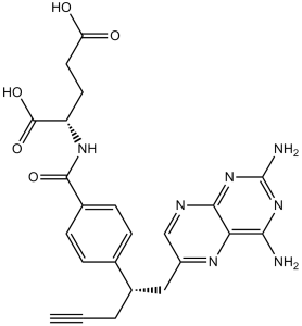
C23H23N7O5
|
|---|---|
| 分子量 |
477.47
|
| 精确质量 |
477.176
|
| CAS号 |
146464-95-1
|
| 相关CAS号 |
(R)-Pralatrexate;1320211-70-8
|
| PubChem CID |
148121
|
| 外观&性状 |
Light yellow to yellow solid powder
|
| 密度 |
1.5±0.1 g/cm3
|
| 熔点 |
215 °C(dec.)
|
| 折射率 |
1.704
|
| LogP |
0.23
|
| tPSA |
207.3
|
| 氢键供体(HBD)数目 |
5
|
| 氢键受体(HBA)数目 |
11
|
| 可旋转键数目(RBC) |
10
|
| 重原子数目 |
35
|
| 分子复杂度/Complexity |
809
|
| 定义原子立体中心数目 |
1
|
| SMILES |
C#CCC(CC1=CN=C2C(=N1)C(=NC(=N2)N)N)C3=CC=C(C=C3)C(=O)N[C@@H](CCC(=O)O)C(=O)O
|
| InChi Key |
OGSBUKJUDHAQEA-WMCAAGNKSA-N
|
| InChi Code |
InChI=1S/C23H23N7O5/c1-2-3-14(10-15-11-26-20-18(27-15)19(24)29-23(25)30-20)12-4-6-13(7-5-12)21(33)28-16(22(34)35)8-9-17(31)32/h1,4-7,11,14,16H,3,8-10H2,(H,28,33)(H,31,32)(H,34,35)(H4,24,25,26,29,30)/t14?,16-/m0/s1
|
| 化学名 |
N -(4-{1-[(2,4-diaminopteridin-6-yl)methyl]but-3-yn-1-yl}benzoyl)-L-glutamic acid
|
| 别名 |
PDX; Pralatrexate; 10-Propargyl-10-deazaaminopterin; trade name: Folotyn.
|
| HS Tariff Code |
2934.99.9001
|
| 存储方式 |
Powder -20°C 3 years 4°C 2 years In solvent -80°C 6 months -20°C 1 month |
| 运输条件 |
Room temperature (This product is stable at ambient temperature for a few days during ordinary shipping and time spent in Customs)
|
| 溶解度 (体外实验) |
|
|||
|---|---|---|---|---|
| 溶解度 (体内实验) |
配方 1 中的溶解度: ≥ 2.5 mg/mL (5.24 mM) (饱和度未知) in 10% DMSO + 40% PEG300 + 5% Tween80 + 45% Saline (这些助溶剂从左到右依次添加,逐一添加), 澄清溶液。
例如,若需制备1 mL的工作液,可将100 μL 25.0 mg/mL澄清DMSO储备液加入到400 μL PEG300中,混匀;然后向上述溶液中加入50 μL Tween-80,混匀;加入450 μL生理盐水定容至1 mL。 *生理盐水的制备:将 0.9 g 氯化钠溶解在 100 mL ddH₂O中,得到澄清溶液。 配方 2 中的溶解度: ≥ 2.5 mg/mL (5.24 mM) (饱和度未知) in 10% DMSO + 90% (20% SBE-β-CD in Saline) (这些助溶剂从左到右依次添加,逐一添加), 澄清溶液。 例如,若需制备1 mL的工作液,可将 100 μL 25.0 mg/mL澄清DMSO储备液加入900 μL 20% SBE-β-CD生理盐水溶液中,混匀。 *20% SBE-β-CD 生理盐水溶液的制备(4°C,1 周):将 2 g SBE-β-CD 溶解于 10 mL 生理盐水中,得到澄清溶液。 请根据您的实验动物和给药方式选择适当的溶解配方/方案: 1、请先配制澄清的储备液(如:用DMSO配置50 或 100 mg/mL母液(储备液)); 2、取适量母液,按从左到右的顺序依次添加助溶剂,澄清后再加入下一助溶剂。以 下列配方为例说明 (注意此配方只用于说明,并不一定代表此产品 的实际溶解配方): 10% DMSO → 40% PEG300 → 5% Tween-80 → 45% ddH2O (或 saline); 假设最终工作液的体积为 1 mL, 浓度为5 mg/mL: 取 100 μL 50 mg/mL 的澄清 DMSO 储备液加到 400 μL PEG300 中,混合均匀/澄清;向上述体系中加入50 μL Tween-80,混合均匀/澄清;然后继续加入450 μL ddH2O (或 saline)定容至 1 mL; 3、溶剂前显示的百分比是指该溶剂在最终溶液/工作液中的体积所占比例; 4、 如产品在配制过程中出现沉淀/析出,可通过加热(≤50℃)或超声的方式助溶; 5、为保证最佳实验结果,工作液请现配现用! 6、如不确定怎么将母液配置成体内动物实验的工作液,请查看说明书或联系我们; 7、 以上所有助溶剂都可在 Invivochem.cn网站购买。 |
| 制备储备液 | 1 mg | 5 mg | 10 mg | |
| 1 mM | 2.0944 mL | 10.4719 mL | 20.9437 mL | |
| 5 mM | 0.4189 mL | 2.0944 mL | 4.1887 mL | |
| 10 mM | 0.2094 mL | 1.0472 mL | 2.0944 mL |
1、根据实验需要选择合适的溶剂配制储备液 (母液):对于大多数产品,InvivoChem推荐用DMSO配置母液 (比如:5、10、20mM或者10、20、50 mg/mL浓度),个别水溶性高的产品可直接溶于水。产品在DMSO 、水或其他溶剂中的具体溶解度详见上”溶解度 (体外)”部分;
2、如果您找不到您想要的溶解度信息,或者很难将产品溶解在溶液中,请联系我们;
3、建议使用下列计算器进行相关计算(摩尔浓度计算器、稀释计算器、分子量计算器、重组计算器等);
4、母液配好之后,将其分装到常规用量,并储存在-20°C或-80°C,尽量减少反复冻融循环。
计算结果:
工作液浓度: mg/mL;
DMSO母液配制方法: mg 药物溶于 μL DMSO溶液(母液浓度 mg/mL)。如该浓度超过该批次药物DMSO溶解度,请首先与我们联系。
体内配方配制方法:取 μL DMSO母液,加入 μL PEG300,混匀澄清后加入μL Tween 80,混匀澄清后加入 μL ddH2O,混匀澄清。
(1) 请确保溶液澄清之后,再加入下一种溶剂 (助溶剂) 。可利用涡旋、超声或水浴加热等方法助溶;
(2) 一定要按顺序加入溶剂 (助溶剂) 。
| NCT Number | Recruitment | interventions | Conditions | Sponsor/Collaborators | Start Date | Phases |
| NCT02594267 | Completed | Drug: Pralatrexate Injection | Peripheral T-Cell Lymphoma (PTCL) | Acrotech Biopharma Inc. | November 10, 2015 | Phase 1 |
| NCT03355768 | Withdrawn | Drug: Romidepsin Drug: Pralatrexate |
Lymphoma, T-Cell, Peripheral | Jennifer Amengual | September 1, 2018 | Phase 3 |
| NCT03598998 | Active, not recruiting | Biological: Pembrolizumab Drug: Pralatrexate |
Anaplastic Large Cell Lymphoma Nodal Peripheral T-Cell Lymphoma With TFH Phenotype |
City of Hope Medical Center | February 4, 2019 | Phase 1 Phase 2 |
| NCT03240211 | Recruiting | Drug: Pembrolizumab Drug: Pralatrexate |
PTCL CTCL |
University of Virginia | February 2, 2022 | Phase 1 |
 |
|---|
 |
 |
InvivoChem的所有产品仅用于作科学研究,不面向患者销售
Copyright 2020 InvivoChem LLC | All Rights Reserved 粤ICP备20063088号-1


 COA
COA
 463611831
