| 规格 | 价格 | 库存 | 数量 |
|---|---|---|---|
| 25mg |
|
||
| 50mg |
|
||
| 100mg |
|
||
| 500mg |
|
||
| 1g |
|

| 靶点 |
Endogenous hormone
|
|---|---|
| 体外研究 (In Vitro) |
睾酮、雄烯二酮和黄体酮衍生物的生物转化研究最为深入的一项研究是在培养的蓝鲍属真菌菌株中进行的。所有检查的底物都经过转化或羟基化,这使研究人员能够分离出生物转化的代谢物。同一作者和他的同事研究了睾酮、雄烯二酮和具有额外C1-C2双键和/或17α-甲基的黄体酮衍生物在培养的蓝鲍属真菌菌株中的转化,作为评估类固醇(包括4A)的真核生物转化的模型。尽管底物包括4-烯-3-氧代体系,但它们在C-17的取代基和/或额外的C1-C2双键的存在方面显示出一些差异。
|
| 体内研究 (In Vivo) |
雄激素或类固醇应在广泛的医疗监督下进行监管,特别是雄烯二酮或4-雄烯3-17-二酮(4A)及其衍生物。它们的代谢产物影响人类和非人类,即真菌、动物和啮齿动物。此外,运动员只考虑睾酮水平的增加和骨骼成熟,而忽略了服用此类补充剂的其他已知和未知后果。此外,其中一些积极影响尚未得到充分的科学证明。据报道,雄烯二酮对雄性和雌性小鼠具有致癌作用,现有的雄烯二酮类致癌数据有限,因此有必要进行更多的研究,以提供更广泛的剂量限制,从而减少或预防此类毒性作用。显然,与雄烯二酮的益处相比,在男性、女性和儿童中补充雄烯二酮类会产生许多毒性作用。这篇综述旨在对雄烯二酮的消费、代谢、健康影响和毒性提供详细的见解。预计随着更多关于雄烯二酮药物补充的研究数据的提供,对人类健康有用剂量的更大控制将成为可能。
|
| 动物实验 |
激素替代疗法是预防绝经后骨质疏松症的一种潜在策略。然而,由于其可能对其他器官产生有害的副作用,该疗法存在诸多弊端。目前尚不清楚雄烯二酮是否会通过改变代谢类固醇激素的肝酶活性来影响生理激素水平。因此,Flynn 对成年雌性大鼠进行了一项研究,在交配前两周,通过灌胃给予雌性大鼠雄烯二酮(0、5、30 或 60 mg/kg/天),并持续至妊娠第 19 天。此外,研究人员还对未妊娠的雌性大鼠进行了相同时间段的雄烯二酮灌胃(0 或 60 mg/kg/天),并在妊娠第 20 天解剖妊娠大鼠的肝脏,以及在治疗五周后解剖未妊娠大鼠的肝脏。将肝微粒体与睾酮孵育后,与对照组相比,6-羟基睾酮、15-羟基睾酮、7-羟基睾酮、16-羟基睾酮和2-羟基睾酮的生成量均显著升高。在30和60 mg/kg/天的剂量水平下均观察到了6-羟基睾酮的生成。另一方面,在非妊娠大鼠中,雄烯二酮(60 mg/kg/天)显著增加了15-、6-、16-和2-羟基睾酮的生成。结果表明,高剂量口服雄烯二酮可增强雌性大鼠肝脏中某些代谢类固醇激素的细胞色素P450的活性。值得注意的是,妊娠和非妊娠雌性大鼠对不同剂量雄烯二酮的反应无显著差异。
|
| 药代性质 (ADME/PK) |
吸收、分布和排泄
口服雄烯二酮的吸收情况似乎存在个体差异,但确实存在一定的吸收。雄烯二酮分布于身体的各种组织……。 /乳汁/ 尚不清楚合成代谢类固醇是否会分布到母乳中。尚未有关于人类问题的记录。服用合成代谢类固醇的女性不应哺乳。/合成代谢类固醇/ 代谢/代谢物 本研究对两种骨螺科腹足动物:斑纹骨螺(Bolinus brandaris)和六角骨螺(Hexaplex trunculus)体内雄激素前体雄烯二酮(AD)的代谢进行了比较研究。从斑纹骨螺中分离出的微粒体组分主要将AD转化为5α-二氢睾酮,而六角骨螺则主要将其代谢为睾酮。在斑纹鳉(Bolinus brandaris)中仅检测到了AD代谢的性别差异,这归因于雄性体内较高的5α-还原酶活性。随后,研究人员考察了有机锡化合物三丁基锡(TBT)和三苯基锡(TPT)对AD代谢的影响。结果发现,仅在雌性中观察到显著的干扰作用,并且两种化合物的作用机制存在差异:TPT在浓度低至100 nM时即可强效抑制斑纹鳉的5α-还原酶活性,而TBT(10 μM)则通过增加17β-羟基类固醇脱氢酶(17β-HSD)的活性来改变短尾鳉(H. trunculus)中AD的代谢。因此,这项研究表明,腹足类动物中雄激素前体AD的代谢在活性和代谢谱方面均存在显著差异,并进一步证实了TBT和TPT能够干扰雄激素合成的关键酶促途径。 骨骼是雄激素的靶器官。这些类固醇在骨细胞内发挥作用的机制仍知之甚少。因此,本研究评估了从16名绝经后妇女(平均年龄69岁,范围56-80岁)和3名接受全髋关节置换术的老年男性(平均年龄71岁,范围69-73岁)的骨骼中培养的成骨细胞样骨细胞中雄烯二酮(女性体内主要的循环雄激素)的代谢情况。每种细胞株均在标准化条件下与不同浓度的[1,2,6,7-(3)H]雄烯二酮(0.05-5 μM)孵育。在所有情况下均生成了5α-还原代谢物和17β-羟基类固醇。切除骨的体积密度与相应培养骨细胞株的雄烯二酮代谢之间无相关性。所有19株细胞株的5α-还原酶活性(雄烯二酮和二氢睾酮之和)的表观Km值为0.7 ± 0.1 μM(平均值 ± 标准误),17β-羟基类固醇脱氢酶(睾酮和二氢睾酮之和)的表观Km值为2.3 ± 0.8 μM(平均值 ± 标准误),这些值与其他雄激素靶器官的报道值相似。我们的结果表明,人成骨细胞样细胞能够将雄烯二酮转化为活性更强的生物雄激素睾酮和二氢睾酮。由于5α-还原酶和17β-羟类固醇脱氢酶的Km值均高于血清雄烯二酮浓度,因此睾酮和二氢睾酮的生成似乎主要取决于底物的可用性。 扩散至腹腔的子宫内膜细胞中芳香化酶表达的上调可能通过局部雌激素合成增强其存活,这可能导致子宫内膜异位症。介导子宫内膜中芳香化酶诱导的因素尚不明确,但类固醇生成因子(SF)-1表达的增加可能发挥作用。本研究旨在确定腹腔液中主要的性类固醇——雄烯二酮(A4)是否调节子宫内膜芳香化酶的表达。这是一项在学术中心进行的细胞/组织培养研究。本研究采用了定量实时PCR、高效液相色谱(HPLC)和染色质免疫沉淀技术。用A4(10 nM)处理培养的人子宫内膜组织块和间质细胞,可显著上调5'端含有外显子IIa的芳香化酶mRNA转录本的表达。在子宫内膜间质细胞和人子宫内膜表面上皮(HES)细胞系中,A4诱导芳香化酶mRNA的表达与SF-1表达的增加相关。在HES细胞中,氚标记的A4代谢为雌二醇、睾酮(T)、二氢睾酮和雄甾二醇。雌二醇和T(而非非芳香化雄激素)均可上调HES细胞中芳香化酶和SF-1 mRNA的表达。染色质免疫沉淀实验表明,A4增强了SF-1向其位于CYP19外显子IIa上游的反应元件(-136 bp)的募集。结合雌激素受体拮抗剂ICI 182,780和芳香化酶抑制剂法卓唑均能抑制A4和T诱导芳香化酶和SF-1 mRNA表达的发现,表明A4和T的诱导作用是通过它们转化为雌激素介导的。子宫内膜细胞暴露于A4可能通过其芳香化为雌激素来增强CYP19基因的表达。 雄烯二酮在肾上腺和性腺中由脱氢表雄酮合成。它经17β-羟基类固醇脱氢酶代谢为睾酮,并经芳香化酶复合物代谢为雌酮。雌酮代谢为雌二醇。 雄烯二酮分布于全身各组织,并代谢为睾酮和雌酮。每剂雄烯二酮产生的睾酮量似乎因人而异。通常情况下,女性口服雄烯二酮后血清睾酮水平的升高幅度大于男性。 雄烯二酮是已知的人体睾酮代谢产物。 雄烯二酮来源于脱氢表雄酮或17-羟孕酮的转化。它可进一步转化为睾酮或雌酮。肾上腺雄烯二酮的生成受促肾上腺皮质激素(ACTH)调控,而性腺雄烯二酮的生成受促性腺激素调控。在男性中,雄烯二酮转化为睾酮需要17β-羟类固醇脱氢酶的催化。在女性中,雄烯二酮转化为雌激素(例如雌酮和雌二醇)需要芳香化酶的催化。 |
| 毒性/毒理 (Toxicokinetics/TK) |
毒性概述
识别和用途:雄烯二酮是一种合成的雄激素类似物,曾用作膳食补充剂。人体研究:有雄烯二酮诱发阳痿和严重少精症的病例报告。一项研究探讨了补充雄烯二酮对10名男性激素水平的影响及其与抗阻运动的相互作用。运动可提高睾酮水平,且不同条件下无差异。补充雄烯二酮后,运动可使血浆雌二醇水平在90分钟内显著升高约83%。因此,补充雄烯二酮不太可能为男性运动员带来任何合成代谢益处,并且在进行高强度抗阻运动时会提高雌激素水平。另一项研究表明,每日口服300毫克雄烯二酮可提高部分健康男性的血清睾酮和雌二醇浓度。然而,补充雄烯二酮会导致雌激素相关化合物和硫酸脱氢表雄酮浓度显著升高,睾酮合成下调,并对35至65岁男性的血脂和冠心病风险产生不利影响。动物研究:在豚鼠最大致敏试验中,皮内诱导浓度为5%,经皮诱导和激发浓度为25%,雄烯二酮未显示出皮肤致敏性。兔子报告有眼刺激症状。雄性大鼠单次口服1000 mg/kg的雄烯二酮会导致死亡,而500 mg/kg剂量下动物存活。与化合物相关的临床症状包括精神萎靡、步态紊乱和蹲伏姿势。在致死剂量下,其他症状包括俯卧位、意识丧失、呼吸紊乱和利尿增加。连续4周,每天一次口服给予雄性和雌性大鼠0、15、50和150 mg/kg剂量的雄烯二酮,结果显示雌性大鼠出现剂量依赖性效应,例如体重增加、子宫、宫颈、垂体和肾上腺萎缩,以及红细胞和血红蛋白数量增加。雄性大鼠则出现胸腺改变。这些效应被认为是类固醇激素典型的内分泌效应。向妊娠猴输注雄烯二酮导致提前出现分娩样子宫肌层活动,并使母体血浆雌激素、催产素和羊膜纤连蛋白浓度升高,与正常足月分娩时的水平相似。在大鼠中,雄烯二酮对单个骨骼或软组织的发育没有特定影响。根据OECD TG 471(在鼠伤寒沙门氏菌TA98、TA1537、TA100、TA102和TA1535中进行的Ames试验),在细菌回复突变试验中,雄烯二酮在最高推荐剂量5.0 mg/平板(无论是否存在代谢活化)下均未显示出致突变性。生态毒性研究:通过水体暴露于雄烯二酮可在相对较短的时间内导致成年雌性食蚊鱼雄性化。环境相关浓度的外源性雄烯二酮可显著调节钝吻鮈的生殖生理,且这种调节具有性别特异性。雄烯二酮可转化为睾酮和雌激素,当摄入足够剂量时,雄烯二酮可引起不良的雄性化和雌性化效应。雄烯二酮被认为是雄激素类固醇的前体,因为睾酮是一种雄激素或男性激素。在男性体内,雄烯二酮转化为睾酮需要17β-羟类固醇脱氢酶的催化。在女性体内,雄烯二酮转化为雌激素(例如雌酮和雌二醇)需要芳香化酶的催化。 相互作用 已知睾丸内睾酮(IT)浓度是血清睾酮浓度的100-200倍;然而,睾丸内睾酮前体的浓度、其睾丸至血清的浓度梯度、对促性腺激素的依赖性以及对人绒毛膜促性腺激素(hCG)刺激的反应尚未得到详细研究。我们假设,使用促性腺激素释放激素(GnRH)拮抗剂后,血清和睾丸内雄烯二酮(ADD)和脱氢表雄酮(DHEA)水平会显著降低,而人绒毛膜促性腺激素(hCG)刺激后,ADD和DHEA水平会升高,但血清DHEA水平不会受到类似抑制。我们使用GnRH拮抗剂阿西林抑制了23名健康男性的促性腺激素水平,并将他们随机分配到四个hCG剂量组:0、15、60或125 IU,隔日皮下注射,持续10天。在基线和治疗10天后采集血液和睾丸内液样本,用于测定血清和睾丸内激素水平。基线睾丸内雄烯二酮(IT ADD)[中位数(第25百分位数,第75百分位数)]为629(308,860)nmol/L,睾丸内脱氢表雄酮(IT DHEA)为564(411,879)nmol/L,分别比其血清浓度高175倍和27倍。阿西林可分别抑制睾丸内ADD和DHEA 98%和82%,而人绒毛膜促性腺激素(hCG)给药可显著升高二者水平。同样,血清ADD被抑制50%,但血清DHEA水平未发生变化。与血清相比,ADD和DHEA在人睾丸中的浓度较高。血清和睾丸内ADD和睾丸内DHEA在GnRH给药后显著受到抑制,而hCG则刺激其升高,但血清DHEA不受此影响,提示大部分循环DHEA并非来源于睾丸。 香豆素或茚满二酮衍生物或抗炎镇痛药(非甾体类或水杨酸盐)在与合成代谢类固醇(尤其是17α-烷基化化合物)合用时,其抗凝血作用可能会增强,这是由于促凝血因子合成或分解代谢的改变导致促凝血因子浓度降低,以及抗凝血剂受体亲和力增加所致;在与合成代谢类固醇合用期间和之后,可能需要根据凝血酶原时间测定结果调整抗凝血剂的剂量。/合成代谢类固醇/ 抗糖尿病药物、磺脲类药物或胰岛素与合成代谢类固醇合用可能会降低血糖浓度;糖尿病患者应密切监测低血糖症状……/合成代谢类固醇/ 同时使用皮质类固醇、糖皮质激素(尤其是具有显著盐皮质激素活性的药物)、长期治疗性促皮质素或含钠药物或食物与合成代谢类固醇合用,可能会增加水肿的可能性;此外,同时使用糖皮质激素或促皮质素与合成代谢类固醇合用,可能会促进严重痤疮的发生。 /合成代谢类固醇/ 有关雄烯二酮(共7种)的更多相互作用(完整)数据,请访问HSDB记录页面。 非人类毒性值 大鼠(雄性)口服LD50:500至1000 mg/kg体重 大鼠(雌性)口服LD50:>500 mg/kg体重 大鼠(雄性和雌性)皮肤给药LD50:>2000 mg/kg体重 |
| 参考文献 |
[1]. Molecules. 2021 Oct; 26(20): 6210.
|
| 其他信息 |
根据美国国家毒理学计划(NTP)的数据,雄烯二酮可能致癌。
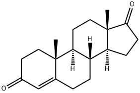
雄甾-4-烯-3,17-二酮是一种3-氧代Δ(4)类固醇,其结构为雄甾-4-烯,在3位和17位被氧代基团取代。它是一种在肾上腺和性腺合成的类固醇激素。它是一种雄激素,也是人类、大型蚤(Daphnia magna)和小鼠的代谢产物。它是一种17-氧代类固醇、雄甾烷类化合物和3-氧代Δ(4)类固醇。 它是一种Δ-4 C19类固醇,不仅在睾丸中产生,也在卵巢和肾上腺皮质中产生。根据组织类型的不同,雄烯二酮可作为睾酮、雌酮和雌二醇的前体。 据报道,飞蝗、智人以及其他有相关数据的生物体内均存在雄烯二酮。 治疗性雄烯二酮是一种强效的雄激素前体,是睾酮的直接前体,可用作补充剂以提高血浆睾酮水平和促进肌肉合成代谢。(NCI) 雄烯二酮是一种类固醇激素,由肾上腺和性腺以17α-羟孕酮或脱氢表雄酮为底物合成,是睾酮的前体。 雄烯二酮是一种δ-4 19碳类固醇,不仅在睾丸中产生,也在卵巢和肾上腺皮质中产生。根据组织类型的不同,雄烯二酮可作为睾酮、雌酮和雌二醇的前体。它是男性和女性性激素的共同前体。部分雄烯二酮也会分泌到血浆中,并可在外周组织中转化为睾酮和雌激素。雄烯二酮来源于脱氢表雄酮或17-羟孕酮的转化。它可进一步转化为睾酮或雌酮。肾上腺雄烯二酮的生成受促肾上腺皮质激素(ACTH)调控,而性腺雄烯二酮的生成受促性腺激素调控。 一种δ-4 C19类固醇,不仅在睾丸中产生,也在卵巢和肾上腺皮质中产生。根据组织类型的不同,雄烯二酮可作为睾酮、雌酮和雌二醇的前体。 作用机制 4-雄烯二酮是一种19碳类固醇激素,由肾上腺和性腺产生,是合成雄激素睾酮和雌激素雌酮、雌二醇的生化途径中的中间步骤。 如果同时摄入足够的热量和蛋白质,合成代谢类固醇可通过促进蛋白质合成代谢和刺激食欲来逆转分解代谢过程和负氮平衡。/合成代谢类固醇/ 治疗用途 /临床试验/ ClinicalTrials.gov 是一个注册库和结果数据库,收录了世界各地由公共和私人资助的人体临床研究。该网站由美国国家医学图书馆 (NLM) 和美国国立卫生研究院 (NIH) 维护。 ClinicalTrials.gov 上的每条记录都提供了研究方案的概要信息,包括以下内容:疾病或病症;干预措施(例如,正在研究的医疗产品、行为或程序);研究的标题、描述和设计;参与要求(资格标准);研究开展地点;研究地点的联系方式;以及其他健康网站相关信息的链接,例如美国国家医学图书馆 (NLM) 的 MedlinePlus(提供患者健康信息)和 PubMed(提供医学领域学术文章的引文和摘要)。数据库中包含雄烯二酮。 药物警告 如果给予足够剂量和足够时间,雄烯二酮及其相关分子可能会在人体内产生雄激素(从而产生合成代谢作用)或雌激素作用。……儿童和青少年尤其容易受到雄烯二酮的不可逆影响,因为它会转化为活性性激素。这些影响包括扰乱正常的性发育,特别是女孩出现男性化症状,表现为严重痤疮、体毛和面部毛发过多、声音变粗、阴蒂永久性增大、月经周期紊乱和不孕。转化为雌激素会导致男孩女性化,表现为乳房增大和睾丸萎缩。女孩长期暴露于过量雌激素可能增加患乳腺癌和子宫癌的风险。最后,男孩和女孩体内过量雄激素和雌激素的共同作用会导致青春期早熟、长骨骨骺过早闭合,从而显著影响成年身高。目前尚无关于长期服用补充雄烯二酮安全性的数据。男性外源性睾酮的不良反应包括痤疮、睾丸萎缩、男性乳房发育症、行为改变以及可能增加患前列腺癌的风险。女性使用外源性睾酮的不良反应包括多毛症、声音低沉、痤疮、阴蒂肥大、闭经、男性型脱发和皮肤粗糙。青少年使用外源性睾酮会导致骨骺过早闭合和成年身高降低。睾酮的其他不良反应包括肝功能衰竭和血小板聚集增加。/睾酮/ 雄烯二酮禁用于前列腺癌、乳腺癌和子宫癌患者。 研究发现,口服雄烯二酮会降低高密度脂蛋白胆固醇(HDL-C)水平,这可能会增加心血管疾病的风险。 有关雄烯二酮(共21条)的更多药物警告(完整)数据,请访问HSDB记录页面。 |
| 分子式 |
C19H26O2
|
|---|---|
| 分子量 |
286.41
|
| 精确质量 |
286.193
|
| CAS号 |
63-05-8
|
| PubChem CID |
6128
|
| 外观&性状 |
Crystals from hexane
|
| 密度 |
1.1±0.1 g/cm3
|
| 沸点 |
431.4±45.0 °C at 760 mmHg
|
| 熔点 |
173-174 °C (crystals from hexane)
; Melting point: 143 °C (alpha form), 173 (beta form)
; Melting point: 172 °C, OECD Guideline 102 (Melting point / Melting Range)
; 170 - 173 °C
|
| 闪点 |
161.1±25.7 °C
|
| 蒸汽压 |
0.0±1.0 mmHg at 25°C
|
| 折射率 |
1.552
|
| LogP |
2.9
|
| tPSA |
34.14
|
| 氢键供体(HBD)数目 |
0
|
| 氢键受体(HBA)数目 |
2
|
| 可旋转键数目(RBC) |
0
|
| 重原子数目 |
21
|
| 分子复杂度/Complexity |
546
|
| 定义原子立体中心数目 |
5
|
| SMILES |
O=C1C([H])([H])C([H])([H])[C@]2([H])[C@]1(C([H])([H])[H])C([H])([H])C([H])([H])[C@]1([H])[C@@]3(C([H])([H])[H])C([H])([H])C([H])([H])C(C([H])=C3C([H])([H])C([H])([H])[C@]12[H])=O
|
| InChi Key |
AEMFNILZOJDQLW-QAGGRKNESA-N
|
| InChi Code |
InChI=1S/C19H26O2/c1-18-9-7-13(20)11-12(18)3-4-14-15-5-6-17(21)19(15,2)10-8-16(14)18/h11,14-16H,3-10H2,1-2H3/t14-,15-,16-,18-,19-/m0/s1
|
| 化学名 |
(8R,9S,10R,13S,14S)-10,13-dimethyl-2,6,7,8,9,11,12,14,15,16-decahydro-1H-cyclopenta[a]phenanthrene-3,17-dione
|
| HS Tariff Code |
2934.99.9001
|
| 存储方式 |
Powder -20°C 3 years 4°C 2 years In solvent -80°C 6 months -20°C 1 month |
| 运输条件 |
Room temperature (This product is stable at ambient temperature for a few days during ordinary shipping and time spent in Customs)
|
| 溶解度 (体外实验) |
DMSO: > 10 mM
|
|---|
| 制备储备液 | 1 mg | 5 mg | 10 mg | |
| 1 mM | 3.4915 mL | 17.4575 mL | 34.9150 mL | |
| 5 mM | 0.6983 mL | 3.4915 mL | 6.9830 mL | |
| 10 mM | 0.3491 mL | 1.7457 mL | 3.4915 mL |
1、根据实验需要选择合适的溶剂配制储备液 (母液):对于大多数产品,InvivoChem推荐用DMSO配置母液 (比如:5、10、20mM或者10、20、50 mg/mL浓度),个别水溶性高的产品可直接溶于水。产品在DMSO 、水或其他溶剂中的具体溶解度详见上”溶解度 (体外)”部分;
2、如果您找不到您想要的溶解度信息,或者很难将产品溶解在溶液中,请联系我们;
3、建议使用下列计算器进行相关计算(摩尔浓度计算器、稀释计算器、分子量计算器、重组计算器等);
4、母液配好之后,将其分装到常规用量,并储存在-20°C或-80°C,尽量减少反复冻融循环。
计算结果:
工作液浓度: mg/mL;
DMSO母液配制方法: mg 药物溶于 μL DMSO溶液(母液浓度 mg/mL)。如该浓度超过该批次药物DMSO溶解度,请首先与我们联系。
体内配方配制方法:取 μL DMSO母液,加入 μL PEG300,混匀澄清后加入μL Tween 80,混匀澄清后加入 μL ddH2O,混匀澄清。
(1) 请确保溶液澄清之后,再加入下一种溶剂 (助溶剂) 。可利用涡旋、超声或水浴加热等方法助溶;
(2) 一定要按顺序加入溶剂 (助溶剂) 。
 CJC-1295 With DAC
 黄嘌呤钠盐
黄嘌呤钠盐
 四乙酰核糖
四乙酰核糖
 钨酸钠二水合物
InvivoChem的所有产品仅用于作科学研究,不面向患者销售
Copyright 2020 InvivoChem LLC | All Rights Reserved 粤ICP备20063088号-1


 COA
COA
 463611831
