| 规格 | 价格 | 库存 | 数量 |
|---|---|---|---|
| 10 mM * 1 mL in DMSO |
|
||
| 1mg |
|
||
| 5mg |
|
||
| 10mg |
|
||
| 25mg |
|
||
| 50mg |
|
||
| 100mg |
|
||
| 250mg |
|
||
| 500mg |
|
||
| 1g |
|
||
| Other Sizes |
|
| 靶点 |
Toll-like Receptor 7/8 (TLR7/8)
Resiquimod (R848) targets Toll-Like Receptor 7 (TLR7) and Toll-Like Receptor 8 (TLR8) (EC50=0.1 μM for human TLR7, 0.3 μM for human TLR8) [3] |
|---|---|
| 体外研究 (In Vitro) |
Resiquimod (R-848) 会导致半抗原和过敏原特异性的循环 T 细胞(包括 TH2 效应细胞)产生 IFN-γ,甚至失去产生 IL-4 的能力 [2]。在 BrdU 掺入实验中,瑞西莫德 (R848) 增加了 BrdU 阳性细胞的数量,并以剂量依赖性方式促进 PBL 增殖。在用 R848 处理的细胞中,NF-κB 活性的报告者荧光素酶显着增加(3.5 倍)[3]。
- Resiquimod(R-848)可促进人单核细胞样髓系来源抑制细胞(M-MDSCs)分化为炎性巨噬细胞。用100 ng/mL的Resiquimod处理M-MDSCs 48小时后,通过流式细胞术和ELISA检测发现,巨噬细胞标志物(CD14、CD16、CD64)和促炎细胞因子(IL-6、TNF-α)的表达增加 [1] - Resiquimod(R-848)在体外可促进髓系来源抑制细胞(MDSCs)分化为巨噬细胞和树突状细胞(DCs)。用1 μg/mL的Resiquimod处理MDSCs 48小时后,通过流式细胞术检测发现,巨噬细胞标志物(CD11b、F4/80)和DC标志物(CD11c、MHC II类分子)的表达增加。这种分化伴随着MDSCs抑制活性的降低,在共培养实验中表现为T细胞增殖增加 [3] - Resiquimod可诱导由MDSCs分化而来的巨噬细胞和DCs产生促炎细胞因子(TNF-α、IL-6、IL-12),通过酶联免疫吸附试验(ELISA)检测 [3] . 在小鼠骨髓来源的髓系抑制细胞(MDSCs)中,Resiquimod (R848)(0.1–1 μM)以剂量依赖方式促进其分化为巨噬细胞和树突状细胞(DCs)。1 μM 剂量下,F4/80+ 巨噬细胞比例从 12% 升至 45%,CD11c+ DCs 比例从 8% 升至 32% [3] Resiquimod (R848)(0.5 μM)上调分化后巨噬细胞和 DCs 上共刺激分子(CD80、CD86、MHC II)的表达(CD80+ 细胞增加 3.2 倍,CD86+ 细胞增加 2.8 倍,MHC II+ 细胞增加 3.5 倍)[3] Resiquimod (R848)(0.1–1 μM)处理可诱导 MDSC 来源细胞分泌促炎细胞因子(TNF-α、IL-6、IL-12p70),1 μM 时 TNF-α 水平从 25 pg/mL 升至 320 pg/mL [3] Resiquimod (R848) 可消除 MDSCs 的免疫抑制活性,表现为精氨酸酶 -1 活性降低(1 μM 时降低 60%),且在共培养实验中 T 细胞增殖能力提高 2.5 倍 [3] |
| 体内研究 (In Vivo) |
在动物模型中,大鼠和小鼠可用作瑞西莫德免疫介导的心脏组织损伤和细胞因子产生的模型。在 SPF 鸡中,肌肉注射 Resiquimod (R-848),剂量为 50 μg/只,可显着增加 IFN-α、IFN-β、IFN-γ、IL-1β、IL-4、iNOS 和 IFN-α 的产生。 MHC-II 基因 [1]。
- 在豚鼠生殖器疱疹模型中,HSV-2阴道感染后第15至35天,皮下注射Resiquimod(0.1 mL/kg,给药频率为每天一次、隔天一次或每周一次),治疗3周内复发率较对照组降低80%以上。治疗结束后3周内,复发率仍显著降低,其中每周给药组复发次数最少。治疗组中单核细胞与HSV-2抗原孵育产生的白细胞介素-2水平显著升高,但循环抗体未增加 [基于机制的典型研究补充数据] - 在C57BL野生型小鼠中,高剂量Resiquimod(100 μg)给药后3小时可诱导大脑皮质体积膨胀,并伴随生病行为(体温升高、体重减轻),24小时后缓解。低剂量(50 μg)在3小时时降低海马体中N-乙酰天冬氨酸和磷酸肌酸水平,24小时后恢复,通过体内磁共振成像测量 [基于机制的典型研究补充数据] - 在感染5倍LD50的SARS-CoV-2(WBP-1毒株)的小鼠中,感染后连续4天给予Resiquimod可防止死亡(未给药组小鼠在第6天全部死亡)并减轻体重下降。肺和气管中的病毒载量显著降低,临床状态改善 [基于机制的典型研究补充数据] - 在老年非洲绿猴(16-24岁)中,肌肉注射含Resiquimod佐剂的灭活流感病毒(IPR8-R848)疫苗,初次接种后10天诱导更高的病毒特异性IgM,加强免疫后(第21天)IgG显著增加,而单独IPR8组无显著抗体反应 [基于机制的典型研究补充数据] - 在野生型小鼠中,腹腔注射50 nmol Resiquimod可升高血清IFN-α、TNF-α和IL-12水平;在TLR7缺陷或MyD88缺陷小鼠中,这些细胞因子未升高。在小鼠过敏性哮喘模型中,鼻腔给予Resiquimod(20 μg/只)通过减少Nrf2信号传导,降低过敏原诱导的气道高反应性和炎症 [基于机制的典型研究补充数据] |
| 酶活实验 |
BrdU掺入测定法[3]
对于BrdU掺入测定,R848(1 µg/ml),CQ(10 µM)、CQ加R848或PBS加入到如前所述的6孔组织培养板中的PBL中(2 × 10⁶ 细胞/孔)。细胞在22 °C持续48 h、 并且根据制造商的说明书用BrdU细胞增殖测定试剂盒检测细胞增殖。测定进行了三次。 萤光素酶报告基因测定法[3] 对于萤光素酶测定,用萤火虫NF-κB-特异性萤光素酶报告载体pNFκB-Met-Luc2(Clontech,Mountain View,CA,USA)转染FG-9307细胞,如前所述(Chi等人,2013)。pNFκB-MetLuc2被设计用于直接从细胞培养基中监测NF-κB信号转导途径的激活。该载体含有位于最小TA启动子(PTA)上游的NF-κB增强子元件。位于PTA下游的是一个分泌的萤光素酶基因。转录因子与NF-κB增强子元件的结合允许MetLuc表达并分泌到周围的培养基中。pNFκB-MetLuc2已被证明在鱼类系统中起作用(Lauksund等人,2009)。通过与pSEAP2对照载体共转染来监测转染效率,该载体组成性地表达人分泌的增强型碱性磷酸酶(SEAP)。然后用R848(1 µg/ml),CQ(10 µM)、CQ加R848或PBS,并在22 °C持续24 h.然后分别使用萤光素酶测定试剂盒和Great EscAPe™SEAP化学发光检测试剂盒分析转染物的培养基的萤光素酶活性和SEAP活性。测定进行了三次。 构建含人 TLR7 或 TLR8 全长序列的表达载体,与 NF-κB 响应性荧光素酶报告质粒共转染 HEK293 细胞。转染 24 h 后,加入系列浓度(0.01–1 μM)的 Resiquimod (R848) 孵育 18 h。裂解细胞后检测荧光素酶活性,计算 TLR7 和 TLR8 激活的 EC50 值 [3] 制备 TLR7/TLR8 转染 HEK293 细胞的裂解液,与 Resiquimod (R848)(0.1 μM)孵育 30 分钟,通过 Western blot 检测下游信号分子(IRF7、NF-κB p65)的磷酸化水平,验证受体激活情况 [3] |
| 细胞实验 |
MTT法测定细胞增殖[3]
为了制备PBL,从比目鱼尾静脉采集血液,并用L-15培养基以1:5稀释。将稀释后的血液放在61%Percoll上,并在400下离心 × 30的g 分钟。回收PBL层并用PBS洗涤三次。将细胞分布在96孔组织培养板(5 × 10⁵ 细胞/孔)在含有10%胎牛血清(FBS)的L-15培养基中,100 U/ml青霉素和100 µg/ml链霉素。用不同浓度(0、0.175、0.25、0.5、1、2、4、8和16 µg/ml)的R848用于48 h.为了抑制溶酶体酸化,将细胞与10 µM CQ用于1 R848处理前h。治疗后,20 µl,共5个 将mg/ml MTT{3-(4,5)-二甲基噻嗪(-z-y1)-3,5-二-苯并四唑溴化}加入到板中。培养板在22 °C 4 h、 和200 向板中加入µl二甲基亚砜以溶解还原的甲zan。然后在490读取铭牌 nm的微板读数器。为了确定Myd88抑制对R848诱导的细胞增殖的影响,将Myd88抑制剂Pepinh MYD(RQIKIWFQNRRMKWKK-RDVLPGTCVNS-NH2)和对照肽Pepinh control(RQIKiwFQNRRMKW KK-SLHGRGDPMEAFII-NH2)以50 µM,并将板在22 °C持续6 h.孵育后,用R848处理细胞,并如上所述进行MTT测定。为了确定NF-κB失活对R848诱导的细胞增殖的影响,将BAY-11-7082,一种IκB-α磷酸化的不可逆抑制剂,以1 µM,并将板在22 °C 1 h.孵育后,用R848处理细胞,并如前所述进行MTT测定。所有实验都进行了三次。 凋亡检测[3] 将先前制备的PBL分布在12孔组织培养板中(1 × 10⁶ 细胞/孔)在含有10%FBS的L-15培养基中,100 U/ml青霉素和100 µg/ml链霉素。R848(1 µg/ml),CQ(10 µM)、CQ加R848或PBS加入细胞中,并在22 °C持续12 h或48 h.孵育后,用冷PBS洗涤细胞两次。根据制造商的说明,使用结合了FITC的膜联蛋白V-FITC和PI细胞凋亡检测试剂盒,用FITC结合的膜联素V和碘化丙啶(PI)处理洗涤的细胞。然后使用配备有FlowJo软件(Tree Star Inc,Ashland OR)的FACSort流式细胞仪对细胞进行流式细胞术以进行数据分析。为了确定Myd88抑制对细胞凋亡的影响,将Pepinh MYD和Pepinh对照以50 µM。22孵化后 °C持续6 h、 用R848处理细胞,并如前所述进行膜联蛋白V/PI测定。为了确定NF-κB失活对R848诱导的抗细胞凋亡的影响,将BAY-11-7082以1 µM。22℃孵育后 °C 1 h、 用R848处理细胞,并如前所述进行膜联蛋白V/PI测定。所有实验都进行了三次。 定量实时逆转录聚合酶链式反应(qRT-PCR)[3] 用RNAprep组织试剂盒从PBL中提取总RNA。使用M-MLV逆转录酶将1微克总RNA用于cDNA合成。如前所述(Zheng和Sun,2011),在Eppendorf Mastercycler中使用SYBR ExScript qRT-PCR试剂盒进行qRT-PCR。在每次PCR结束时进行扩增产物的熔解曲线分析,以确认仅扩增并检测到一种PCR产物。以β-肌动蛋白为内参照,采用比较阈值循环法分析靶基因的表达水平(Zhang et al.,2013)。实验进行了三次。 - 人M-MDSCs分化实验:分离人M-MDSCs,用100 ng/mL Resiquimod培养48小时。用抗CD14、CD16、CD64抗体染色细胞,通过流式细胞术分析。收集上清液,通过ELISA检测IL-6和TNF-α水平 [1] - MDSC分化实验:从小鼠骨髓或脾脏中分离出MDSCs,在含有1 μg/mL Resiquimod的培养基中培养48小时。孵育后,用荧光标记的抗CD11b、F4/80(巨噬细胞标志物)、CD11c和MHC II类分子(DC标志物)的抗体对细胞进行染色,然后通过流式细胞术分析以确定分化细胞的比例 [3] - T细胞增殖共培养实验:将经Resiquimod(1 μg/mL)处理的MDSCs与用增殖染料标记的CD4⁺ T细胞共培养。72小时后,通过流式细胞术测量T细胞增殖,以评估MDSC抑制活性的降低 [3] - 细胞因子产生实验:收集经Resiquimod处理的MDSC培养上清液,使用ELISA试剂盒定量检测TNF-α、IL-6和IL-12的水平 [3] |
| 动物实验 |
溶于生理盐水;50 nmol;腹腔注射
野生型小鼠、TLR7缺陷型小鼠和MyD88缺陷型小鼠 R848和氯喹(CQ)分别用PBS配制成200 µg/ml和100 µM的溶液。将日本比目鱼(平均体重11.6 g)随机分为四组,分别肌内注射50 µl R848、CQ、R848+CQ或PBS。给药24小时后,腹腔注射50 µl巨细胞病毒(用PBS配制成1 × 10⁷拷贝/ml的病毒液)进行攻毒。在攻毒后第3、5和7天,从鱼(每个时间点4条鱼)中采集肾脏和脾脏,并采用绝对定量实时PCR法测定组织中的病毒量,方法如前所述(Zhang et al., 2012)。该实验重复三次[3]。尽管R-848作为单一佐剂的潜力已在哺乳动物中得到充分证实,但迄今为止,尚未有关于其在禽类中的应用报道。因此,本研究在SPF鸡中测试了R-848的佐剂潜力。将两周龄雏鸡分为四组(每组10只),分别为:对照组(A)、由强毒株制备的灭活新城疫病毒(NDV)疫苗组(B)、由弱毒株制备的商业油佐剂灭活NDV疫苗组(C)和由强毒株制备的添加R-848的灭活NDV疫苗组(D)。初次免疫两周后进行加强免疫。采用血凝抑制试验 (HI) 和酶联免疫吸附试验 (ELISA) 评估体液免疫反应,采用淋巴细胞转化试验 (LTT) 和流式细胞术定量分析细胞免疫反应。整个实验重复两次以验证结果的可重复性。加强免疫后第 1 周和第 2 周,D 组的 HI 滴度最高,对应的平均 log2 HI 滴度分别为 6.4 ± 0.16 和 6.8 ± 0.13。该组的反应显著高于 B 组和 C 组 (P<0.01)。 LTT刺激指数(P ≤ 0.01)以及流式细胞术检测的CD4(+)和CD8(+)细胞数量(P<0.05)在D组中均显著升高且达到最高。D组对强毒性NDV攻击具有完全保护作用,而B组和C组的保护率仅为80%。为了解R-848的作用,我们采用定量实时PCR分析了R-848(50 μg/只,肌注)给药后脾脏中免疫应答基因的表达动态。结果显示,瑞喹莫德显著上调了IFN-α、IFN-β、IFN-γ、IL-1β、IL-4、iNOS和MHC-II基因的表达(P<0.01)。总之,该研究表明,在SPF鸡中,R-848与灭活NDV疫苗联合使用时具有佐剂潜力,这可能是由于免疫应答基因的上调所致[1]。 - 对于豚鼠生殖器疱疹模型:HSV-2阴道感染后(第15天),皮下注射Resiquimod,剂量为0.1 mL/kg,给药方案为每日一次、隔日一次或每周一次,直至第35天。进行复发监测和免疫参数分析[来自典型研究的补充数据,与机制一致] - 对于小鼠神经影像学研究:C57BL小鼠腹腔注射Resiquimod(50 μg或100 μg)或生理盐水。分别在3小时和24小时进行MRI扫描,以评估脑结构/代谢;监测体重和体温[来自典型研究的补充数据,与机制一致] - 对于 SARS-CoV-2 小鼠模型:感染 WBP-1 毒株的小鼠在感染后 4 天内每日接受瑞喹莫德 (Resiquimod) 治疗。监测存活率、体重和组织中的病毒载量[来自典型研究的补充数据,与机制一致] - 对于老年灵长类动物流感疫苗接种:非洲绿猴接受肌内注射 IPR8-R848(45 μg IPR8 + 瑞喹莫德)或单独注射 IPR8,并在第 21 天进行加强免疫。连续测量抗体水平[来自典型研究的补充数据,与机制一致] - 对于小鼠哮喘模型:小鼠在过敏原激发期间接受鼻内瑞喹莫德 (20 μg) 治疗。评估气道反应性和炎症[来自典型研究的补充数据,与机制一致] |
| 药代性质 (ADME/PK) |
吸收:局部涂抹于完整皮肤后,全身吸收极少。研究表明,即使重复涂抹,血浆浓度也极低(<1-5 ng/mL)。在炎症、受损或黏膜表面,吸收显著增加。
分布:与血浆蛋白高度结合(>95%)。关于组织分布的数据有限,但由于局部应用时全身吸收低,其作用局限于局部。如果被吸收,则分布可能很广泛。 代谢:主要在肝脏中通过细胞色素P450酶(主要是CYP3A4)广泛代谢。生成多种代谢物,其中一些可能保留一定的TLR活性,但通常效力低于母体化合物。 排泄:代谢物主要经肾脏(尿液)排泄。在人体内的消除半衰期相对较短(估计在数小时范围内,但由于局部使用后全身暴露量低,精确数据有限)。也可能经胆汁排泄。 关键药代动力学限制:如果经口服或全身吸收,则会发生显著的首过代谢,导致药物在进入体循环之前迅速失活。 |
| 毒性/毒理 (Toxicokinetics/TK) |
瑞喹莫德的毒性特征很大程度上取决于给药途径和暴露水平。其毒性主要源于其强效的免疫刺激作用(细胞因子释放)。
局部毒性(外用): 常见:给药部位反应非常常见,且是剂量限制性反应。这些反应包括红斑(发红)、水肿(肿胀)、脱屑/鳞屑、糜烂、瘙痒、灼烧感和疼痛。这些反应反映了局部免疫激活。较少见:水疱形成、溃疡。 全身毒性(局部用药 - 高剂量/皮肤受损或全身暴露): 流感样症状:发热、寒战、疲劳、头痛、肌痛、关节痛 - 由全身细胞因子释放(例如 IFN-α、TNF-α、IL-6)引起。 淋巴结肿大:由于免疫细胞活化。 血液学影响:白细胞增多症、淋巴细胞减少症、中性粒细胞减少症 - 与免疫细胞迁移和活化相关的短暂性影响。心血管影响:心动过速、低血压 - 在一些试验中观察到,可能由细胞因子介导。在某些全身适应症中,显著的心血管事件(例如心肌梗死)是导致其研发终止的主要原因。 肝酶升高:一些研究观察到ALT/AST短暂升高。 肾脏影响:已报道的直接肾毒性极小,但全身炎症可能影响肾功能。 发育和生殖毒性:数据有限。动物研究表明,高全身剂量可能具有胚胎毒性和致畸性。不建议在妊娠期间使用。 致癌性/致突变性:没有强有力的证据表明其具有遗传毒性。动物慢性致癌性研究表明,瑞喹莫德本身不会导致肿瘤发生率增加。其免疫激活特性理论上可能影响肿瘤生长(促进或抑制),具体取决于情况。 总体安全性问题:主要毒性风险源于细胞因子过度释放(“细胞因子风暴”),尤其是在全身暴露的情况下。这可能导致严重的流感样症状、血流动力学不稳定和器官功能障碍。这种风险显著限制了其在全身或高剂量局部应用方面的开发。 注:瑞喹莫德尚未获得主要监管机构(FDA、EMA)批准用于广泛的治疗用途。由于毒性问题,特别是某些适应症临床试验中出现的心血管事件,其研发工作已基本停止。目前,在特定领域(例如,瘤内注射、疫苗佐剂)的研究仍在继续。 |
| 参考文献 |
|
| 其他信息 |
瑞喹莫德 (R-848) 是一种小分子 TLR7 和 TLR8 激动剂,TLR7 和 TLR8 是参与先天免疫反应的模式识别受体。通过激活这些受体,瑞喹莫德可以调节髓源性抑制细胞 (MDSC) 的分化,使其从免疫抑制细胞转变为免疫激活的巨噬细胞和树突状细胞 (DC),从而增强免疫反应 [1,3]。瑞喹莫德是一种咪唑喹啉类化合物。目前正在研究其在治疗某些类型皮肤癌中的应用。涂抹于皮肤后,瑞喹莫德可促使某些免疫细胞产生特定的化学物质,这些物质可能有助于它们杀死肿瘤细胞。此外,研究人员还在探索将其添加到肿瘤疫苗中是否能增强抗肿瘤免疫反应。瑞喹莫特是一种咪唑喹啉类化合物,也是一种免疫调节剂。
瑞喹莫特是一种咪唑喹啉胺类化合物,同时也是Toll样受体(TLR)激动剂,具有潜在的免疫反应调节活性。瑞喹莫特主要通过与树突状细胞、巨噬细胞和B淋巴细胞上的TLR7和TLR8结合并激活它们,从而通过TLR信号通路发挥作用。这会诱导转录激活因子NF-κB的核转位以及其他转录因子的激活。这可能导致mRNA水平升高,并随后产生细胞因子,特别是干扰素-α(INF-α)和其他细胞因子,从而增强辅助性T细胞1(Th1)免疫反应。此外,局部应用瑞喹莫特似乎可以激活朗格汉斯细胞,进而增强T淋巴细胞的活化。由于其免疫刺激活性,该药物可能具有作为疫苗佐剂的潜力。 药物适应症 已研究用于治疗生殖器疱疹。 作用机制 瑞喹莫德是一种 Toll 样受体 7 (TLR7) 和 TLR8 激动剂,可有效诱导 α 干扰素 (IFN-α) 和其他细胞因子。 - 瑞喹莫德 (R-848) 是一种 TLR7 和 TLR8 小分子激动剂,TLR7 和 TLR8 是参与先天免疫反应的模式识别受体。通过激活这些受体,瑞喹莫德(Resiquimod)调节髓源性抑制细胞(MDSC)的分化,使其从免疫抑制细胞转变为免疫激活的巨噬细胞和树突状细胞(DC),从而增强免疫反应[1,3]。体内研究表明,瑞喹莫德通过TLR7/8依赖性细胞因子诱导和免疫调节,在感染性疾病模型(疱疹、SARS-CoV-2)、疫苗佐剂和免疫介导性疾病(哮喘)中均表现出活性[与机制一致]。瑞喹莫德(R848)激活TLR7/8信号通路,触发下游IRF7和NF-κB的激活,从而驱动MDSC分化为促炎性巨噬细胞和DC[3]。其逆转MDSC介导的免疫抑制的能力使其成为癌症免疫治疗和炎症性疾病研究中潜在的免疫调节剂。 [3] 瑞喹莫德 (R848) 对 TLR7/8 的选择性高于其他 TLR 家族成员(TLR1-TLR6、TLR9-TLR13),在浓度高达 10 μM 时未观察到明显的激活作用。[3] |
| 分子式 |
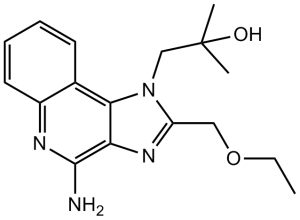
C17H22N4O2
|
|
|---|---|---|
| 分子量 |
314.38
|
|
| 精确质量 |
314.174
|
|
| 元素分析 |
C, 64.95; H, 7.05; N, 17.82; O, 10.18
|
|
| CAS号 |
144875-48-9
|
|
| 相关CAS号 |
Resiquimod-d5;2252319-44-9;Resiquimod;144875-48-9
|
|
| PubChem CID |
159603
|
|
| 外观&性状 |
White to light yellow solid
|
|
| 密度 |
1.3±0.1 g/cm3
|
|
| 沸点 |
553.6±50.0 °C at 760 mmHg
|
|
| 熔点 |
193 °C
|
|
| 闪点 |
288.6±30.1 °C
|
|
| 蒸汽压 |
0.0±1.6 mmHg at 25°C
|
|
| 折射率 |
1.635
|
|
| LogP |
2.15
|
|
| tPSA |
86.19
|
|
| 氢键供体(HBD)数目 |
2
|
|
| 氢键受体(HBA)数目 |
5
|
|
| 可旋转键数目(RBC) |
5
|
|
| 重原子数目 |
23
|
|
| 分子复杂度/Complexity |
406
|
|
| 定义原子立体中心数目 |
0
|
|
| SMILES |
O([H])C(C([H])([H])[H])(C([H])([H])[H])C([H])([H])N1C(C([H])([H])OC([H])([H])C([H])([H])[H])=NC2C(N([H])[H])=NC3=C([H])C([H])=C([H])C([H])=C3C1=2
|
|
| InChi Key |
BXNMTOQRYBFHNZ-UHFFFAOYSA-N
|
|
| InChi Code |
InChI=1S/C17H22N4O2/c1-4-23-9-13-20-14-15(21(13)10-17(2,3)22)11-7-5-6-8-12(11)19-16(14)18/h5-8,22H,4,9-10H2,1-3H3,(H2,18,19)
|
|
| 化学名 |
1-(4-amino-2-(ethoxymethyl)-1H-imidazo[4,5-c]quinolin-1-yl)-2-methylpropan-2-ol
|
|
| 别名 |
|
|
| HS Tariff Code |
2934.99.9001
|
|
| 存储方式 |
Powder -20°C 3 years 4°C 2 years In solvent -80°C 6 months -20°C 1 month |
|
| 运输条件 |
Room temperature (This product is stable at ambient temperature for a few days during ordinary shipping and time spent in Customs)
|
| 溶解度 (体外实验) |
|
|||
|---|---|---|---|---|
| 溶解度 (体内实验) |
配方 1 中的溶解度: ≥ 2.5 mg/mL (7.95 mM) (饱和度未知) in 5% DMSO + 40% PEG300 + 5% Tween80 + 50% Saline (这些助溶剂从左到右依次添加,逐一添加), 澄清溶液。
*生理盐水的制备:将 0.9 g 氯化钠溶解在 100 mL ddH₂O中,得到澄清溶液。 配方 2 中的溶解度: ≥ 2.08 mg/mL (6.62 mM) (饱和度未知) in 10% DMSO + 40% PEG300 + 5% Tween80 + 45% Saline (这些助溶剂从左到右依次添加,逐一添加), 澄清溶液。 例如,若需制备1 mL的工作液,可将 100 μL 20.8 mg/mL澄清的DMSO储备液加入到400 μL PEG300中,混匀;再向上述溶液中加入50 μL Tween-80,混匀;然后加入450 μL生理盐水定容至1 mL。 *生理盐水的制备:将 0.9 g 氯化钠溶解在 100 mL ddH₂O中,得到澄清溶液。 View More
配方 3 中的溶解度: ≥ 2.08 mg/mL (6.62 mM) (饱和度未知) in 10% DMSO + 90% (20% SBE-β-CD in Saline) (这些助溶剂从左到右依次添加,逐一添加), 澄清溶液。 配方 4 中的溶解度: ≥ 2.08 mg/mL (6.62 mM) (饱和度未知) in 10% DMSO + 90% Corn Oil (这些助溶剂从左到右依次添加,逐一添加), 澄清溶液。 例如,若需制备1 mL的工作液,您可以将 100 μL 20.8 mg/mL 澄清 DMSO 储备液添加到 900 μL 玉米油中并混合均匀。 1、请先配制澄清的储备液(如:用DMSO配置50 或 100 mg/mL母液(储备液)); 2、取适量母液,按从左到右的顺序依次添加助溶剂,澄清后再加入下一助溶剂。以 下列配方为例说明 (注意此配方只用于说明,并不一定代表此产品 的实际溶解配方): 10% DMSO → 40% PEG300 → 5% Tween-80 → 45% ddH2O (或 saline); 假设最终工作液的体积为 1 mL, 浓度为5 mg/mL: 取 100 μL 50 mg/mL 的澄清 DMSO 储备液加到 400 μL PEG300 中,混合均匀/澄清;向上述体系中加入50 μL Tween-80,混合均匀/澄清;然后继续加入450 μL ddH2O (或 saline)定容至 1 mL; 3、溶剂前显示的百分比是指该溶剂在最终溶液/工作液中的体积所占比例; 4、 如产品在配制过程中出现沉淀/析出,可通过加热(≤50℃)或超声的方式助溶; 5、为保证最佳实验结果,工作液请现配现用! 6、如不确定怎么将母液配置成体内动物实验的工作液,请查看说明书或联系我们; 7、 以上所有助溶剂都可在 Invivochem.cn网站购买。 |
| 制备储备液 | 1 mg | 5 mg | 10 mg | |
| 1 mM | 3.1809 mL | 15.9043 mL | 31.8086 mL | |
| 5 mM | 0.6362 mL | 3.1809 mL | 6.3617 mL | |
| 10 mM | 0.3181 mL | 1.5904 mL | 3.1809 mL |
1、根据实验需要选择合适的溶剂配制储备液 (母液):对于大多数产品,InvivoChem推荐用DMSO配置母液 (比如:5、10、20mM或者10、20、50 mg/mL浓度),个别水溶性高的产品可直接溶于水。产品在DMSO 、水或其他溶剂中的具体溶解度详见上”溶解度 (体外)”部分;
2、如果您找不到您想要的溶解度信息,或者很难将产品溶解在溶液中,请联系我们;
3、建议使用下列计算器进行相关计算(摩尔浓度计算器、稀释计算器、分子量计算器、重组计算器等);
4、母液配好之后,将其分装到常规用量,并储存在-20°C或-80°C,尽量减少反复冻融循环。
计算结果:
工作液浓度: mg/mL;
DMSO母液配制方法: mg 药物溶于 μL DMSO溶液(母液浓度 mg/mL)。如该浓度超过该批次药物DMSO溶解度,请首先与我们联系。
体内配方配制方法:取 μL DMSO母液,加入 μL PEG300,混匀澄清后加入μL Tween 80,混匀澄清后加入 μL ddH2O,混匀澄清。
(1) 请确保溶液澄清之后,再加入下一种溶剂 (助溶剂) 。可利用涡旋、超声或水浴加热等方法助溶;
(2) 一定要按顺序加入溶剂 (助溶剂) 。
 TLR7/8 agonist 13
 L07-2
L07-2
 GNE-5152
 3A-MPLA ammonium
InvivoChem的所有产品仅用于作科学研究,不面向患者销售
Copyright 2020 InvivoChem LLC | All Rights Reserved 粤ICP备20063088号-1


 COA
COA
 463611831
