| 规格 | 价格 | 库存 | 数量 |
|---|---|---|---|
| 500mg |
|
||
| 1g |
|
||
| 5g |
|
||
| 10g |
|
||
| 25g |
|
||
| Other Sizes |
|

| 靶点 |
Mitochondrial electron transport chain complex I
|
|---|---|
| 体外研究 (In Vitro) |
鱼藤酮影响 ENS 信号传导的方式涉及丝裂原激活多肽 (MAPK)、Toll 样受体、Wnt 和 Ras 的密切参与 [2]。
鱼藤酮抑制线粒体呼吸链复合体I可诱导多种细胞死亡。然而,其机制仍然难以捉摸。由于活性氧(ROS)在细胞凋亡中起重要作用,并且鱼藤酮抑制线粒体呼吸链复合体I被认为能够提高线粒体ROS的产生,我们研究了鱼藤酮诱导的细胞凋亡与线粒体活性氧之间的关系。鱼藤酮能够在HL-60细胞的分离线粒体和培养细胞中诱导线粒体复合体I底物支持的线粒体ROS产生。通过DNA断裂、细胞色素c释放和caspase 3活性证实鱼藤酮诱导的细胞凋亡。鱼藤酮诱导的细胞凋亡与鱼藤酮诱导的线粒体ROS产生之间存在定量相关性。抗氧化剂(谷胱甘肽、n -乙酰半胱氨酸和维生素C)可抑制鱼藤酮诱导的细胞凋亡。过量表达镁超氧化物歧化酶的HT1080细胞对鱼藤酮诱导的细胞凋亡的抗性比对照细胞更强,这也证实了鱼藤酮诱导的线粒体ROS在细胞凋亡中的作用。这些结果表明鱼藤酮能够通过增加线粒体活性氧的产生量来诱导细胞凋亡。[5] |
| 体内研究 (In Vivo) |
鱼藤酮可用于动物模型,以开发帕金森氏症的调节模型。鱼藤酮会导致兴奋性神经递质显着增加;在 PD 动物的小脑中检测到谷氨酸和天冬氨酸以及兴奋性 GABA、甘氨酸和牛磺酸显着减少 [1]。鱼藤酮(1.5、2 或 2.5 mg/kg)导致黑质中 α-突触核蛋白剂量需求增加。此外,在剂量为 2 和 2.5 mg/kg 时,鱼藤酮可诱导黑质中酪氨酸激酶免疫反应性神经元数量和纹状体中多巴胺数量显着减少 [4]。
|
| 酶活实验 |
呼吸测量[5]
如前所述,用克拉克氧电极测量耗氧量。简单地说,将1 × 10⁷个HL-60细胞用不同浓度鱼藤酮处理30分钟。然后收集细胞并在含有0.3 m甘露醇、10 mm HEPES钾(pH 7.4)、5 mm磷酸钾(pH 7.4)和1 mm MgCl2的培养基中重悬。这些细胞被注射到一个呼吸室,然后被密封。呼吸室的总容积为1.6 ml。假设初始氧浓度为6.8 mg/l,测量并计算呼吸作用为氧浓度的变化率。细胞呼吸转化为对照的百分比。 ATP测定[5] 对于ATP的测量,使用市售的荧光素-荧光素酶测定试剂盒。简单地说,将HL-60细胞用不同浓度的鱼藤酮处理24 h,然后将其收集在1 ml的Eppendorf管中。用冰冷的PBS单次洗涤后,用试剂盒提供的体细胞atp释放试剂裂解细胞。加入荧光素底物和荧光素酶,在Perkin Elmer 3B荧光仪上评估生物发光。内标法测定全细胞ATP含量。细胞ATP水平转化为未处理细胞(对照)的百分比。 流式细胞术测定细胞超氧化物生成[5] 细胞超氧化物生成的测量如前所述,进行了一些修改。HL-60细胞用不同浓度的鱼藤酮处理30 min,收集细胞,250 × g汉克平衡盐溶液(Hank’s balanced salt solution, HBSS)洗涤,将细胞重悬于含有10 μm氢乙啶(hydroethidine, HE)的HBSS中,37℃孵养10 min。HT1080纤维瘤细胞用鱼藤酮处理30 min,然后取出培养液,HBSS洗涤一次。将细胞用含0.25%胰蛋白酶的HBSS孵育5分钟,再用含10 μm氢乙胺(HE)的HBSS重悬,37℃孵育10分钟。将所有细胞悬液置于12 × 75-mm管中检测。流式细胞术在Beckman-Coulter XL流式细胞仪上进行。采用610 nm长通滤光片采集乙锭荧光。 DNA碎片[5] DNA片段分析按照前面描述的方法进行,并进行了一些修改。在抗氧化剂存在或不存在的情况下,用不同浓度鱼藤酮处理HL-60细胞。细胞(2 × 10⁷)用PBS(4°C, pH 7.4)洗涤一次,250 × g离心5分钟。然后用0.5 ml裂解缓冲液(10 mmTris-HCL, pH 7.4, 10 mm EDTA, 0.5%十二烷基硫酸钠)在冰上处理10分钟。37℃条件下,RNase A(终浓度100 μg/ml)作用1 h后,细胞在100 μg/ml蛋白酶k的存在下,50℃条件下孵育4 h,在溶液中加入50 μl 3 m醋酸钠(pH 5.2)和1 ml冷(4℃)100%乙醇沉淀DNA。然后收集DNA并溶解在TE缓冲液(10 mm Tris pH 8.0, EDTA 1 mm)中。在含有10 μg/ml溴化乙啶的1.2%琼脂糖凝胶上加载10 - 20 μl的DNA进行分析。电泳在0.5 × Tris硼酸EDTA缓冲液(18mm Tris碱基(pH 8.0), 18mm硼酸和1mm EDTA)中进行,在70 V下电泳2小时。在紫外光下观察DNA并拍照。 |
| 细胞实验 |
Cell Counting Kit-8 [2]
采用细胞计数试剂盒-8 (Cell Counting Kit-8, CCK8)检测鱼藤酮对ENS细胞活力的影响。96孔板每孔共接种2000个细胞,培养24 h,取出培养液,分别于浓度为0.01、0.02、0.07、0.21、0.62、1.85、5.56、16.67、50 μM的Rotenone/鱼藤酮环境下培养ENS细胞48 h,以未处理鱼藤酮的细胞为对照。然后,每孔加入10µL CCK-8,孵育3 h,用酶标仪检测450 nm处吸光度。 总RNA制备[2] 将0.3 μMRotenone/鱼藤酮处理/不处理48 h的细胞按TRIZOL法制备总RNA,用DNaseI去除总RNA中污染的DNA。采用NanoDrop 1000检测RNA浓度,每个样品取1 μg RNA,通过1%琼脂糖凝胶电泳分析RNA完整性,剩余RNA保存于- 80°C,用于RNA测序和实时聚合酶链反应(RT-PCR)。 |
| 动物实验 |
本研究旨在探讨帕金森病(PD)大鼠模型小脑中神经化学的变化。将大鼠分为两组:对照组和纹状体内注射鱼藤酮诱导的PD大鼠模型组。与对照组相比,PD大鼠模型小脑中兴奋性氨基酸神经递质谷氨酸和天冬氨酸显著升高,而抑制性氨基酸神经递质γ-氨基丁酸(GABA)、甘氨酸和牛磺酸显著降低。这与脂质过氧化、一氧化氮和肿瘤坏死因子-α显著升高以及还原型谷胱甘肽显著降低相关。此外,PD大鼠模型小脑中乙酰胆碱酯酶活性显著降低,而Na+,K+-ATP酶活性显著升高。此外,帕金森病(PD)大鼠模型的小脑切片显示浦肯野细胞明显坏死、细胞形态不规则受损、胞质收缩、坏死以及神经元周围空泡化。目前的研究结果表明,兴奋性和抑制性氨基酸平衡的紊乱可能在PD的发病机制中发挥作用。根据目前的神经化学和组织病理学变化,在PD的治疗中应考虑小脑的病变。[1]帕金森病(PD)是一种主要影响运动系统的神经退行性疾病,其病因是黑质致密部多巴胺能神经元的死亡。目前PD的研究方向是寻找可以单独使用或与左旋多巴联合使用的新型分子,以预防长期服用左旋多巴期间出现的左旋多巴诱导运动障碍(LID)样症状。因此,本研究旨在探讨诺丽果(Morinda citrifolia)是否对鱼藤酮诱导的帕金森病(PD)具有治疗作用,尤其关注线粒体功能障碍介导的内源性细胞凋亡。方法:雄性Sprague-Dawley大鼠立体定位注射鱼藤酮(SNPc和VTA各3 µg),并同时给予诺丽果乙酸乙酯提取物和左旋多巴治疗。[3] 近年来,皮下注射鱼藤酮因其便捷、简单且能有效模拟动物模型中的帕金森病(PD)特征而备受关注。然而,文献报道的剂量范围较广,难以客观评价该技术的有效性。本研究旨在确定建立帕金森病模型所需的最佳皮下注射鱼藤酮剂量。我们连续5周,每天皮下注射三种剂量的鱼藤酮(1.5、2或2.5 mg/kg)给雄性Wistar大鼠。鱼藤酮导致黑质中α-突触核蛋白呈剂量依赖性增加。此外,2 mg/kg和2.5 mg/kg剂量的鱼藤酮显著降低了黑质中酪氨酸羟化酶免疫反应性神经元的数量以及纹状体中的多巴胺水平。然而,2.5 mg/kg剂量组的死亡率高达46.7%,而2 mg/kg剂量组的死亡率仅为6.7%;2.5 mg/kg剂量组的高死亡率限制了其应用。每日注射2 mg/kg剂量5周后,未观察到对体重的不利影响。此外,与接受载体或生理盐水的对照组相比,2mg/kg 组大鼠从水平杆和网格墙上下来的潜伏期更长,站立次数减少,从转棒上跌落的潜伏期更短。透射电镜观察到的线粒体损伤在该剂量下也很明显。综上所述,我们的数据表明,每日皮下注射 2mg/kg 鱼藤酮可促进大鼠体内 α-突触核蛋白的形成,并重现帕金森病的典型特征,同时保持较低的死亡率。[4]
|
| 药代性质 (ADME/PK) |
吸收、分布和排泄
胃肠道吸收率低且不完全。在动物实验中,静脉注射鱼藤酮的毒性比口服高数百倍。脂肪和油脂可增加吸收。 雄性和雌性Sprague Dawley大鼠分别服用纯化鱼藤酮(纯度99.23%)。……采用口服和静脉注射(iv)剂量的(14C)-鱼藤酮(0.01至5.0 mg/kg),对雌雄大鼠进行了初步排泄平衡、排泄平衡、药代动力学和肠肝循环研究。采用薄层色谱法分析尿液和粪便中的代谢物。……粪便是静脉注射和口服给药的主要排泄途径,尿液中回收少量(14C)。静脉注射后,雌雄大鼠的排泄在48小时内几乎完全,口服给药后,雄性大鼠的排泄也几乎完全。口服给药后72小时,雌性动物的排泄基本完成。组织中14C的滞留量较低。药代动力学数据可用双室模型描述。数据与肠肝循环一致。…… 早期用鱼藤酮喂养的兔子和狗的实验表明,该毒物存在滞留和/或代谢。尿液中未检测到完整的鱼藤酮,但给药后至少8天内粪便中仍含有鱼藤酮。小鼠在接受 (14)C 标记的鱼藤酮治疗 48 小时后,情况略有不同:20% 的 (14)C 存在于尿液中,0.3% 被呼出,5% 残留在体内,其余存在于粪便中。 当追踪 (14)C 标记的鱼藤酮在小鼠不同器官中的代谢和分布时,发现 21.6% 的放射性存在于小肠中,19.5% 存在于尿液中,4.4% 存在于肝脏中。 小鼠和大鼠分别口服或腹腔注射 5'β-[3-甲氧基-(14)C]鱼藤酮 50 小时后,呼出 (14)CO2 的比例分别为 27% 和 12.5%。 /5'β-[3-甲氧基-(14)C]鱼藤酮/ 代谢/代谢物 雄性和雌性Sprague Dawley大鼠分别服用纯化的鱼藤酮(纯度99.23%)。……在尿液和粪便中检测到9种代谢物。其中4种与标准品中的(14)C标记杂质共色谱分离。在粪便或尿液中均未检测到母体化合物。主要代谢物(S0)极性很强,在雄性大鼠粪便中占(14)C排泄量的40.82%至72.99%,在雌性大鼠中占33.48%至65.76%。雄性大鼠尿液中类似的极性代谢物占(14)C排泄量的69.67%至93.37%,在雌性大鼠中占43.51%至94.88%。仅有一种来自大鼠的代谢物(S8)与已知标准品(鱼藤酮)共迁移。其含量约占给药剂量的0.23%(占尿液中(14)CX的8.2%,占尿液中排泄剂量的2.79%)。 这些物种体内和体外由鱼藤酮生成的主要代谢物基本相同,按哺乳动物毒性递减的顺序排列如下:8'-羟基鱼藤酮、6αβ,12αβ-鱼藤酮、6αβ,12αα-鱼藤酮和6',7'-二氢-6',7'-二羟基鱼藤酮。羟基化代谢物对昆虫和大鼠肝脏线粒体的抑制活性也降低。这些研究还观察到水溶性结合物的形成,其在哺乳动物组织中的含量高于昆虫组织。还鉴定出一种由3-O-去甲基化产生的酚类代谢物。 体外实验表明,鱼藤酮在脊椎动物和昆虫组织中会发生多种细胞色素P-450催化的氧化反应。氧化反应发生在异丙烯基的甲基和双键上,以及B环和C环之间酮基的邻位碳原子上,从而生成一对对映异构体,即鱼藤酮I和II。 鱼藤酮也可完全氧化为二氧化碳(CO2),因此,给小鼠和大鼠服用的鱼藤酮分别有高达13%和27%的量通过呼气排出体外。摄入的鱼藤酮约有 20% 可作为大鼠和小鼠尿液代谢物排出体外。 有关鱼藤酮(共 9 种代谢物)的更多代谢/代谢物(完整)数据,请访问 HSDB 记录页面。 生物半衰期 ……鱼藤酮在蓝鳃太阳鱼的头部、内脏和尸体中的半衰期分别约为 22 天、11 天和 28 天,已鉴定的主要代谢物为鱼藤酮和 6',7'-二氢-6',7'-二羟基鱼藤酮。 |
| 毒性/毒理 (Toxicokinetics/TK) |
毒性概述
鱼藤酮通过干扰线粒体中的电子传递链发挥作用。具体而言,它抑制复合物 I 中铁硫中心向泛醌的电子转移。这会阻止 NADH 转化为可用的细胞能量 (ATP)。(L1274) 非人类毒性值 兔口服 LD50 1500 mg/kg 小鼠口服 LD50 350 mg/kg 大鼠口服 LD50 25 mg/kg(油溶液) 大鼠口服 LD50 60 mg/kg 有关鱼藤酮(共 9 项)的更多非人类毒性值(完整数据),请访问 HSDB 记录页面。 |
| 参考文献 |
|
| 其他信息 |
治疗用途
兽用:体外寄生虫杀灭剂 鱼藤酮也用于治疗人类疥疮和头虱,以及牲畜和宠物身上的各种体外寄生虫。/既往用途/ 药物(兽用):在兽医学中,鱼藤酮以粉末形式用于控制鸡和其他家禽身上的寄生螨虫,以及犬、猫和马身上的虱子和蜱虫。 |
| 分子式 |
C23H22O6
|
|---|---|
| 分子量 |
394.42
|
| 精确质量 |
394.141
|
| 元素分析 |
C, 70.04; H, 5.62; O, 24.34
|
| CAS号 |
83-79-4
|
| 相关CAS号 |
83-79-4
|
| PubChem CID |
6758
|
| 外观&性状 |
White to light yellow solid powder
|
| 密度 |
1.3±0.1 g/cm3
|
| 沸点 |
559.8±50.0 °C at 760 mmHg
|
| 熔点 |
159-164 °C(lit.)
|
| 闪点 |
244.6±30.2 °C
|
| 蒸汽压 |
0.0±1.5 mmHg at 25°C
|
| 折射率 |
1.591
|
| LogP |
4.65
|
| tPSA |
63.22
|
| 氢键供体(HBD)数目 |
0
|
| 氢键受体(HBA)数目 |
6
|
| 可旋转键数目(RBC) |
3
|
| 重原子数目 |
29
|
| 分子复杂度/Complexity |
664
|
| 定义原子立体中心数目 |
3
|
| SMILES |
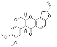
O=C1[C@H]2C3C=C(OC)C(OC)=CC=3OC[C@H]2OC2C3C[C@H](C(=C)C)OC=3C=CC1=2
|
| InChi Key |
JUVIOZPCNVVQFO-HBGVWJBISA-N
|
| InChi Code |
InChI=1S/C23H22O6/c1-11(2)16-8-14-15(28-16)6-5-12-22(24)21-13-7-18(25-3)19(26-4)9-17(13)27-10-20(21)29-23(12)14/h5-7,9,16,20-21H,1,8,10H2,2-4H3/t16-,20-,21+/m1/s1
|
| 化学名 |
(1S,6R,13S)-16,17-dimethoxy-6-prop-1-en-2-yl-2,7,20-trioxapentacyclo[11.8.0.03,11.04,8.014,19]henicosa-3(11),4(8),9,14,16,18-hexaen-12-one
|
| 别名 |
Ro-KO; Rotenone; Cube root; 83-79-4; Dactinol; Paraderil; Barbasco; Tubatoxin; (-)-Rotenone; Derris; Ronone; HSDB 1762; bCube-Pulver
|
| HS Tariff Code |
2934.99.9001
|
| 存储方式 |
Powder -20°C 3 years 4°C 2 years In solvent -80°C 6 months -20°C 1 month 注意: 请将本产品存放在密封且受保护的环境中(例如氮气保护),避免吸湿/受潮。 |
| 运输条件 |
Room temperature (This product is stable at ambient temperature for a few days during ordinary shipping and time spent in Customs)
|
| 溶解度 (体外实验) |
DMSO: ~50 mg/mL (~126.8 mM)
H2O: < 0.1 mg/mL |
|---|---|
| 溶解度 (体内实验) |
配方 1 中的溶解度: ≥ 2.5 mg/mL (6.34 mM) (饱和度未知) in 10% DMSO + 40% PEG300 + 5% Tween80 + 45% Saline (这些助溶剂从左到右依次添加,逐一添加), 澄清溶液。
例如,若需制备1 mL的工作液,可将100 μL 25.0 mg/mL澄清DMSO储备液加入到400 μL PEG300中,混匀;然后向上述溶液中加入50 μL Tween-80,混匀;加入450 μL生理盐水定容至1 mL。 *生理盐水的制备:将 0.9 g 氯化钠溶解在 100 mL ddH₂O中,得到澄清溶液。 配方 2 中的溶解度: 2.5 mg/mL (6.34 mM) in 10% DMSO + 90% (20% SBE-β-CD in Saline) (这些助溶剂从左到右依次添加,逐一添加), 悬浊液; 超声助溶。 例如,若需制备1 mL的工作液,可将 100 μL 25.0 mg/mL澄清DMSO储备液加入900 μL 20% SBE-β-CD生理盐水溶液中,混匀。 *20% SBE-β-CD 生理盐水溶液的制备(4°C,1 周):将 2 g SBE-β-CD 溶解于 10 mL 生理盐水中,得到澄清溶液。 View More
配方 3 中的溶解度: ≥ 2.5 mg/mL (6.34 mM) (饱和度未知) in 10% DMSO + 90% Corn Oil (这些助溶剂从左到右依次添加,逐一添加), 澄清溶液。 配方 4 中的溶解度: 2.5 mg/mL (6.34 mM) in 5% DMSO + 95% (20% SBE-β-CD in Saline) (这些助溶剂从左到右依次添加,逐一添加), 悬浊液; 超声助溶。 *20% SBE-β-CD 生理盐水溶液的制备(4°C,1 周):将 2 g SBE-β-CD 溶解于 10 mL 生理盐水中,得到澄清溶液。 1、请先配制澄清的储备液(如:用DMSO配置50 或 100 mg/mL母液(储备液)); 2、取适量母液,按从左到右的顺序依次添加助溶剂,澄清后再加入下一助溶剂。以 下列配方为例说明 (注意此配方只用于说明,并不一定代表此产品 的实际溶解配方): 10% DMSO → 40% PEG300 → 5% Tween-80 → 45% ddH2O (或 saline); 假设最终工作液的体积为 1 mL, 浓度为5 mg/mL: 取 100 μL 50 mg/mL 的澄清 DMSO 储备液加到 400 μL PEG300 中,混合均匀/澄清;向上述体系中加入50 μL Tween-80,混合均匀/澄清;然后继续加入450 μL ddH2O (或 saline)定容至 1 mL; 3、溶剂前显示的百分比是指该溶剂在最终溶液/工作液中的体积所占比例; 4、 如产品在配制过程中出现沉淀/析出,可通过加热(≤50℃)或超声的方式助溶; 5、为保证最佳实验结果,工作液请现配现用! 6、如不确定怎么将母液配置成体内动物实验的工作液,请查看说明书或联系我们; 7、 以上所有助溶剂都可在 Invivochem.cn网站购买。 |
| 制备储备液 | 1 mg | 5 mg | 10 mg | |
| 1 mM | 2.5354 mL | 12.6768 mL | 25.3537 mL | |
| 5 mM | 0.5071 mL | 2.5354 mL | 5.0707 mL | |
| 10 mM | 0.2535 mL | 1.2677 mL | 2.5354 mL |
1、根据实验需要选择合适的溶剂配制储备液 (母液):对于大多数产品,InvivoChem推荐用DMSO配置母液 (比如:5、10、20mM或者10、20、50 mg/mL浓度),个别水溶性高的产品可直接溶于水。产品在DMSO 、水或其他溶剂中的具体溶解度详见上”溶解度 (体外)”部分;
2、如果您找不到您想要的溶解度信息,或者很难将产品溶解在溶液中,请联系我们;
3、建议使用下列计算器进行相关计算(摩尔浓度计算器、稀释计算器、分子量计算器、重组计算器等);
4、母液配好之后,将其分装到常规用量,并储存在-20°C或-80°C,尽量减少反复冻融循环。
计算结果:
工作液浓度: mg/mL;
DMSO母液配制方法: mg 药物溶于 μL DMSO溶液(母液浓度 mg/mL)。如该浓度超过该批次药物DMSO溶解度,请首先与我们联系。
体内配方配制方法:取 μL DMSO母液,加入 μL PEG300,混匀澄清后加入μL Tween 80,混匀澄清后加入 μL ddH2O,混匀澄清。
(1) 请确保溶液澄清之后,再加入下一种溶剂 (助溶剂) 。可利用涡旋、超声或水浴加热等方法助溶;
(2) 一定要按顺序加入溶剂 (助溶剂) 。
|
|
|
|
|
|
|
|
InvivoChem的所有产品仅用于作科学研究,不面向患者销售
Copyright 2020 InvivoChem LLC | All Rights Reserved 粤ICP备20063088号-1


 463611831
