| 规格 | 价格 | 库存 | 数量 |
|---|---|---|---|
| 10 mM * 1 mL in DMSO |
|
||
| 1mg |
|
||
| 2mg |
|
||
| 5mg |
|
||
| 10mg |
|
||
| 25mg |
|
||
| 50mg |
|
||
| 100mg |
|
||
| 250mg |
|
||
| 500mg |
|
||
| Other Sizes |
|

| 靶点 |
MMP-2 (Ki = 13.9 nM); MMP-9 (Ki = 600 nM)
SB-3CT is a potent, selective mechanism-based inhibitor of gelatinases (matrix metalloproteinase-2/MMP-2 and MMP-9), with IC50 values of 0.3 nM for MMP-2 and 0.8 nM for MMP-9 in cell-free enzyme assays [2] - It shows no significant inhibition of other MMP subtypes (MMP-1, MMP-3, MMP-7) or human serine proteases (trypsin, plasmin) at concentrations up to 10 μM, confirming high gelatinase selectivity [2] |
|---|---|
| 体外研究 (In Vitro) |
SB-3CT在体外直接抑制Matrigel中的骨髓内皮细胞侵袭和小管形成。细胞测定:将PC3细胞接种在完全培养基中的35毫米培养皿中(5×10⁴个细胞/培养皿)。第二天,将培养基替换为仅补充有 1% DMSO(媒介物)或 1% DMSO 中的 SB-3CT(终浓度 0.1-50 μM)的完全培养基。在不同的时间,用胰蛋白酶收获细胞并计数。
在重组MMP-2/MMP-9酶反应中:1 nM SB-3CT 抑制MMP-2介导的明胶降解约98%,抑制MMP-9介导的明胶降解约95%(荧光明胶实验)[2] - 在人前列腺癌PC-3细胞(高表达MMP-9)中:5 μM SB-3CT 处理72小时可抑制细胞增殖约60%(MTT法),减少细胞侵袭约80%(Matrigel Transwell实验),下调MMP-9蛋白水平约75%(Western blot)[3] - 在氧糖剥夺(OGD,模拟缺血)处理的大鼠脑微血管内皮细胞(BMECs)中:2 μM SB-3CT 处理24小时可减少细胞凋亡约55%(Annexin V-FITC/PI染色),保留紧密连接蛋白ZO-1表达约65%(免疫荧光)[4] - 在脂多糖(LPS)激活的小鼠原代小胶质细胞中:1 μM SB-3CT 处理18小时可减少TNF-α分泌约60%、IL-1β分泌约55%(ELISA),机制为抑制MMP-9介导的促炎细胞因子激活[1] |
| 体内研究 (In Vivo) |
在 L-CI.5s T 细胞淋巴瘤模型中,SB-3CT(5-50 mg/kg/d,腹腔注射)可有效抑制肝转移并提高小鼠的存活率。 SB-3CT(50 mg/kg/d,腹膜内注射)可抑制骨转移模型中的人前列腺癌生长、骨质溶解和血管生成。在栓塞引起的“永久性”局灶性脑缺血的小鼠模型中,SB-3CT 可以抵消神经元层粘连蛋白的降解,保护神经元免受缺血性细胞死亡,并改善栓塞性 MCA 闭塞后的神经行为结果。
在重度创伤性脑损伤(TBI,控制性皮质撞击模型)的雄性Sprague-Dawley大鼠中:TBI后1小时静脉注射3 mg/kg SB-3CT,72小时后 cerebral 病灶体积较溶剂对照组减少约40%;免疫组化显示小胶质细胞激活(Iba-1⁺细胞)减少约50%[1] - 在PC-3前列腺癌骨转移裸鼠(胫骨内注射1×10⁵个细胞)中:每日一次口服10 mg/kg SB-3CT,持续28天,骨内肿瘤体积减少约55%,溶骨病灶面积减少约60%(显微CT成像);血浆MMP-9水平降低约70%(ELISA)[3] - 在栓塞性局灶性脑缺血(大脑中动脉阻塞/MCAO模型)的C57BL/6小鼠中:MCAO后30分钟静脉注射2 mg/kg SB-3CT,24小时后梗死体积减少约35%,神经功能缺损评分改善约40%[4] |
| 酶活实验 |
荧光猝灭底物 MOCAcPLGLA2pr(Dnp)-AR-NH2 用于测量 MMP-2、MMP-9 和 MMP-7 的酶活性。使用 PTI 荧光分光计测量荧光。比色皿室的温度设置为 25 °C。
MMP-2/MMP-9明胶酶活性检测流程(基于[2]):人重组MMP-2/MMP-9在激活缓冲液(50 mM Tris-HCl pH 7.5,10 mM CaCl₂,0.05% Brij-35)中用对氨基苯汞乙酸(APMA)激活。激活后的酶与荧光明胶底物(DQ-gelatin)及SB-3CT(0.01~10 nM)混合于反应缓冲液中,37°C孵育2小时。检测激发波长485 nm/发射波长535 nm处的荧光强度,相对于溶剂对照组计算抑制率,采用四参数逻辑回归确定IC50[2] - MMP-2/MMP-9选择性实验流程(基于[2]):重组MMP-1、MMP-3、MMP-7按与MMP-2/9相同的激活方案制备。每种酶与其特异性荧光肽底物(MMP-1:Mca-Pro-Leu-Gly-Leu-Dpa-Ala-Arg-NH₂;MMP-3:Mca-Arg-Pro-Lys-Pro-Tyr-Ala-Nva-Trp-Met-Lys(Dnp)-NH₂)及10 μM SB-3CT 混合,37°C孵育2小时后检测荧光;对非明胶酶MMPs无显著抑制(<5%)[2] |
| 细胞实验 |
细胞增殖试验[3]
将PC3细胞接种在完全培养基中的35 mm培养皿中(5×10⁴个细胞/皿)。第二天,将培养基替换为单独添加1%DMSO(载体)或1%DMSO中的SB-3CT终浓度0.1-50μM)的完全培养基。在不同时间,用胰蛋白酶收获细胞并计数。 SB-3CT对BMEC-1细胞存活率的影响[3] 将BMEC-1细胞接种在完全培养基中的96孔培养板(104个细胞/孔)中。24小时后,将培养基替换为无血清、无酚红的培养基,补充有载体(1%DMSO)或SB-3CT(终浓度为1 nM-50μM)。72小时后,根据制造商的说明,向每个孔中加入10μL WST-1,并在450nm处测量光密度。 毛细管样小管形成试验[3] 用300μL冰冷的Matrigel溶液(10mg/mL)涂覆24孔板。然后将平板在37°C下孵育30分钟,以进行Matrigel聚合,然后在添加了不同量的SB-3CT(0.1-1μM)或载体(1%DMSO)的完全培养基存在下,将5×104 BMEC-1细胞放置在Matrigel涂层孔上。在37°C下孵育过夜后,使用Olympus®DP12显微相机以10倍放大率拍摄每个孔中随机选择的三个区域的数码照片。使用Adobe Photoshop 7.0计算毛细管状结构所占的面积。 内皮细胞侵袭试验[3] 将悬浮在含有0.1%牛血清白蛋白的Medium-199中的BMEC-1细胞接种到涂有25μg/过滤器Matrigel的Transwell插入物(8μM孔径)上(每个插入物接种2×105个细胞),补充有SB-3CT(0.1-1μM)或1%DMSO(载体)。将补充有5%FBS的培养基作为化学引诱剂放置在下腔室中。在37°C下孵育24小时后,迁移到过滤器下侧的细胞用Diff-Quik®染色,并在200倍放大倍数下计数。 PC-3细胞侵袭实验流程(基于[3]):人前列腺癌PC-3细胞在含10%胎牛血清(FBS)的RPMI 1640培养基中培养至80%汇合。胰酶消化后,用无血清RPMI 1640重悬,以5×10⁴细胞/孔接种于含SB-3CT(1~10 μM)的Matrigel包被Transwell上室,下室加入含10% FBS的RPMI 1640(趋化因子)。48小时后去除上室未侵袭细胞,甲醇固定、结晶紫染色后显微镜下计数侵袭细胞;处理72小时后通过MTT法(570 nm吸光度)评估细胞增殖[3] - BMEC氧糖剥夺(OGD)模型实验流程(基于[4]):大鼠BMECs在DMEM/F12培养基+10% FBS中培养。为诱导OGD,将细胞转移至无糖DMEM中,在低氧培养箱(1% O₂、5% CO₂、94% N₂)中孵育4小时。复氧(21% O₂)期间加入SB-3CT(0.5~5 μM)处理24小时。Annexin V-FITC/PI染色流式细胞术检测凋亡,抗ZO-1抗体免疫染色观察紧密连接[4] |
| 动物实验 |
五周龄雄性CB-17.SCID小鼠[3]
50 mg/kg 腹腔注射;隔日一次;持续五周 原位明胶酶谱分析[3] 将HT1080肿瘤皮下移植到SCID小鼠体内,并在处死前连续两天腹腔注射(ip)1 mL载体(10% DMSO的PBS溶液)或1 mL含1.25 mg SB-3CT的10% DMSO溶液(相当于50 mg/kg小鼠体重)。按照先前描述的方法,将8 μm厚的未固定冰冻切片肿瘤组织与100 μg/mL DQ™-明胶和1 μg/mL DAPI(Molecular Probes公司)孵育1小时,进行原位明胶酶谱分析。 PC3人骨肿瘤的建立及实验治疗[3] 如前所述29,将四分之一人胎儿股骨碎片皮下植入SCID小鼠体内。四周后,如前所述29,将1 × 10⁵个PC3细胞经小鼠皮肤直接注射到先前植入骨的骨髓中。肿瘤细胞接种24小时后,每隔一天腹腔注射小鼠载体(10% DMSO)或SB-3CT(溶于10% DMSO,50 mg/kg小鼠体重)。每个实验组包含9只动物。 肿瘤细胞接种五周后,处死小鼠,取出骨植入物,称重,在10%中性缓冲福尔马林中固定过夜,然后使用Lo-Rad M-IV乳腺X线摄影仪采用放大标本技术进行X射线成像。使用柯达2000显影屏和X线胶片显影图像。为进行组织形态计量学和组织学分析,骨肿瘤样本先用10%乙二胺四乙酸(EDTA)(pH 6.5)的PBS溶液脱钙,然后脱水、浸润并进行石蜡包埋。本研究采用已报道的方法合成了Mobashery实验室发现的SB-3CT。小鼠被分为四组:载体对照组和SB-3CT治疗组,SB-3CT治疗组分别在栓塞性大脑中动脉(MCA)闭塞后接受1天或7天的治疗。将SB-3CT(12.5 mg/mL)新鲜溶解于25% DMSO/65% PEG-200/10%水中,并通过带有0.2 μm、直径13 mm的无菌疏水性PTFE膜的Acrodisc针筒式过滤器进行过滤。小鼠在栓塞性缺血后2小时腹腔注射2 μL/g体重的该溶液(相当于25 mg/kg),4小时后再次注射。在重复给药条件下,在栓塞性缺血后2小时和4小时腹腔注射相同剂量的SB-3CT,随后从缺血后第1天到第6天每天注射一次。先前的研究表明,腹腔注射SB-3CT不会改变平均动脉血压、pH值、PCO2和PO2[4]。 大鼠TBI模型(引自[1]):雄性Sprague-Dawley大鼠(250-300 g)麻醉后进行可控皮层冲击(CCI)以诱导严重TBI(冲击深度:2.5 mm,速度:4 m/s)。 TBI后1小时,大鼠接受静脉注射SB-3CT(3 mg/kg,溶于10% DMSO + 90%生理盐水)或载体。TBI后72小时,处死大鼠;收集脑组织,切片,并用2,3,5-氯化三苯基四氮唑(TTC)染色以测量病灶体积。采用抗Iba-1抗体进行免疫组织化学染色以评估小胶质细胞活化[1]。裸鼠PC-3骨转移模型(引自[3]):将6-8周龄的雌性裸鼠麻醉,并将1×10⁵个PC-3细胞(悬浮于0.05 mL PBS中)注射到左侧胫骨髓腔内。接种后7天,将小鼠分为两组:(1)SB-3CT组:每日一次灌胃给予10 mg/kg溶于5% DMSO + 95%玉米油的SB-3CT;(2)载体组:5% DMSO + 95%玉米油。28天后,处死小鼠;收集胫骨进行微型CT成像(用于量化溶骨性病变)和肿瘤体积测量。采用ELISA法分析血浆中MMP-9的含量[3]。 - 小鼠MCAO模型(引自[4]):将雄性C57BL/6小鼠(20-25 g)麻醉,用尼龙线结扎大脑中动脉(MCA)60分钟,以诱导局灶性脑缺血。 MCAO后30分钟,小鼠接受静脉注射SB-3CT(2 mg/kg,溶于5%乙醇+95%生理盐水)或载体。再灌注24小时后,处死小鼠;将脑组织切片并用TTC染色以测量梗死体积。处死前评估神经功能缺损评分(0-5分)[4] |
| 毒性/毒理 (Toxicokinetics/TK) |
在人/大鼠/小鼠细胞(PC-3、BMEC、小胶质细胞)中:浓度高达 10 μM 的 SB-3CT 处理 72 小时未见明显的细胞毒性(细胞存活率 >90% vs. 溶剂对照组,MTT 法)[1,3,4]
- 在大鼠(TBI 模型,3 mg/kg 静脉注射)和小鼠(MCAO 模型,2 mg/kg 静脉注射;PC-3 模型,10 mg/kg 口服)中:在治疗剂量下未检测到明显的体重减轻(>初始体重的 5%)或肝脏、肾脏或脾脏的组织病理学异常[1,3,4] |
| 参考文献 |
|
| 其他信息 |
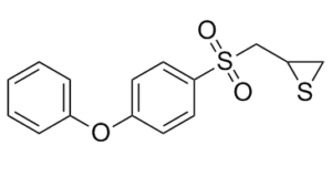
2-[(4-苯氧基苯基)磺酰甲基]硫杂环丙烷是一种芳香醚。
SB-3CT 是一种合成的、基于机制的选择性明胶酶 (MMP-2/9) 抑制剂,其特征在于与 MMP 活性位点不可逆结合,使其成为 MMP-2/9 介导疾病临床前研究的宝贵工具 [2] - 其治疗潜力主要集中在神经系统疾病(TBI、脑缺血)和癌症转移(前列腺癌骨转移),通过抑制 MMP-2/9 介导的细胞外基质降解、炎症和血管生成 [1,3,4] - 摘要中未提供临床开发(I/II 期)或 FDA 批准信息;它主要用作研究试剂,用于研究 MMP-2/9 生物学 [1,2,3,4] - 开发了一种水溶性 SB-3CT 衍生物,用于改善神经系统模型(例如 TBI)中的静脉给药,其 MMP-2/9 抑制效力与母体化合物相似 [1] |
| 分子式 |
C15H14O3S2
|
|
|---|---|---|
| 分子量 |
306.40
|
|
| 精确质量 |
306.038
|
|
| 元素分析 |
C, 58.80; H, 4.61; O, 15.67; S, 20.93
|
|
| CAS号 |
292605-14-2
|
|
| 相关CAS号 |
|
|
| PubChem CID |
9883002
|
|
| 外观&性状 |
White to pink solid powder
|
|
| 密度 |
1.3±0.1 g/cm3
|
|
| 沸点 |
501.4±46.0 °C at 760 mmHg
|
|
| 熔点 |
101 °C
|
|
| 闪点 |
257.1±29.0 °C
|
|
| 蒸汽压 |
0.0±1.2 mmHg at 25°C
|
|
| 折射率 |
1.628
|
|
| LogP |
3.36
|
|
| tPSA |
77.05
|
|
| 氢键供体(HBD)数目 |
0
|
|
| 氢键受体(HBA)数目 |
4
|
|
| 可旋转键数目(RBC) |
5
|
|
| 重原子数目 |
20
|
|
| 分子复杂度/Complexity |
401
|
|
| 定义原子立体中心数目 |
0
|
|
| SMILES |
S1C([H])([H])C1([H])C([H])([H])S(C1C([H])=C([H])C(=C([H])C=1[H])OC1C([H])=C([H])C([H])=C([H])C=1[H])(=O)=O
|
|
| InChi Key |
LSONWRHLFZYHIN-UHFFFAOYSA-N
|
|
| InChi Code |
InChI=1S/C15H14O3S2/c16-20(17,11-14-10-19-14)15-8-6-13(7-9-15)18-12-4-2-1-3-5-12/h1-9,14H,10-11H2
|
|
| 化学名 |
2-[(4-phenoxyphenyl)sulfonylmethyl]thiirane
|
|
| 别名 |
SB3CT; SB3-CT; 2-[(4-phenoxyphenyl)sulfonylmethyl]thiirane; 2-((4-phenoxyphenylsulfonyl)methyl)thiirane; 2-(((4-Phenoxyphenyl)sulfonyl)methyl)thiirane; (4-phenoxyphenylsulfonyl)methylthiirane; CHEMBL483857; Thiirane, 2-[[(4-phenoxyphenyl)sulfonyl]methyl]-; SB-3CT
|
|
| HS Tariff Code |
2934.99.9001
|
|
| 存储方式 |
Powder -20°C 3 years 4°C 2 years In solvent -80°C 6 months -20°C 1 month 注意: 请将本产品存放在密封且受保护的环境中,避免吸湿/受潮。 |
|
| 运输条件 |
Room temperature (This product is stable at ambient temperature for a few days during ordinary shipping and time spent in Customs)
|
| 溶解度 (体外实验) |
|
|||
|---|---|---|---|---|
| 溶解度 (体内实验) |
配方 1 中的溶解度: 5 mg/mL (16.32 mM) in 10% DMSO 20% Cremophor EL + 70% ddH2O (这些助溶剂从左到右依次添加,逐一添加), 悬浮液;超声助溶。
配方 2 中的溶解度: 2.5 mg/mL (8.16 mM) in 10% DMSO + 40% PEG300 + 5% Tween80 + 45% Saline (这些助溶剂从左到右依次添加,逐一添加), 悬浊液; 超声助溶。 例如,若需制备1 mL的工作液,可将 100 μL 25.0 mg/mL澄清DMSO储备液加入到400 μL PEG300中,混匀;然后向上述溶液中加入50 μL Tween-80,混匀;加入450 μL生理盐水定容至1 mL。 *生理盐水的制备:将 0.9 g 氯化钠溶解在 100 mL ddH₂O中,得到澄清溶液。 View More
配方 3 中的溶解度: ≥ 2.5 mg/mL (8.16 mM) (饱和度未知) in 10% DMSO + 90% (20% SBE-β-CD in Saline) (这些助溶剂从左到右依次添加,逐一添加), 澄清溶液。 配方 4 中的溶解度: ≥ 2.5 mg/mL (8.16 mM) (饱和度未知) in 10% DMSO + 90% Corn Oil (这些助溶剂从左到右依次添加,逐一添加), 澄清溶液。 例如,若需制备1 mL的工作液,可将100 μL 25.0 mg/mL 澄清 DMSO 储备液加入900 μL 玉米油中,混合均匀。 配方 5 中的溶解度: 4% DMSO+corn oil: 10mg/mL 1、请先配制澄清的储备液(如:用DMSO配置50 或 100 mg/mL母液(储备液)); 2、取适量母液,按从左到右的顺序依次添加助溶剂,澄清后再加入下一助溶剂。以 下列配方为例说明 (注意此配方只用于说明,并不一定代表此产品 的实际溶解配方): 10% DMSO → 40% PEG300 → 5% Tween-80 → 45% ddH2O (或 saline); 假设最终工作液的体积为 1 mL, 浓度为5 mg/mL: 取 100 μL 50 mg/mL 的澄清 DMSO 储备液加到 400 μL PEG300 中,混合均匀/澄清;向上述体系中加入50 μL Tween-80,混合均匀/澄清;然后继续加入450 μL ddH2O (或 saline)定容至 1 mL; 3、溶剂前显示的百分比是指该溶剂在最终溶液/工作液中的体积所占比例; 4、 如产品在配制过程中出现沉淀/析出,可通过加热(≤50℃)或超声的方式助溶; 5、为保证最佳实验结果,工作液请现配现用! 6、如不确定怎么将母液配置成体内动物实验的工作液,请查看说明书或联系我们; 7、 以上所有助溶剂都可在 Invivochem.cn网站购买。 |
| 制备储备液 | 1 mg | 5 mg | 10 mg | |
| 1 mM | 3.2637 mL | 16.3185 mL | 32.6371 mL | |
| 5 mM | 0.6527 mL | 3.2637 mL | 6.5274 mL | |
| 10 mM | 0.3264 mL | 1.6319 mL | 3.2637 mL |
1、根据实验需要选择合适的溶剂配制储备液 (母液):对于大多数产品,InvivoChem推荐用DMSO配置母液 (比如:5、10、20mM或者10、20、50 mg/mL浓度),个别水溶性高的产品可直接溶于水。产品在DMSO 、水或其他溶剂中的具体溶解度详见上”溶解度 (体外)”部分;
2、如果您找不到您想要的溶解度信息,或者很难将产品溶解在溶液中,请联系我们;
3、建议使用下列计算器进行相关计算(摩尔浓度计算器、稀释计算器、分子量计算器、重组计算器等);
4、母液配好之后,将其分装到常规用量,并储存在-20°C或-80°C,尽量减少反复冻融循环。
计算结果:
工作液浓度: mg/mL;
DMSO母液配制方法: mg 药物溶于 μL DMSO溶液(母液浓度 mg/mL)。如该浓度超过该批次药物DMSO溶解度,请首先与我们联系。
体内配方配制方法:取 μL DMSO母液,加入 μL PEG300,混匀澄清后加入μL Tween 80,混匀澄清后加入 μL ddH2O,混匀澄清。
(1) 请确保溶液澄清之后,再加入下一种溶剂 (助溶剂) 。可利用涡旋、超声或水浴加热等方法助溶;
(2) 一定要按顺序加入溶剂 (助溶剂) 。
|
|---|
|
|
|
|---|
InvivoChem的所有产品仅用于作科学研究,不面向患者销售
Copyright 2020 InvivoChem LLC | All Rights Reserved 粤ICP备20063088号-1


 COA
COA


 463611831
