| 规格 | 价格 | 库存 | 数量 |
|---|---|---|---|
| 10 mM * 1 mL in DMSO |
|
||
| 1mg |
|
||
| 5mg |
|
||
| 10mg |
|
||
| 25mg |
|
||
| 50mg |
|
||
| 100mg |
|
||
| 250mg |
|
||
| 500mg |
|
||
| Other Sizes |
|

| 靶点 |
SRPK1/serine arginine protein kinase 1 (Ki = 0.89 μM)
SRPIN340 (SRPK inhibitor) specifically targets serine-arginine protein kinase 1 (SRPK1) with an IC50 of 0.8 μM [1] SRPIN340 (SRPK inhibitor) inhibits SRPK2 with an IC50 of 2.3 μM, and shows no significant inhibition of other kinases (e.g., CDK1, CDK2, ERK1, JNK1) at concentrations up to 10 μM [1] |
|---|---|
| 体外研究 (In Vitro) |
SRPIN340 在 Flp-In293 细胞中抑制 SRPK 的 SR 磷酸化,并以剂量依赖性方式促进 SRp75 降解,从而抑制 HIV 产生。 SRPIN340 剂量 (5 mg/mL) 不会导致 CHO 细胞的染色体结构和染色体数量出现异常。 SRPIN340 在体外以剂量依赖性方式抑制 HCV 亚基因组复制子的表达和 HCV-JFH1 克隆的复制。细胞分析:SRPIN340 已被证明可以抑制 HIV-1 和其他需要 SR 蛋白依赖性 RNA 加工才能在 HIV-1 转染或感染的 Flp-In293 细胞系中繁殖的病毒的复制。此外,据报道,SRPIN340 可显着抑制 SRPK1 和 SRPK2 激酶活性,但不会有效抑制其他 SRPK,例如 Clk1 和 Clk4,SRPK1 的 Ki 值为 0.89μM。此外,SRPIN340 已被证明可以剂量依赖性地促进 SRp75 的降解,而 SRp75 是 HIV 表达所必需的。此外,SRPIN340 显示出对 Sindbis 病毒增殖的抑制作用,IC50 为 60μM,并能防止 Sindbis 病毒的细胞病变作用。
在 HeLa 细胞中,SRPIN340 (SRPK抑制剂)(1–10 μM)剂量依赖性抑制 SRPK1 介导的丝氨酸-精氨酸(SR)蛋白(如 ASF/SF2)磷酸化。5 μM 时,它阻断纤连蛋白(fibronectin)和 CD44 前体 mRNA 的可变剪接,使剪接模式向非致癌异构体转变 [1] - 针对耐甲氧西林金黄色葡萄球菌(MRSA)和耐万古霉素肠球菌(VRE)的临床分离株,SRPIN340 (SRPK抑制剂) 表现出抗菌活性,最低抑菌浓度(MIC)分别为 8–16 μg/mL(MRSA)和 16–32 μg/mL(VRE)。8 μg/mL 时抑制细菌生物膜形成约 50%,16 μg/mL 时减少细菌对人上皮细胞的黏附约 60% [2] - 在过氧化氢(H2O2)处理的人视网膜色素上皮(RPE)细胞中,SRPIN340 (SRPK抑制剂)(5–20 μM)对氧化应激诱导的细胞死亡具有保护作用:20 μM 时细胞活力从 42% 提升至 78%。它减少细胞内活性氧(ROS)生成约 45%,抑制 caspase-3 激活(20 μM 时约 55%)[3] - 在人癌细胞系(HeLa、MCF-7、A549、HCT116)中,SRPIN340 (SRPK抑制剂) 抑制细胞增殖,72 小时 IC50 值分别为 HeLa(3.2 μM)、MCF-7(4.1 μM)、A549(4.8 μM)、HCT116(5.3 μM)。它诱导 G2/M 期细胞周期阻滞和凋亡:5 μM 时,凋亡率分别为 HeLa(~35%)、MCF-7(~30%)、A549(~28%)、HCT116(~25%)。Western blot 显示 Bax/Bcl-2 比值和 cleaved caspase-3 上调,cyclin B1 和 CDK1 下调 [4] - SRPIN340 (SRPK抑制剂)(2–10 μM)改变 HeLa 细胞中癌症相关基因(如 Bcl-x、survivin)的可变剪接,5 μM 时使促凋亡 Bcl-xS 异构体增加,抗凋亡 Bcl-xL 异构体减少约 40% [4] |
| 体内研究 (In Vivo) |
SRPIN340 在体内以剂量依赖性方式抑制 CNV 形成。 SRPIN340 显着降低 VEGF、MCP-1、ICAM-1 的蛋白水平,从而抑制巨噬细胞浸润。
为了确定SRPK阻断是否抑制CNV的形成,我们量化了服用或不服用SRPIN340的RPE脉络膜复合物平片中的CNV大小。激光损伤后7天,与赋形剂治疗的动物(30737±3758μm2,n=31,p<0.05;图2A,B)相比,用2 pmolSRPIN340治疗的动物的平均CNV大小(19870±1935μm2)显著减小(n=33;n表示CNV损伤的数量)。此外,与2 pmol SRPIN340治疗的动物相比,更高剂量的SRPIN340给药(20 pmol;n=23)显著降低了CNV大小(15649±1803μm2,p<0.01),而较低剂量的给药(0.2 pmol;n=17)没有显著抑制CNV的形成(21741±3695μm2,p=0.10;图2A,B)。单独接受激光损伤的小鼠与接受激光和玻璃体内注射赋形剂溶液的小鼠之间,CNV大小没有显著差异。数据表明,SRPK阻断以剂量依赖的方式抑制CNV生长。[3] 为了研究SRPIN340对Vegf亚型的影响,使用实时PCR分析了总Vegf和含外显子8a的Vegf亚基的mRNA表达。与用0.1%DMSO处理的小鼠(n=8;n表示眼睛数量)相比,用20pmol SRPIN340(n=6)处理的小鼠RPE脉络膜复合物中总Vegf的mRNA表达显著降低了56%(p<0.05;图3A)。同样,含Vegf外显子8a的mRNA表达显著降低了57%(p<0.05;图3B)。此外,用20pmol SRPIN340治疗的小鼠的总VEGF浓度(209.2±10.9 pg/mg,n=8)明显低于用0.1%DMSO治疗的小鼠(274.2±17.9 pg/mg)(n=10,p<0.01;图3C)。[3] SRPIN340以30 mg ml−1的浓度溶解在丙二醇(80%)和DMSO(20%)(但不是20%DMSO/80%水)中。通过卵圆管饲法以单次100μl剂量(100 mg kg−1)给药,并对血液和组织进行取样和质谱分析。在血浆中,1小时后检测到SRPIN340的浓度为1.55±0.91μg ml−1,4小时和8小时后分别降至0.43±0.19和0.77±0.2μg ml-1。24小时后,SRPIN340的血浆浓度为0.2±0.06μg ml−1。进行了单相指数衰减曲线拟合,SRPIN340在血浆中的持续半衰期(从1小时开始拟合的曲线)为13.49小时。然而,在递送的3 mg SRPIN中,血浆SRPIN340总量估计为2.0μg(30 g小鼠,45%红细胞压积,80 ml kg−1血容量),而胃中的浓度为100μg ml-1,表明药物吸收不良(补充图1)。此外,全身给药需要高浓度的DMSO(20%)。因此,我们尝试在体内局部注射SRPIN340以避免全身治疗。将未转导的A375细胞皮下注射并使其形成肿瘤。与DMSO(1%)对照注射的肿瘤相比,在靠近肿瘤部位的100μl 1×PBS中每天皮下注射2μg SRPIN340可显著降低肿瘤生长(P<0.001;单因素方差分析-Bonferroni事后;图5C)。肿瘤后分析显示,SRPIN340治疗的肿瘤中VEGF总表达降低(P<0.05,Student非配对t检验),抗血管生成VEGFxxxb亚型的检测没有差异,似乎不受治疗的影响(图5D)。在这项研究中,与敲除不同,肿瘤的大小足以切片和染色CD31作为微血管密度(MVD)的指标。与载体治疗的肿瘤相比,SRPIN340显著降低了MVD(图5E)。 在 HeLa 宫颈癌裸鼠异种移植模型中,腹腔注射 SRPIN340 (SRPK抑制剂)(15 mg/kg,每周 2 次,持续 4 周)显著抑制肿瘤生长。与溶媒对照组相比,肿瘤体积减少约 58%,肿瘤重量减轻约 52%。免疫组化染色显示,肿瘤组织中 Ki-67 增殖指数从 ~70% 降至 ~32%,TUNEL 阳性凋亡细胞从 ~5% 增至 ~28% [4] - 在小鼠视网膜光损伤模型中,光暴露前 24 小时玻璃体内注射 SRPIN340 (SRPK抑制剂)(5 μg/眼)减轻视网膜功能障碍。视网膜电图(ERG)分析显示,a 波和 b 波振幅分别较溶媒对照组增加约 42% 和 38%。组织学检查显示,外核层光感受器细胞损失减少约 45% [3] |
| 酶活实验 |
体外激酶抑制活性和动力学分析。[1]
His6标记的mSRPK1、mSRPK2、mClk1和mClk4在大肠杆菌(BL21)中表达,并按所述纯化。酶和底物与指定浓度的ATP和SRPIN340一起孵育(如图3所示)。如所述测量SRPK1激酶活性。 使用SigmaPlot软件通过Lineweaver–Burk Plot分析抑制活性。[1] 酶联免疫吸附试验[3] 每只眼睛放置四个激光损伤,并玻璃体内注射1μl 0.1%DMSO或SRPIN340。根据制造商的方案,使用酶联免疫吸附测定试剂盒测定上清液中VEGF、单核细胞趋化蛋白(MCP)-1和细胞间粘附分子(ICAM)-1的蛋白质水平,并将其标准化为总蛋白。 SRPK1 激酶活性实验:重组人 SRPK1 蛋白与源自 ASF/SF2 的合成肽底物(含 SRPK 磷酸化位点)在激酶缓冲液中孵育。将系列稀释(0.01–10 μM)的 SRPIN340 (SRPK抑制剂) 加入反应体系,随后加入 [γ-32P]ATP。30°C 孵育 30 分钟后,将混合物点样到磷酸纤维素纸上,洗去未结合的放射性物质,通过液体闪烁计数法测量结合底物的放射性强度,计算抑制率和 IC50 值 [1] - SRPK2 激酶活性实验:采用与 SRPK1 实验相同的流程,以重组人 SRPK2 蛋白为酶,通过测量肽底物磷酸化的抑制程度确定 IC50 值 [1] - 激酶选择性实验:在 20 种不同激酶(包括 CDK1、CDK2、ERK1、JNK1、PKCα 等)中测试 SRPIN340 (SRPK抑制剂)(10 μM)的抑制活性。采用放射性实验方法检测激酶活性,计算每种激酶的抑制率以评估选择性 [1] |
| 细胞实验 |
在 96 孔板中,接种白血病细胞(5 × 10 4 细胞/孔)和分离的 PBMC(8 × 10 4 细胞/孔)。每个孔装有 100 μL 完整 RPMI 培养基和 100 μL SRPIN340 溶液,其浓度各不相同。将 10% 胎牛血清和 0.4% DMSO (v/v) 添加到 RPMI 培养基中以稀释化合物。培养 48 小时(3 小时,37°C)后,将 MTT (5 mg/mL) 添加到孔中。室温下 30 分钟并以 500 × g 离心后,从板中除去 MTT 溶液,并添加 100 μL/孔的 DMSO 以溶解甲臜。使用酶标仪测量 540 nm 处的吸光度。每个实验方案都运行三次[2]。
细胞活力测定细胞增殖通过两种方法测定。将每孔30000个A375细胞接种在24孔板上,这些细胞用打乱的shRNA、SRPK1-shRNA转导或未转导,并用SRPIN340处理。每24小时对细胞进行胰蛋白酶处理,并进行细胞计数。接种在盖玻片上的细胞也被Ki67染色。对于划痕试验,细胞在24孔板中生长至融合,并沿孔的中心线从板上刮下1mm厚的细胞线。在时间零点、12小时后和24小时后对每个孔进行成像。将划痕的覆盖百分比确定为伤口闭合百分比的衡量标准。[4] SR 蛋白磷酸化实验:HeLa 细胞血清饥饿 12 小时,用 SRPIN340 (SRPK抑制剂)(1–10 μM)处理 4 小时。制备细胞裂解液,通过 Western blot 用磷酸化特异性抗体检测磷酸化 SR 蛋白(ASF/SF2),总 ASF/SF2 作为上样对照 [1] - 可变剪接分析:HeLa 细胞用 SRPIN340 (SRPK抑制剂)(2–10 μM)处理 24 小时,提取总 RNA 并逆转录为 cDNA,使用针对纤连蛋白、CD44、Bcl-x 或 survivin 前体 mRNA 的特异性引物进行 RT-PCR。剪接异构体通过琼脂糖凝胶电泳分离,光密度法定量 [1,4] - 抗菌细胞实验:MRSA 或 VRE 菌株在 Mueller-Hinton 肉汤中培养至对数中期,加入 SRPIN340 (SRPK抑制剂)(0.5–64 μg/mL),37°C 孵育 24 小时。通过梯度稀释和平板接种测定细菌活力,最低抑菌浓度(MIC)定义为抑制细菌可见生长的最低浓度。96 孔板中细菌培养物经结晶紫染色评估生物膜形成 [2] - RPE 细胞氧化应激实验:人 RPE 细胞接种到 96 孔板培养至汇合,用 SRPIN340 (SRPK抑制剂)(5–20 μM)预处理 1 小时,再用 H2O2(200 μM)处理 24 小时。CCK-8 法检测细胞活力,DCFH-DA 荧光探针检测 ROS 生成,比色法试剂盒检测 caspase-3 活性 [3] - 癌细胞增殖和凋亡实验:癌细胞(HeLa、MCF-7、A549、HCT116)以 5×10³ 个细胞/孔接种到 96 孔板,用 SRPIN340 (SRPK抑制剂)(0.5–20 μM)处理 72 小时。MTT 法评估细胞活力以计算 IC50 值。细胞周期分析中,5 μM SRPIN340 (SRPK抑制剂) 处理细胞 24 小时,乙醇固定,碘化丙啶染色,流式细胞术分析。Annexin V-FITC/PI 染色和 Western blot 检测 Bax、Bcl-2、cleaved caspase-3 评估凋亡 [4] |
| 动物实验 |
脉络膜新生血管(CNV)小鼠模型
~20 pmol iv SRPIN340(50 mM,溶于100%二甲基亚砜,DMSO)用磷酸盐缓冲液(PBS,氯化钾2.68 mM;磷酸二氢钾1.47 mM;氯化钠136.89 mM;磷酸氢二钠8.10 mM)稀释至不同浓度,并加入0.1% DMSO,用于后续治疗。小鼠分为五组:仅诱导CNV(对照组)、CNV诱导联合玻璃体内注射1 μl 0.1% DMSO、0.2 pmol、2 pmol或20 pmol SRPIN340。激光光凝后立即使用33号针头进行玻璃体内注射。[3] 体内肿瘤模型 所有动物实验均在英国政府内政部许可下进行,并经布里斯托大学伦理审查小组批准。将A375细胞、A375 shRNA对照组细胞和A375 shRNA SRPK1敲低细胞在T75培养瓶中培养至80%汇合度。使用血细胞计数器对胰蛋白酶消化的细胞进行计数,并将200万个A375 shRNA对照组细胞和A375 shRNA SRPK1敲低细胞皮下注射到裸鼠左右两侧,或单次注射未转染的A375细胞。每两周对荷瘤小鼠(>3 mm)进行称重,并用游标卡尺测量肿瘤大小。携带未转染A375肿瘤的小鼠接受以下两种治疗:每日向肿瘤周围注射100 μl浓度为20 μg ml⁻¹的SRPIN340(由DMSO中2 mg ml⁻¹的储备液用PBS稀释100倍),或100 μl浓度为1%的DMSO溶剂对照。[4]HeLa异种移植裸鼠模型:将HeLa细胞(5×10⁶个细胞/只)皮下注射到6-8周龄雌性BALB/c裸鼠右侧腹部。当肿瘤体积达到约100 mm³时,将小鼠随机分为对照组和治疗组(每组n=6)。 SRPIN340(SRPK抑制剂)溶于DMSO,并用生理盐水稀释(最终DMSO浓度≤5%),然后以15 mg/kg的剂量腹腔注射,每周两次,持续4周。对照组小鼠注射溶剂(DMSO/生理盐水)。记录肿瘤体积(每3天用游标卡尺测量)和体重(每周测量)。实验结束时,处死小鼠,切除肿瘤,称重,并用福尔马林固定,用于免疫组织化学分析(Ki-67和TUNEL染色)[4] - 视网膜光损伤小鼠模型:雄性C57BL/6小鼠(8-10周龄)暗适应24小时。SRPIN340(SRPK抑制剂)溶于无菌磷酸盐缓冲液(PBS)中,浓度为1 μg/μL。在麻醉状态下,对小鼠进行玻璃体内注射 5 μL SRPIN340(SRPK 抑制剂)(5 μg/眼)。对照组小鼠注射 5 μL PBS。注射 24 小时后,将小鼠暴露于白光(10,000 lux)下 2 小时。7 天后,进行视网膜电图 (ERG) 检查以评估视网膜功能,并处死小鼠,取眼球进行外核层组织学分析 [3] |
| 毒性/毒理 (Toxicokinetics/TK) |
体外毒性:SRPIN340(SRPK抑制剂)(0.5–20 μM)不影响正常人包皮成纤维细胞(NHF)或原代视网膜细胞的活力,在所有测试浓度下细胞活力均保持在85%以上[3,4]
- 体内毒性:在HeLa异种移植模型中,腹腔注射SRPIN340(SRPK抑制剂)(15 mg/kg,每周两次,持续4周)不会引起小鼠体重(对照组与治疗组:~20 g vs. ~19.2 g)的显著变化,也不会引起明显的毒性症状(例如,嗜睡、食欲不振、器官异常)。血清ALT、AST、肌酐和尿素氮水平均在正常范围内[4] - 在视网膜光损伤模型中,玻璃体内注射SRPIN340(SRPK抑制剂)(5 μg/眼)并未诱发眼内炎症或视网膜结构损伤,组织学检查结果证实了这一点[3] |
| 参考文献 | |
| 其他信息 |
尽管病毒基因组通常很小,但它编码种类繁多的蛋白质。病毒利用宿主RNA加工机制提供可变剪接能力,以表达这种蛋白质组的多样性。富含丝氨酸-精氨酸(SR)的蛋白及其激活激酶是这种可变剪接机制的核心。在本研究中,我们以HIV基因组为模型。我们发现HIV表达会降低SR蛋白的总体水平和活性。然而,我们也发现,当SR蛋白之一SRp75被SR蛋白激酶(SRPK)2磷酸化时,HIV表达会显著增加(20倍)。因此,SRPK2抑制剂以及可能的功能相关激酶(例如SRPK1)的抑制剂可能成为有效的抗病毒药物。在此,我们进一步验证了这一假设,并证明HIV表达会下调Flp-In293细胞中的SR蛋白,从而导致这些细胞中HIV表达水平很低。然而,SRPK2功能的增强会促进HIV的表达。此外,我们引入了SR蛋白磷酸化抑制剂340(SRPIN340),它优先抑制SRPK1和SRPK2,并下调SRp75。尽管SRPIN340(或其衍生物)是一种异烟酰胺类化合物,但仍需进一步优化以提高其特异性和降低细胞毒性,但我们在此证明,SRPIN340能够抑制辛德毕斯病毒在噬斑试验中的增殖,并能不同程度地抑制HIV的产生。因此,我们发现SRPK(一种在细胞RNA加工机制中广为人知的激酶)至少被某些病毒用于增殖,并由此推测SRPIN340或其衍生物可能有助于抑制病毒性疾病。[1] 信使RNA的剪接受丝氨酸-精氨酸富集(SR)蛋白家族成员的位点特异性结合调控,而SR蛋白激酶(SRPK)1和2通过磷酸化其RS结构域来调控SR蛋白的整体活性。我们曾报道,专门设计的SRPK抑制剂在体外和体内均能有效抑制多种DNA和RNA病毒。本文中,我们发现SRPK抑制剂SRPIN340以剂量依赖的方式抑制丙型肝炎病毒(HCV)亚基因组复制子的表达以及HCV-JFH1克隆的体外复制。抑制作用与宿主细胞的抗增殖或非特异性细胞毒性无关。SRPK1或SRPK2的过表达导致HCV复制增强,而SRPK的小干扰RNA(siRNA)敲低则显著抑制HCV复制。免疫细胞化学显示,SRPK与HCV核心蛋白和NS5A蛋白在核周区域存在一定程度的共定位。我们的研究结果表明,SRPK 是 HCV 复制所必需的宿主因子,而这些激酶的功能性抑制剂可能构成一类新型的抗 HCV 病毒药物。[2]
目的:利用小鼠模型研究丝氨酸/精氨酸富集蛋白激酶 (SRPK) 特异性抑制剂 SRPIN340 在减轻脉络膜新生血管 (CNV) 形成方面的应用。[3] 方法:对 C57BL/6J 小鼠进行激光光凝诱导 CNV,随后进行玻璃体内注射 SRPIN340 或载体。治疗 7 天后,采用平铺法评估 CNV 的大小。采用酶联免疫吸附试验(ELISA)检测视网膜色素上皮-脉络膜复合体中血管内皮生长因子(VEGF)和炎症相关分子(如单核细胞趋化蛋白(MCP)-1和细胞间黏附分子(ICAM)-1)的蛋白水平。采用实时定量PCR检测总VEGF、含外显子8a的VEGF亚型以及F4/80(巨噬细胞特异性标志物)的表达水平。[3] 结果:SRPIN340以剂量依赖的方式抑制CNV的形成。与溶剂对照组相比,SRPIN340显著降低了VEGF、MCP-1和ICAM-1的蛋白水平,从而抑制了巨噬细胞的浸润。此外,SRPIN340抑制了总Vegf和含有外显子8a的Vegf亚型的基因表达水平。[3] 结论:SRPIN340是SRPK的特异性抑制剂,可抑制Vegf表达并减弱CNV形成。我们的数据表明,SRPIN340 有可能作为一种新型化学疗法应用于新生血管性年龄相关性黄斑变性。[3] SRPIN340(SRPK 抑制剂) 是一种选择性小分子 SRPK1 和 SRPK2 抑制剂,是通过高通量筛选可阻断 SRPK 介导的 SR 蛋白磷酸化的化合物而发现的。[1] - 其核心作用机制是抑制 SRPK 催化的 SR 蛋白磷酸化,从而调节参与细胞增殖、凋亡和其他生物学过程的基因的 RNA 选择性剪接。[1,4] - SRPIN340(SRPK 抑制剂) 通过抑制生物膜形成和细菌黏附,对耐药革兰氏阳性菌(MRSA、VRE)具有抗菌活性。[2] - 它通过减少氧化应激和 caspase 依赖性损伤,对视网膜光损伤发挥神经保护作用。细胞凋亡[3] - SRPIN340(SRPK抑制剂)的抗肿瘤活性与G2/M期细胞周期阻滞、诱导细胞凋亡以及调节癌症相关基因的选择性剪接有关[4] - SRPIN340(SRPK抑制剂)在癌症、耐药细菌感染和视网膜退行性疾病方面显示出潜在的治疗应用价值[1,2,3,4] |
| 分子式 |
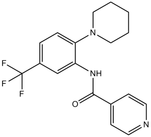
C18H18F3N3O
|
|
|---|---|---|
| 分子量 |
349.35
|
|
| 精确质量 |
349.14
|
|
| 元素分析 |
C, 61.88; H, 5.19; F, 16.31; N, 12.03; O, 4.58
|
|
| CAS号 |
218156-96-8
|
|
| 相关CAS号 |
|
|
| PubChem CID |
2797577
|
|
| 外观&性状 |
White to light yellow solid powder
|
|
| 密度 |
1.3±0.1 g/cm3
|
|
| 沸点 |
395.9±42.0 °C at 760 mmHg
|
|
| 闪点 |
193.3±27.9 °C
|
|
| 蒸汽压 |
0.0±0.9 mmHg at 25°C
|
|
| 折射率 |
1.578
|
|
| LogP |
4.15
|
|
| tPSA |
45.23
|
|
| 氢键供体(HBD)数目 |
1
|
|
| 氢键受体(HBA)数目 |
6
|
|
| 可旋转键数目(RBC) |
3
|
|
| 重原子数目 |
25
|
|
| 分子复杂度/Complexity |
445
|
|
| 定义原子立体中心数目 |
0
|
|
| SMILES |
FC(C1C([H])=C([H])C(=C(C=1[H])N([H])C(C1C([H])=C([H])N=C([H])C=1[H])=O)N1C([H])([H])C([H])([H])C([H])([H])C([H])([H])C1([H])[H])(F)F
|
|
| InChi Key |
DWFGGOFPIISJIT-UHFFFAOYSA-N
|
|
| InChi Code |
InChI=1S/C18H18F3N3O/c19-18(20,21)14-4-5-16(24-10-2-1-3-11-24)15(12-14)23-17(25)13-6-8-22-9-7-13/h4-9,12H,1-3,10-11H2,(H,23,25)
|
|
| 化学名 |
N-[2-piperidin-1-yl-5-(trifluoromethyl)phenyl]pyridine-4-carboxamide
|
|
| 别名 |
|
|
| HS Tariff Code |
2934.99.9001
|
|
| 存储方式 |
Powder -20°C 3 years 4°C 2 years In solvent -80°C 6 months -20°C 1 month |
|
| 运输条件 |
Room temperature (This product is stable at ambient temperature for a few days during ordinary shipping and time spent in Customs)
|
| 溶解度 (体外实验) |
|
|||
|---|---|---|---|---|
| 溶解度 (体内实验) |
配方 1 中的溶解度: ≥ 2.5 mg/mL (7.16 mM) (饱和度未知) in 10% DMSO + 90% (20% SBE-β-CD in Saline) (这些助溶剂从左到右依次添加,逐一添加), 澄清溶液。
例如,若需制备1 mL的工作液,可将100 μL 25.0 mg/mL澄清DMSO储备液加入900 μL 20% SBE-β-CD生理盐水溶液中,混匀。 *20% SBE-β-CD 生理盐水溶液的制备(4°C,1 周):将 2 g SBE-β-CD 溶解于 10 mL 生理盐水中,得到澄清溶液。 配方 2 中的溶解度: 1% DMSO +30% polyethylene glycol+1% Tween 80 : 8 mg/mL 请根据您的实验动物和给药方式选择适当的溶解配方/方案: 1、请先配制澄清的储备液(如:用DMSO配置50 或 100 mg/mL母液(储备液)); 2、取适量母液,按从左到右的顺序依次添加助溶剂,澄清后再加入下一助溶剂。以 下列配方为例说明 (注意此配方只用于说明,并不一定代表此产品 的实际溶解配方): 10% DMSO → 40% PEG300 → 5% Tween-80 → 45% ddH2O (或 saline); 假设最终工作液的体积为 1 mL, 浓度为5 mg/mL: 取 100 μL 50 mg/mL 的澄清 DMSO 储备液加到 400 μL PEG300 中,混合均匀/澄清;向上述体系中加入50 μL Tween-80,混合均匀/澄清;然后继续加入450 μL ddH2O (或 saline)定容至 1 mL; 3、溶剂前显示的百分比是指该溶剂在最终溶液/工作液中的体积所占比例; 4、 如产品在配制过程中出现沉淀/析出,可通过加热(≤50℃)或超声的方式助溶; 5、为保证最佳实验结果,工作液请现配现用! 6、如不确定怎么将母液配置成体内动物实验的工作液,请查看说明书或联系我们; 7、 以上所有助溶剂都可在 Invivochem.cn网站购买。 |
| 制备储备液 | 1 mg | 5 mg | 10 mg | |
| 1 mM | 2.8625 mL | 14.3123 mL | 28.6246 mL | |
| 5 mM | 0.5725 mL | 2.8625 mL | 5.7249 mL | |
| 10 mM | 0.2862 mL | 1.4312 mL | 2.8625 mL |
1、根据实验需要选择合适的溶剂配制储备液 (母液):对于大多数产品,InvivoChem推荐用DMSO配置母液 (比如:5、10、20mM或者10、20、50 mg/mL浓度),个别水溶性高的产品可直接溶于水。产品在DMSO 、水或其他溶剂中的具体溶解度详见上”溶解度 (体外)”部分;
2、如果您找不到您想要的溶解度信息,或者很难将产品溶解在溶液中,请联系我们;
3、建议使用下列计算器进行相关计算(摩尔浓度计算器、稀释计算器、分子量计算器、重组计算器等);
4、母液配好之后,将其分装到常规用量,并储存在-20°C或-80°C,尽量减少反复冻融循环。
计算结果:
工作液浓度: mg/mL;
DMSO母液配制方法: mg 药物溶于 μL DMSO溶液(母液浓度 mg/mL)。如该浓度超过该批次药物DMSO溶解度,请首先与我们联系。
体内配方配制方法:取 μL DMSO母液,加入 μL PEG300,混匀澄清后加入μL Tween 80,混匀澄清后加入 μL ddH2O,混匀澄清。
(1) 请确保溶液澄清之后,再加入下一种溶剂 (助溶剂) 。可利用涡旋、超声或水浴加热等方法助溶;
(2) 一定要按顺序加入溶剂 (助溶剂) 。
|
|---|
|
|
 Biotin-SRPK2 Recombinant Human Active Protein Kinase
 Biotin-SRPK1 Recombinant Human Active Protein Kinase
 SRPK1 Recombinant Human Active Protein Kinase
 Srpk1-IN-1
InvivoChem的所有产品仅用于作科学研究,不面向患者销售
Copyright 2020 InvivoChem LLC | All Rights Reserved 粤ICP备20063088号-1


 COA
COA


 463611831
