| 规格 | 价格 | 库存 | 数量 |
|---|---|---|---|
| 1mg |
|
||
| 5mg |
|
||
| 10mg |
|
||
| 25mg |
|
||
| 50mg |
|
||
| 100mg |
|
||
| 250mg |
|
||
| 500mg |
|
||
| Other Sizes |
|
| 靶点 |
Aquaporin 4 (AQP4) (IC50 = 3.1 μM)
|
|---|---|
| 体外研究 (In Vitro) |
在体外抑制AQP4介导的水转运,通过转染AQP4的HEK293细胞在低渗缓冲液中的肿胀实验显示,TGN-020(浓度未明确)剂量依赖性减少细胞肿胀,表明AQP4被抑制[1]
- 在原代大鼠星形胶质细胞培养中,TGN-020(10-100 μM)处理24小时后,显著降低GFAP(星形胶质细胞活化标志物)和PCNA(细胞增殖标志物)的表达[4] 一个配体用于 E3 泛素连接酶,另一个配体用于靶蛋白;这两个配体通过接头连接形成 PROTAC。 PROTAC 利用细胞内泛素-蛋白酶体系统选择性降解靶蛋白[3]。 |
| 体内研究 (In Vivo) |
TGN-020(0.02 mg/μL;2微升玻璃体内注射)可以减轻九周龄雄性Wistar大鼠视网膜的视网膜水肿,这些大鼠已接受STZ诱发糖尿病[2]。 TGN-020(100 mg/kg;腹腔注射;SCI 后立即给予单剂量)可降低水肿程度,抑制 GFAP、PCNA 和 AQP4 的表达,并增强第 3、7、14、21 和 21 天的功能恢复。 28 以下 SCI。在患有 SCI 的成年雌性 Sprague-Dawley 大鼠(180-220 g,9-10 周龄)中,TGN-020 抑制神经胶质疤痕的形成并增加 GAP-43 的表达[4]。
在糖尿病视网膜中,TGN-020抑制了VEGF的免疫反应性和蛋白水平。AQP4免疫反应性高于对照组视网膜,AQP4的表达与GFAP共定位。与VEGF类似,TGN-020也抑制AQP4和GFAP。在伊文思蓝试验中,TGN-020减少了糖尿病视网膜的渗漏。在培养的Müller细胞中,暴露于TGN-020与暴露于贝伐单抗一样,可以抑制高糖条件下细胞体积和细胞内ROS产生的增加。 结论:TGN-020可能对糖尿病视网膜水肿有抑制作用。[2] TGN-020与脊髓损伤(SCI)后第3天脊髓水肿的缓解[4] 我们测量了组织的含水量,以反映SCI部位的水肿程度。SCI后第3天,SCI组和TGN-020组T10脊髓局灶性压迫导致可见损伤,而假手术组则不明显。脊髓损伤中心出血和坏死明显,脊髓损伤组的损伤区域明显多于TGN-020组(图2A)。受伤脊髓的照片与各组脊髓含水量的计算结果相当一致(图2B)。SCI组(76.09±0.93)和TGN-020组(73.07±0.87)第3天损伤部位邻近节段的组织含水量明显高于假手术组(69.02±0.45)(p<0.01)。 0.05),表明TGN-020可以显著降低SCI引起的含水量增加。 TGN-020和SCI后第3天AQP4表达降低[4] 与假手术组相比,SCI组(p<0.01)和TGN-020组(p<0.05)在压缩损伤后3天,AQP4在损伤部位中心及其周围的表达显著增加(图3A、B)。在TGN-020组中,AQP4的表达明显低于SCI组(p<0.05)。用GFAP对AQP4进行免疫荧光染色的结果与蛋白质印迹的结果一致(图3C,D)。在第3天,SCI组和TGN-020组的AQP4荧光强度均显著高于假手术组(p < 0.01), TGN-020组的AQP4表达明显低于SCI组(p<0.05)。这些数据表明,TGN-020可以下调AQP4的表达,从而减轻脊髓水肿的形成。 TGN-020与SCI后第3天星形胶质细胞增殖的抑制作用[4] GFAP是一种细胞骨架中间丝蛋白,被用作特定的星形胶质细胞标志物。增殖细胞核抗原(PCNA)是众所周知的细胞增殖标志物。为了研究AQP4是否与SCI星形胶质细胞增殖有关,我们在SCI后3天检测了PCNA和GFAP的表达。Western印迹表明,假手术组GFAP水平较低,几乎没有PCNA表达(图4A-C)。然而,值得注意的是,与假手术组相比,SCI组和TGN-020组在第3天损伤部位中心周围的GFAP和PCNA表达显著增加(p < 0.01). 在第3天,TGN-020组的GFAP和PCNA表达明显低于SCI组(p < 0.05). TGN-020与SCI后4周胶质瘢痕形成和轴突再生[4] 为了研究TGN-020给药是否可以在SCI后4周保护脊髓组织免受损伤,我们计算了空腔面积的大小,以评估星形胶质细胞瘢痕的形成。4周时,假手术组的脊髓正常,但SCI组和TGN-020组的脊髓都出现了破坏,包括非常大和不规则的空洞(图5A、B)。TGN-020组的空洞面积明显小于SCI组(4.2±0.6 mm2 vs.3.0±0.4 mm2,p<0.01)。这些观察结果表明,TGN-020显著减少了SCI后的继发性脊髓组织退化。 TGN-020与脊髓损伤后4周的神经元存活率[4] SCI诱导的神经元坏死和凋亡导致神经元丢失。脊髓损伤后4周,在横断面上进行尼氏染色,以研究TGN-020的潜在神经保护作用。检测到大量具有延伸细胞体的尼氏阳性神经元,主要位于正常脊髓的腹角(图6B)。因此,在受伤脊髓的前角检测到运动神经元。如图6A所示,在假手术组中,神经元表现出完整的颗粒状形态,以及大而多的尼氏小体,表明神经细胞中有大量的蛋白质合成。在SCI组中,神经元表现出萎缩的细胞体、固缩的核和不规则的形态,病变周围的细胞内甲苯胺蓝染色减少。此外,存活的运动神经元数量明显低于假手术组(图6C,p < 0.01). 在TGN-020组中,存活的运动神经元数量也明显低于假手术组(图6C,p < 0.01), 但神经元形态比SCI组好,细胞质染色更深,尼氏颗粒的损失明显减少(图6C,p<0.01)。这些结果表明,TGN-020给药抑制了神经元的损失。 TGN-020与SCI后功能性运动恢复的促进作用[4] 为了评估TGN-020对SCI后大鼠功能性运动恢复的影响,通过Basso-Beattie-Bresnahan(BBB)量表在第1、3、7、14、21和28天评估行为结果,该量表已被广泛用于评估大鼠SCI后后肢运动功能的恢复情况。假手术组在所有时间点的评分均为21分,反映了正常的运动功能,而脊髓损伤组和TGN-020组的BBB评分在压缩损伤后1天降低,然后随着时间的推移逐渐增加到不同程度(图7)。这表明脊髓损伤后运动功能逐渐恢复。在TGN-020组中,SCI后第3天至第28天的所有时间点,BBB评分均显著高于SCI组(第7天p<0.01;第3、14、12和28天的p<0.05)。这些结果表明,TGN-020给药可以显著促进SCI后大鼠运动功能的恢复。 - 在糖尿病视网膜病变小鼠模型中,TGN-020(0.02 mg/μL,玻璃体内注射)显著减轻视网膜水肿,并降低视网膜中AQP4的表达[2] - 在脊髓压迫损伤大鼠模型中,损伤后立即腹腔注射TGN-020(100 mg/kg)可减少脊髓水肿,抑制星形胶质细胞活化(GFAP表达降低),并抑制胶质瘢痕形成(硫酸软骨素蛋白聚糖表达减少)[4] - 在cuprizone诱导的多发性硬化小鼠模型中,TGN-020(200 mg/kg,每日腹腔注射)减少星形胶质细胞增生,促使小胶质细胞从M1型向M2型极化,并抑制NLRP3炎性小体活化,从而促进髓鞘再生和运动功能恢复[4] |
| 细胞实验 |
通过流式细胞术测量细胞体积和ROS产生的细胞内水平[2]
为了检测TR-MUL5细胞体积的变化和高糖条件下细胞内ROS的产生,将细胞在高糖(25 mM)或生理浓度的低血糖(5.5 mM)培养基中孵育2-3天。然后,在生理和高糖条件下,在有或没有TGN-020(100 nM)或贝伐单抗过夜的情况下,使用乙锭荧光流式细胞术分析TR-MUL5的体积变化和细胞内ROS水平。使用荧光探针氢乙啶测量TR-MUL5细胞中的超氧化物水平。氢乙啶被超氧化物氧化形成乙锭,这是一种荧光产物,然后被保留在细胞内,从而可以半定量估计细胞内的超氧化物水平。通过胰蛋白酶消化收集细胞,并在800 g下离心5分钟。用PBS洗涤后,将细胞在37°C下重新悬浮在无酚红的含羟乙基乙胺(1μg/mL)的DMEM中30分钟。将细胞密度调节至2.0×105个细胞/mL。使用488 nm激发和590至610 nm发射波长的流式细胞术分析Müller细胞体积和细胞内超氧化物水平的变化。EC800上的采集和分析软件用于采集和量化荧光强度。我们之前描述了使用EC800流式细胞仪测定TR-MUL5细胞体积变化的细节。 培养的Müller细胞的免疫抑制[2] 为了确定TGN-020对TR-MUL5细胞中VEGF和AQP4表达的影响,通过免疫细胞化学检测了在含有和不含TGN-020>或生理浓度的低血糖(5.5mM)培养基的高糖(25mM)培养基中孵育的细胞。用4%甲醛固定后,将细胞与兔多克隆抗AQP4和小鼠单克隆抗VEGF的一抗在4°C下孵育过夜。用PBS冲洗并阻断后,将这些细胞在室温下在Alexa 594或Alexa 488中孵育2小时,Alexa 594或Alexa 488与1:500稀释的适当二抗结合。细胞核用4′,6-二脒基-2-苯基吲哚染色。用荧光显微镜对处理后的样品进行拍照。 - AQP4功能实验:转染AQP4的HEK293细胞暴露于低渗缓冲液中,通过光学显微镜观察细胞肿胀。TGN-020(0.1-10 μM)剂量依赖性减少细胞肿胀,证明AQP4抑制[1] - 星形胶质细胞活化实验:原代大鼠星形胶质细胞经TGN-020(10-100 μM)处理24小时后,通过免疫细胞化学检测GFAP表达。TGN-020显著减少GFAP阳性细胞数量和染色强度[4] |
| 动物实验 |
玻璃体内注射[2]
使用汉密尔顿注射器和30号针头,对链脲佐菌素(STZ)诱导的糖尿病大鼠进行2微升玻璃体内注射,注射物为TGN-020(0.02 mg/μL)、贝伐珠单抗(0.025 mg/μL)(美国加州旧金山基因泰克公司)或仅注射载体(磷酸盐缓冲液(PBS),pH 7.4)。动物接受全身麻醉,并在玻璃体内注射后48小时进行灌注固定。 伊文思蓝检测[2] 采用伊文思蓝检测法检测STZ诱导的糖尿病大鼠在有或无TGN-020存在下的视网膜血管通透性。将100 μL伊文思蓝(20 mg/mL)PBS溶液经尾静脉注射。10分钟后,小心取出视网膜,并在4% PFA PBS溶液中固定过夜。制备视网膜平铺标本,并使用激光扫描共聚焦显微镜,在594 nm激发波长下进行落射荧光分析,评估渗漏情况。 动物和实验分组[4] 我们尽一切努力减少动物的痛苦。随后将动物随机分为以下三组:假手术组(n = 35),仅进行椎板切除术而不进行脊髓压迫;脊髓损伤组(n = 35),在T10节段进行5分钟的35 g压迫;TGN-020组(n = 35),在脊髓损伤后立即腹腔注射TGN-020(100 mg/kg)。每组随机均分为四个亚组(每组n = 5或6和8),用于以下实验:(A)脊髓含水量测定;(B)蛋白质印迹法;(C)免疫荧光分析、苏木精-伊红染色和尼氏染色;以及(D)运动功能测试。所有组别的大鼠均在损伤后3天或4周处死。 2.2.手术操作及TGN-020给药[4] 基本手术操作和压迫损伤均按先前所述进行。所有手术均在无菌条件下进行。简而言之,大鼠用10%水合氯醛(0.33 mL/kg,腹腔注射)麻醉。麻醉生效后,剃除手术区域的毛发,并用75%乙醇消毒。在T9-T12节段胸中段脊髓上方做3 cm长的背侧纵切口,然后去除周围椎旁软组织以暴露脊髓,保留硬膜完整。用金属压迫器(35 g,5 min)轻轻压迫T10节段脊髓,造成中度损伤(图1)。术后,用4-0丝线分层缝合伤口。术后,将溶于10%二甲基亚砜(DMSO)的TGN-020(100 mg/kg,腹腔注射)给予TGN-020组。注射前将DMSO浓度调整至0.1%。假手术组和脊髓损伤组同时腹腔注射等体积的DMSO。动物苏醒后放回各自的笼子。术后恢复期,连续三天腹腔注射头孢曲松钠(50 mg/kg)。每天手动按压膀胱三次,直至自然排尿反射恢复,并自由摄取食物和水。出现任何异常神经系统症状的动物将被排除在实验之外。 - 糖尿病视网膜病变模型:用链脲佐菌素(STZ)诱导9周龄雄性Wistar大鼠发生糖尿病。每周一次,玻璃体内注射TGN-020(0.02 mg/μL,2 μL),持续4周。采用光学相干断层扫描(OCT)评估视网膜水肿,并采用免疫组织化学分析AQP4表达[2] -脊髓损伤模型:成年雌性Sprague-Dawley大鼠(180-220 g)接受脊髓压迫损伤。损伤后立即腹腔注射TGN-020(100 mg/kg)。采用BBB运动功能评分量表评估功能恢复情况,并在第3天采集脊髓组织进行组织学分析[4] |
| 参考文献 |
|
| 其他信息 |
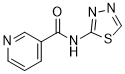
本研究考察了18种化合物对水通道蛋白4 (AQP4) 的体外抑制作用及其计算机模拟对接能。超过半数的受试化合物在体外功能测定中显示出抑制活性,其中包括5-HT(1B/1D)受体激动剂舒马曲坦和利扎曲坦。此外,本研究中使用的化合物在20 μM浓度下的抑制活性与其计算机模拟对接能呈显著正相关(r²=0.64),这与之前的研究结果一致。随后发现三种化合物,即2-(烟酰胺基)-1,3,4-噻二唑、舒马曲坦和利扎曲坦,对AQP4的抑制IC(50)值分别为3、11和2 μM。[1]
目的:探讨选择性水通道蛋白4 (AQP4) 抑制剂2-(烟酰胺基)-1,3,4-噻二唑 (TGN-020) 对糖尿病视网膜血管内皮生长因子 (VEGF) 表达、活性氧 (ROS) 生成以及视网膜水肿的影响。方法:对链脲佐菌素诱导的糖尿病大鼠进行玻璃体内注射贝伐珠单抗、TGN-020或磷酸盐缓冲液 (PBS)。对视网膜切片进行抗胶质纤维酸性蛋白 (GFAP)、抗水通道蛋白4 (AQP4) 和抗血管内皮生长因子 (VEGF) 的免疫染色。采用蛋白质印迹法测定收集的视网膜中 VEGF 的蛋白水平。此外,在糖尿病大鼠的平铺视网膜标本中,观察了在有或无 TGN-020 存在下,伊文思蓝对视网膜血管的渗漏情况。在生理和高糖条件下,采用流式细胞术分析乙锭荧光,测定了在有或无 TGN-020 或贝伐珠单抗存在下,大鼠视网膜 Müller 细胞(TR-MUL5;转基因大鼠 Müller 细胞)的体积变化和细胞内活性氧 (ROS) 水平。结果:在糖尿病视网膜中,TGN-020 抑制了 VEGF 的免疫反应性和蛋白水平。AQP4 的免疫反应性高于对照组视网膜,且 AQP4 的表达与 GFAP 共定位。与 VEGF 类似,TGN-020 也抑制了 AQP4 和 GFAP 的表达。在伊文思蓝染色试验中,TGN-020 降低了糖尿病视网膜的渗漏。在培养的 Müller 细胞中,高糖条件下细胞体积的增加和细胞内 ROS 的产生,在 TGN-020 的作用下与贝伐珠单抗的作用相当,均受到抑制。结论:TGN-020 可能对糖尿病视网膜水肿具有抑制作用。[2] 目前,小分子抑制剂是靶向细胞内蛋白的主要治疗方法,但其开发和临床应用仍面临诸多挑战。蛋白水解靶向嵌合体 (PROTAC) 利用细胞内泛素-蛋白酶体系统选择性地降解靶蛋白。近年来,高活性小分子 PROTAC 的报道屡见不鲜。本文综述了小分子PROTACs的新兴特性,例如诱导快速、深度和持续的降解,有效抑制下游信号通路,展现出更高的靶向选择性,以及克服对小分子抑制剂的耐药性。在肿瘤异种移植模型中,小分子PROTACs能够显著抑制肿瘤进展。此外,本文还介绍了PROTAC技术的最新进展,例如同源PROTACs。与传统小分子药物相比,小分子PROTACs的显著优势以及令人鼓舞的临床前数据表明,小分子PROTAC技术具有极大地促进靶向治疗药物研发的潜力。[3] 目的:寻找能够抑制水肿和胶质瘢痕形成并提高神经元存活率的药物对于改善脊髓损伤(SCI)后的预后至关重要。本研究使用强效选择性水通道蛋白4 (AQP4) 抑制剂2-(烟酰胺)-1,3,4-噻二唑 (TGN-020),探讨其对Sprague-Dawley大鼠脊髓损伤(SCI)的影响。主要方法:使用无菌压板(35 g,5 min)压迫T10节段脊髓,诱导中度损伤。随后腹腔注射TGN-020 (100 mg/kg)或等体积的10%二甲基亚砜(DMSO)。分别于SCI后1、3、7、14、21和28天,采用Basso-Beattie-Bresnahan开放场运动评分量表评估神经功能。于SCI后3天测定脊髓含水量,评估脊髓水肿程度。在脊髓损伤(SCI)后3天和4周,通过蛋白质印迹法和免疫荧光染色法检测了AQP4、胶质纤维酸性蛋白(GFAP)、增殖细胞核抗原(PCNA)和生长相关蛋白-43(GAP-43)的表达水平。分别采用尼氏染色和苏木精-伊红染色法测定了存活神经元的数量和胶质瘢痕的大小。主要结果:我们的研究结果表明,TGN-020在SCI后第3、7、14、21和28天促进了功能恢复,并在SCI后第3天减轻了水肿程度,并抑制了AQP4、GFAP和PCNA的表达。此外,脊髓损伤后4周的观察显示,TGN-020抑制了胶质瘢痕的形成并上调了GAP-43的表达。意义:TGN-020可以减轻脊髓水肿,抑制胶质瘢痕的形成,并促进轴突再生,从而对大鼠的恢复产生有益作用。[4] - TGN-020是一种小分子AQP4抑制剂,在涉及水肿和胶质细胞活化的神经系统疾病(例如糖尿病视网膜病变和脊髓损伤)中具有潜在的治疗应用价值。[2][4] - 它通过阻断AQP4通道发挥作用,从而减少水的流入和随后的组织肿胀。此外,TGN-020抑制星形胶质细胞增殖和小胶质细胞介导的炎症,从而有助于神经保护。[4] |
| 分子式 |
C8H6N4OS
|
|
|---|---|---|
| 分子量 |
206.22
|
|
| 精确质量 |
206.026
|
|
| 元素分析 |
C, 46.59; H, 2.93; N, 27.17; O, 7.76; S, 15.55
|
|
| CAS号 |
51987-99-6
|
|
| 相关CAS号 |
1313731-99-5
|
|
| PubChem CID |
4173511
|
|
| 外观&性状 |
White to off-white solid powder
|
|
| LogP |
1.569
|
|
| tPSA |
99.5
|
|
| 氢键供体(HBD)数目 |
1
|
|
| 氢键受体(HBA)数目 |
5
|
|
| 可旋转键数目(RBC) |
2
|
|
| 重原子数目 |
14
|
|
| 分子复杂度/Complexity |
214
|
|
| 定义原子立体中心数目 |
0
|
|
| SMILES |
C1=CC(=CN=C1)C(=O)NC2=NN=CS2
|
|
| InChi Key |
AGEGZHOPKZFKBP-UHFFFAOYSA-N
|
|
| InChi Code |
InChI=1S/C8H6N4OS/c13-7(6-2-1-3-9-4-6)11-8-12-10-5-14-8/h1-5H,(H,11,12,13)
|
|
| 化学名 |
|
|
| 别名 |
|
|
| HS Tariff Code |
2934.99.9001
|
|
| 存储方式 |
Powder -20°C 3 years 4°C 2 years In solvent -80°C 6 months -20°C 1 month |
|
| 运输条件 |
Room temperature (This product is stable at ambient temperature for a few days during ordinary shipping and time spent in Customs)
|
| 溶解度 (体外实验) |
|
|||
|---|---|---|---|---|
| 溶解度 (体内实验) |
配方 1 中的溶解度: 25 mg/mL (121.23 mM) in 15% Cremophor EL + 85% Saline (这些助溶剂从左到右依次添加,逐一添加), 悬浮液;超声助溶。
*生理盐水的制备:将 0.9 g 氯化钠溶解在 100 mL ddH₂O中,得到澄清溶液。 配方 2 中的溶解度: ≥ 1.67 mg/mL (8.10 mM) (饱和度未知) in 10% DMSO + 90% Corn Oil (这些助溶剂从左到右依次添加,逐一添加), 澄清溶液。 例如,若需制备1 mL的工作液,可将 100 μL 16.7 mg/mL 澄清 DMSO 储备液加入到 900 μL 玉米油中并混合均匀。 View More
配方 3 中的溶解度: 5 mg/mL (24.25 mM) in 0.5% MC 0.5% Tween-80 (这些助溶剂从左到右依次添加,逐一添加), 悬浊液; 超声助溶。 1、请先配制澄清的储备液(如:用DMSO配置50 或 100 mg/mL母液(储备液)); 2、取适量母液,按从左到右的顺序依次添加助溶剂,澄清后再加入下一助溶剂。以 下列配方为例说明 (注意此配方只用于说明,并不一定代表此产品 的实际溶解配方): 10% DMSO → 40% PEG300 → 5% Tween-80 → 45% ddH2O (或 saline); 假设最终工作液的体积为 1 mL, 浓度为5 mg/mL: 取 100 μL 50 mg/mL 的澄清 DMSO 储备液加到 400 μL PEG300 中,混合均匀/澄清;向上述体系中加入50 μL Tween-80,混合均匀/澄清;然后继续加入450 μL ddH2O (或 saline)定容至 1 mL; 3、溶剂前显示的百分比是指该溶剂在最终溶液/工作液中的体积所占比例; 4、 如产品在配制过程中出现沉淀/析出,可通过加热(≤50℃)或超声的方式助溶; 5、为保证最佳实验结果,工作液请现配现用! 6、如不确定怎么将母液配置成体内动物实验的工作液,请查看说明书或联系我们; 7、 以上所有助溶剂都可在 Invivochem.cn网站购买。 |
| 制备储备液 | 1 mg | 5 mg | 10 mg | |
| 1 mM | 4.8492 mL | 24.2460 mL | 48.4919 mL | |
| 5 mM | 0.9698 mL | 4.8492 mL | 9.6984 mL | |
| 10 mM | 0.4849 mL | 2.4246 mL | 4.8492 mL |
1、根据实验需要选择合适的溶剂配制储备液 (母液):对于大多数产品,InvivoChem推荐用DMSO配置母液 (比如:5、10、20mM或者10、20、50 mg/mL浓度),个别水溶性高的产品可直接溶于水。产品在DMSO 、水或其他溶剂中的具体溶解度详见上”溶解度 (体外)”部分;
2、如果您找不到您想要的溶解度信息,或者很难将产品溶解在溶液中,请联系我们;
3、建议使用下列计算器进行相关计算(摩尔浓度计算器、稀释计算器、分子量计算器、重组计算器等);
4、母液配好之后,将其分装到常规用量,并储存在-20°C或-80°C,尽量减少反复冻融循环。
计算结果:
工作液浓度: mg/mL;
DMSO母液配制方法: mg 药物溶于 μL DMSO溶液(母液浓度 mg/mL)。如该浓度超过该批次药物DMSO溶解度,请首先与我们联系。
体内配方配制方法:取 μL DMSO母液,加入 μL PEG300,混匀澄清后加入μL Tween 80,混匀澄清后加入 μL ddH2O,混匀澄清。
(1) 请确保溶液澄清之后,再加入下一种溶剂 (助溶剂) 。可利用涡旋、超声或水浴加热等方法助溶;
(2) 一定要按顺序加入溶剂 (助溶剂) 。
PET images. (a) WT and (b) KO mice. |
Ex vivo PET images of the brain. (a) WT and (b) KO mouse brains. (c) MRI images of the corresponding brain slices.ACS Chem Neurosci. 2011 Oct 19; 2(10): 568–571. |
Time course analysis of SUV. Data from WT (n= 6, ○) and KO (n= 4, |
 1-(Azetidin-3-yl)piperidine
 1-(Piperidin-4-ylmethyl)piperidine
 Adamantan-C-amide-PEG2-C2-amine
 4-(4-Boc-piperazinemethyl)phenylboronic acid pinacol ester
InvivoChem的所有产品仅用于作科学研究,不面向患者销售
Copyright 2020 InvivoChem LLC | All Rights Reserved 粤ICP备20063088号-1


 COA
COA


 ) models are shown along with the standard error of means. (a) Brain, (b) skeletal muscle, and (c) heart.
 463611831
