| 规格 | 价格 | 库存 | 数量 |
|---|---|---|---|
| 5mg |
|
||
| 10mg |
|
||
| 25mg |
|
||
| 50mg |
|
||
| 100mg |
|
||
| 250mg |
|
||
| 500mg |
|
||
| Other Sizes |
|
| 靶点 |
DNA synthesis; Microbial Metabolite
|
|---|---|
| 体外研究 (In Vitro) |
微摩尔量的尿石素 A 会导致细胞闭合和自噬。在人 sw620 结直肠中,尿石素 A 抑制 DNA 合成和细胞周期的发育 [2]。 T24 和 Caco-2 细胞的生长受到尿石素 A 的抑制,尿石素 A 还具有抗增殖作用。在 G2/M 和 S 期,与对照细胞相比,尿石素 A 呈剂量和时间依赖性,24 小时和 48 小时分别为 50 和 100 μM。 IC 50 值依次为 43.9 和 49 μM [3]。在 50 和 100 μM 时,它会导致细胞凋亡 [4]。 HepG2 细胞对尿石素 A 表现出强烈的抗增殖活性。当尿石素 A 促进细胞死亡时,TCF/LEF 快速激活显着下调,β-catenin、c-Myc 和 cyclin D1 的表达减少。此外,尿石素 A 上调 caspase-3、p38-MAPK 和 p53 的表达,同时抑制 NF-κB、p65 和其他神经介质 [5]。
已知其肠道代谢产物黄花酸(EA)、尿石蛋白能有效抑制癌症细胞增殖。本研究探讨了Urolithin A(UA)对HepG2肝癌细胞系的抗增殖和抗氧化作用。在暴露12-36小时后,使用CCK测定法测定UA(0-500μM)对HepG2细胞的抗增殖作用。通过实时PCR和Western blot评估对β-catenin和其他表达因子的影响。我们发现UA对HepG2细胞显示出强大的抗增殖活性。当UA诱导细胞死亡时,发现β-catenin、c-Myc和Cyclin D1的表达降低,TCF/LEF转录激活显著下调。UA还增加了p53、p38 MAPK和caspase-3的蛋白表达,但抑制了NF-κB p65和其他炎症介质的表达。此外,UA和EA处理提供的抗氧化测定与细胞内ROS水平的降低以及细胞内SOD和GSH-Px活性的增加有关。这些结果表明,UA可以抑制细胞增殖,降低癌症的氧化应激状态,从而成为肝癌预防和治疗的有效成分。[2] 自噬是一种进化上保守的途径,其中细胞质内容物被降解和再循环。这项研究发现,亚摩尔浓度的Urolithin A(一种主要的多酚代谢产物)诱导了SW620结直肠癌(CRC)细胞的自噬。暴露于Urolithin A也会剂量依赖性地降低细胞增殖、延迟细胞迁移和基质金属蛋白酶-9(MMP-9)活性。此外,Atg5-siRNA对自噬的抑制,Z-VAD-FMK对胱天蛋白酶的抑制,抑制了Urolithin A刺激的细胞死亡和抗转移作用。微摩尔浓度Urolithin A可诱导自噬和凋亡。Urolithin A抑制细胞周期进程和DNA合成。这些结果表明,饮食中摄入Urolithin A可以诱导自噬并抑制人结直肠癌细胞转移。因此,Urolithins可能有助于CRC治疗,并提供一种替代或辅助化疗药物来对抗这种疾病。[3] IsoUro-A以时间和剂量依赖的方式抑制Caco-2细胞的增殖,尽管其显著低于Urolitin A/Uro-A(48小时时IC50=69.7±4.5和49.2±3.8μM)。这两种尿锂素都将Caco-2细胞周期阻滞在S期和G2/M期,并在以前在人类结肠组织中发现的浓度下诱导细胞凋亡。值得注意的是,Caco-2细胞葡萄糖醛酸化IsoUro-A的效率高于Uro-A(48小时后转化率分别为~50%和~20%)。Uro-A和IsoUro-A葡糖苷酸都没有发挥抗增殖作用。此外,Caco-2细胞的生长抑制率高于正常细胞。 结论:在癌症细胞中,IsoUro-A具有很强的抗增殖活性,但在9位广泛的葡萄糖醛酸化作用降低了这种活性。需要进一步的研究来阐明Uro-A和IsoUro-A的体外构效关系是否在人体中起作用[6]。 |
| 体内研究 (In Vivo) |
此外,在血管壁中输送尿石素 A 1 小时后,爪水肿量减少。此外,在将尿石素A添加到血管壁后一小时,处理过的细胞干表现出高氧自由基抗氧化能力(ORAC)评分,其余未形成的水平与A相当[6]。
研究表明,Urolithin A改善了APP/PS1小鼠的认知障碍,防止了神经元凋亡,并增强了神经发生。此外,UA减轻了皮质和海马中的Aβ沉积、斑块周围微胶质细胞增生和星形胶质细胞增生。我们还发现,UA影响了关键的细胞信号通路,特别是通过增强脑AMPK激活、降低P65NF-κB和P38MAPK的激活以及抑制Bace1和APP降解。 结论:结果表明,UA通过保护神经元免于死亡和通过抗炎信号触发APP/PS1小鼠的神经发生来提供认知保护,表明UA可能是治疗AD的有前景的治疗药物 [1]. Urolithin A是大鼠和人类在饮用石榴汁或纯鞣花单宁香叶素后产生的主要代谢产物。在这项研究中,我们研究了Urolithin A对角叉菜胶诱导的小鼠足肿胀的抗炎作用。口服尿石素A1小时后,足肿胀体积减少。此外,经治疗的小鼠血浆在口服Urolithin A后1小时表现出显著的氧自由基抗氧化能力(ORAC)评分,非结合形式的血浆水平很高。这些结果表明,血浆中的Urolithin A水平、血浆ORAC评分和抗炎作用之间存在很强的相关性,可能有助于解释鞣剂单宁对炎症性疾病的保护机制[4]。 |
| 细胞实验 |
细胞培养和治疗[2]
HepG2细胞系购自美国典型培养物保藏中心。细胞在37°C的DMEM培养基中维持,该培养基补充了10%胎牛血清、100单位/mL青霉素和100μg/mL硫酸链霉素,在5%CO2、95%加湿的气氛中。为了进行CCK测定,按照相对于细胞IC50的比例制备了连续稀释的Urolithin A/UA,并分别孵育12、24和36小时。为了进行实时定量PCR和蛋白质印迹分析,细胞用不同浓度的UA处理24小时,并加入DMSO培养基中。每个孔中DMSO的最终浓度为0.6%,在我们的研究中没有观察到毒性,如前所述(Aiken等人,2004)。添加DMSO的组(与药物处理组体积相同)用作阴性对照。 细胞内ROS产生、SOD、MDA和GSH-Px水平的测定[2] 通过使用如上所述的过氧化氢介导的氧化应激模型,并进行适当的修改,评估了Urolithin A/UA对ROS产生、SOD、MDA和GSH-Px水平的影响(Sohn等人,2013)。简而言之,用UA和EA处理12小时后,用PBS轻轻洗涤HepG2细胞3次,并在37°C下用50 mM H2O2孵育3小时,形成细胞内急性氧化状态。使用BCA蛋白检测试剂盒测量蛋白质浓度。 RNA提取和逆转录[2] 用Urolithin A/UA的IC50处理后,根据制造商的建议,使用Trizol试剂从细胞中提取总RNA。在用无RNase的DNase I去除DNA污染后,根据制造商的方案,使用MMLV逆转录酶和oligo-dT进行第一链cDNA合成。将获得的cDNA储存在-80°C下以供进一步使用。 室迁移试验[3] Transwell聚碳酸酯膜插件(8µm孔径,直径10mm)用于腔室迁移分析。将含有或不含有Urolithin A的SW620细胞悬浮在无血清培养基中(5× 10⁵ 细胞/mL)并添加到上部隔室,而含有10%FBS的L-15培养基添加到下部隔室。4小时后,用棉头敷料器去除Transwell聚碳酸酯膜上侧的未侵袭细胞。用甲醇固定膜底面上的入侵细胞,并用0.5%结晶紫染色。 蛋白质印迹[3] SW620细胞在有或没有1.5µMUrolithin A的情况下孵育,收获,用冰冷的PBS洗涤并裂解30分钟。使用BCA检测试剂盒测定蛋白质浓度,每个样本使用50µg蛋白质进行蛋白质印迹分析,并使用以下抗体:LC3-I/II和抗小鼠HRP-偶联IgG二抗(1:2000)。所有样本均已标准化为β-actin。 流式细胞术[3] SW620细胞用碘化丙啶(PI)标记用于细胞周期分析。用Urolithin A处理24、48或72小时后,通过胰蛋白酶消化收获细胞,并将其悬浮在PBS(0.1 M,pH 7.4)中。将细胞在-20°C的70%乙醇中固定至少30分钟。在分析之前,细胞在冷PBS中洗涤两次,并重新悬浮在200µL PBS(0.25 mg/mL RNase A,0.1mg/mL PI)。在37°C的黑暗中孵育30分钟后,通过流式细胞术分析样本,并通过Cell Quest软件计算直方图。 |
| 动物实验 |
Mice (28 weeks old) were orally administered 300 mg/kg Urolithin A/UA dissolved in 0.5% carboxymethylcellulose at the same time each day for 14 days. Control mice (APP/PS1 transgenic mice and wild-type mice) were orally administered the same quantity of 0.5% carboxymethylcellulose (vehicle).[1]
Morris water maze [1] After Urolithin A/UA treatment, the spatial learning and memory of mice were assessed by the Morris water maze. Briefly, the maze consisted of a stainless steel pool (120 cm in diameter and 50 cm in height) with a submerged escape-platform (10 cm in diameter) placed 1 cm below the water surface. The water temperature was maintained at 24 ± 1 °C. The spatial learning task consisted of four consecutive days of testing with four trials per day. In each trial, the time required to find the hidden platform was recorded as the escape latency. The mice were given a maximum of 60 s to find the hidden platform. If a mouse failed to locate the platform within 60 s, the session was terminated, a maximum escape-latency score of 60 s was assigned, and the mouse was manually guided to the hidden platform (10 s). To test spatial memory, a single probe trial was conducted 24 h after the last trial of the fourth day. The submerged platform was removed and the mice were placed into the pool from the quadrant opposite to the quadrant where the platform used to be (target quadrant). The mice were allowed to freely swim for 60 s. The time spent in the target quadrant and numbers of crossings through this quadrant were recorded. Swimming speed was also recorded. Morris water maze was used to detect the cognitive function. Terminal deoxynucleotidyl transferase-mediated dUTP nick end labeling (TUNEL) assay was performed to detect neuronal apoptosis. Immunohistochemistry analyzed the response of glia, Aβ deposition, and neurogenesis. The expression of inflammatory mediators were measured by enzyme-linked immunosorbent assay (ELISA) and quantitative real-time polymerase chain reaction (qRT-PCR). The modulating effects of Urolithin A on cell signaling pathways were assayed by Western blotting.[1] |
| 参考文献 | |
| 其他信息 |
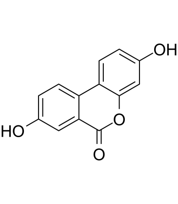
Urolithin A is a member of coumarins. It has a role as a geroprotector.
Urolithin A is a metabolite of ellagic acid. It has been demonstrated to stimulate mitophagy and improve muscle health in old animals and in preclinical models of aging. urolithin A has been reported in Punica granatum and Trogopterus xanthipes with data available. In summary, our results in an AD mouse model demonstrated the protective effects of UA on AD pathology by its targeting of multiple pathological processes such as reactive gliosis, inflammatory signaling, AB plaque formation, and apoptosis. Our findings indicate that UA may serve as a promising therapeutic agent for AD.[1] In summary, the data in this study suggests that UA inhibits cell proliferation on HepG2 cells through suppressing β-catenin signaling and its congenerous mediators. Removing accumulation of β-catenin can increase the TP53 gene and its downstream gene expression by amending the p38-MAPK signaling, and suppressing the expression of NF-κB and other related inflammatory mediators. UA could also reduce the oxidative stress status in HepG2 cells. In Pfundstein et al.’s study, the data showed that the plasma concentrations of metabolism of ellagitannins in the volunteers elevated to 100–200 μM after walnut consumption (Pfundstein et al., 2014). This suggests that the concentration used in our study could be applied in an in vivo model. Moreover, urolithins are metabolites of polyphenols in diet and it seems that reasonable guidance of consuming polyphenols-rich diet could benefit individuals undergoing liver cancer chemotherapy, though an in vivo animal or human model is still further needed to confirm the beliefs. [2] In summary, we showed for the first time that treatment of colon cancer SW620 cells with dietary submicromolar urolithin A can induce autophagy and inhibit CRC cell growth and metastasis. Our in vitro findings provide novel insights into understanding the anti‐tumor functions of dietary urolithin A in CRC. [3] Purpose: Urolithins, metabolites produced by the gut microbiota from ellagic acid, have been acknowledged with cancer chemopreventive activity. Although urolithin A (Uro-A) has been reported to be the most active one, 10-50 % of humans can also produce the isomer isourolithin A (IsoUro-A). However, no biological activity for IsoUro-A has been reported so far. Herein, we describe for the first time the antiproliferative effect of IsoUro-A, compared to Uro-A, against both human colon cancer (Caco-2) and normal (CCD18-Co) cell lines. Methods: Cell proliferation was evaluated by MTT and Trypan blue exclusion assays. Cell cycle was analyzed by flow cytometry and apoptosis measured by the Annexin V/PI method. Finally, urolithins metabolism was analyzed by HPLC-DAD-MS/MS. [6] |
| 分子式 |
C13H8O4
|
|---|---|
| 分子量 |
228.2002
|
| 精确质量 |
228.042
|
| 元素分析 |
C, 68.42; H, 3.53; O, 28.04
|
| CAS号 |
1143-70-0
|
| PubChem CID |
5488186
|
| 外观&性状 |
Light yellow to brown solid powder
|
| 密度 |
1.516g/cm3
|
| 沸点 |
527.9ºC at 760 mmHg
|
| 闪点 |
214.2ºC
|
| 蒸汽压 |
9.24E-12mmHg at 25°C
|
| 折射率 |
1.717
|
| LogP |
2.357
|
| tPSA |
70.67
|
| 氢键供体(HBD)数目 |
2
|
| 氢键受体(HBA)数目 |
4
|
| 可旋转键数目(RBC) |
0
|
| 重原子数目 |
17
|
| 分子复杂度/Complexity |
317
|
| 定义原子立体中心数目 |
0
|
| InChi Key |
RIUPLDUFZCXCHM-UHFFFAOYSA-N
|
| InChi Code |
InChI=1S/C13H8O4/c14-7-1-3-9-10-4-2-8(15)6-12(10)17-13(16)11(9)5-7/h1-6,14-15H
|
| 化学名 |
3,8-dihydroxybenzo[c]chromen-6-one
|
| 别名 |
urolithin A; 1143-70-0; 3,8-dihydroxy-6H-benzo[c]chromen-6-one; 3,8-Dihydroxyurolithin; 3,8-dihydroxybenzo[c]chromen-6-one; 6H-Dibenzo[b,d]pyran-6-one, 3,8-dihydroxy-; 3,8-Hydroxydibenzo-alpha-pyrone; 3,8-Dihydroxy-6H-dibenzo(b,d)pyran-6-one;
|
| HS Tariff Code |
2934.99.9001
|
| 存储方式 |
Powder -20°C 3 years 4°C 2 years In solvent -80°C 6 months -20°C 1 month |
| 运输条件 |
Room temperature (This product is stable at ambient temperature for a few days during ordinary shipping and time spent in Customs)
|
| 溶解度 (体外实验) |
DMSO : ~30 mg/mL (~131.46 mM)
|
|---|---|
| 溶解度 (体内实验) |
配方 1 中的溶解度: ≥ 2.5 mg/mL (10.96 mM) (饱和度未知) in 10% DMSO + 40% PEG300 + 5% Tween80 + 45% Saline (这些助溶剂从左到右依次添加,逐一添加), 澄清溶液。
例如,若需制备1 mL的工作液,可将100 μL 25.0 mg/mL澄清DMSO储备液加入到400 μL PEG300中,混匀;然后向上述溶液中加入50 μL Tween-80,混匀;加入450 μL生理盐水定容至1 mL。 *生理盐水的制备:将 0.9 g 氯化钠溶解在 100 mL ddH₂O中,得到澄清溶液。 配方 2 中的溶解度: 2.5 mg/mL (10.96 mM) in 10% DMSO + 90% (20% SBE-β-CD in Saline) (这些助溶剂从左到右依次添加,逐一添加), 悬浊液; 超声助溶。 例如,若需制备1 mL的工作液,可将 100 μL 25.0 mg/mL澄清DMSO储备液加入900 μL 20% SBE-β-CD生理盐水溶液中,混匀。 *20% SBE-β-CD 生理盐水溶液的制备(4°C,1 周):将 2 g SBE-β-CD 溶解于 10 mL 生理盐水中,得到澄清溶液。 View More
配方 3 中的溶解度: ≥ 2.5 mg/mL (10.96 mM) (饱和度未知) in 10% DMSO + 90% Corn Oil (这些助溶剂从左到右依次添加,逐一添加), 澄清溶液。 配方 4 中的溶解度: 5 mg/mL (21.91 mM) in 0.5% CMC/saline water (这些助溶剂从左到右依次添加,逐一添加), 悬浊液; 超声助溶。 *生理盐水的制备:将 0.9 g 氯化钠溶解在 100 mL ddH₂O中,得到澄清溶液。 1、请先配制澄清的储备液(如:用DMSO配置50 或 100 mg/mL母液(储备液)); 2、取适量母液,按从左到右的顺序依次添加助溶剂,澄清后再加入下一助溶剂。以 下列配方为例说明 (注意此配方只用于说明,并不一定代表此产品 的实际溶解配方): 10% DMSO → 40% PEG300 → 5% Tween-80 → 45% ddH2O (或 saline); 假设最终工作液的体积为 1 mL, 浓度为5 mg/mL: 取 100 μL 50 mg/mL 的澄清 DMSO 储备液加到 400 μL PEG300 中,混合均匀/澄清;向上述体系中加入50 μL Tween-80,混合均匀/澄清;然后继续加入450 μL ddH2O (或 saline)定容至 1 mL; 3、溶剂前显示的百分比是指该溶剂在最终溶液/工作液中的体积所占比例; 4、 如产品在配制过程中出现沉淀/析出,可通过加热(≤50℃)或超声的方式助溶; 5、为保证最佳实验结果,工作液请现配现用! 6、如不确定怎么将母液配置成体内动物实验的工作液,请查看说明书或联系我们; 7、 以上所有助溶剂都可在 Invivochem.cn网站购买。 |
| 制备储备液 | 1 mg | 5 mg | 10 mg | |
| 1 mM | 4.3821 mL | 21.9106 mL | 43.8212 mL | |
| 5 mM | 0.8764 mL | 4.3821 mL | 8.7642 mL | |
| 10 mM | 0.4382 mL | 2.1911 mL | 4.3821 mL |
1、根据实验需要选择合适的溶剂配制储备液 (母液):对于大多数产品,InvivoChem推荐用DMSO配置母液 (比如:5、10、20mM或者10、20、50 mg/mL浓度),个别水溶性高的产品可直接溶于水。产品在DMSO 、水或其他溶剂中的具体溶解度详见上”溶解度 (体外)”部分;
2、如果您找不到您想要的溶解度信息,或者很难将产品溶解在溶液中,请联系我们;
3、建议使用下列计算器进行相关计算(摩尔浓度计算器、稀释计算器、分子量计算器、重组计算器等);
4、母液配好之后,将其分装到常规用量,并储存在-20°C或-80°C,尽量减少反复冻融循环。
计算结果:
工作液浓度: mg/mL;
DMSO母液配制方法: mg 药物溶于 μL DMSO溶液(母液浓度 mg/mL)。如该浓度超过该批次药物DMSO溶解度,请首先与我们联系。
体内配方配制方法:取 μL DMSO母液,加入 μL PEG300,混匀澄清后加入μL Tween 80,混匀澄清后加入 μL ddH2O,混匀澄清。
(1) 请确保溶液澄清之后,再加入下一种溶剂 (助溶剂) 。可利用涡旋、超声或水浴加热等方法助溶;
(2) 一定要按顺序加入溶剂 (助溶剂) 。