| 规格 | 价格 | 库存 | 数量 |
|---|---|---|---|
| 5mg |
|
||
| 10mg |
|
||
| 25mg |
|
||
| 50mg |
|
||
| 100mg |
|
||
| 250mg |
|
||
| 500mg |
|
||
| Other Sizes |
|

| 靶点 |
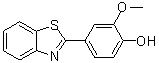
YL-109 (chemical structure: 2-(4-Hydroxy-3-methoxyphenyl)-benzothiazole) exerts its antitumor effects by inducing the expression of the ubiquitin ligase CHIP (Carboxyl terminus of Hsc70-interacting protein). [1]
|
|---|---|
| 体外研究 (In Vitro) |
在乳腺癌细胞中,YL-109(0.001-10 μM;96 小时或 24 小时)可抑制细胞运动、增殖和侵袭性[1]。在 MDA-MB-231 细胞中,YL-109 (1 μM) 会提高 CHIP mRNA 和蛋白质水平[1]。
1. 抑制乳腺癌细胞增殖:YL-109处理24、48、72小时后,剂量依赖性抑制人乳腺癌细胞系MDA-MB-231(三阴性)和MCF-7(ER阳性)的增殖(MTT法)。增殖抑制率随药物浓度升高和处理时间延长而增加,在浓度≥10 μM时表现出显著抑制作用。[1] 2. 抑制细胞迁移和侵袭:YL-109(10、20 μM)显著降低MDA-MB-231细胞的迁移和侵袭能力(Transwell实验和划痕愈合实验)。药物处理组的迁移/侵袭细胞数显著少于溶媒对照组,划痕愈合率也显著降低。[1] 3. 诱导乳腺癌细胞凋亡:YL-109(20 μM)诱导MDA-MB-231和MCF-7细胞凋亡(Annexin V-FITC/PI双染色结合流式细胞术)。凋亡率显著高于对照组,同时伴随促凋亡蛋白(Bax、Cleaved-Caspase 3、Cleaved-Caspase 9)上调和抗凋亡蛋白Bcl-2下调(Western blot检测)。[1] 4. 调控CHIP及致癌蛋白表达:YL-109剂量依赖性上调乳腺癌细胞中CHIP的蛋白和mRNA表达(Western blot和实时PCR);同时下调HER2、EGFR、Akt、STAT3等致癌蛋白的表达(Western blot),这些蛋白均为CHIP介导的泛素化降解底物。[1] 5. CHIP敲低验证作用机制:向MDA-MB-231细胞转染CHIP特异性siRNA后,YL-109的抗肿瘤作用(增殖抑制、迁移/侵袭抑制、凋亡诱导)显著减弱;同时逆转了YL-109介导的HER2、EGFR、Akt、STAT3下调,证实CHIP诱导是YL-109作用的关键机制。[1] 6. 增强CHIP介导的泛素化:YL-109(20 μM)促进MDA-MB-231细胞中HER2和EGFR的泛素化(抗泛素抗体免疫共沉淀实验),表明YL-109通过诱导CHIP介导致癌蛋白的泛素-蛋白酶体降解。[1] |
| 体内研究 (In Vivo) |
在体内,YL-109(15 mg/kg;每 2 天皮下注射一次)可抑制肿瘤生长以及乳腺癌细胞的转移[1]。
1. 裸鼠异种移植模型中抑制肿瘤生长:[1] - 雌性BALB/c裸鼠皮下接种MDA-MB-231细胞(1×10⁶个/只)构建异种移植肿瘤,当肿瘤体积达到~100 mm³时,以10 mg/kg和20 mg/kg剂量的YL-109每2天腹腔注射1次,持续3周。 - YL-109剂量依赖性抑制肿瘤生长:10 mg/kg和20 mg/kg组的最终肿瘤体积和重量显著小于溶媒对照组,20 mg/kg剂量抗肿瘤效果最强,肿瘤生长抑制率约60%。 - 肿瘤组织免疫组化(IHC)染色显示,YL-109处理上调CHIP表达,下调HER2、EGFR、p-Akt、p-STAT3表达;TUNEL染色证实药物处理组肿瘤组织中凋亡细胞增多。 2. 裸鼠模型中抑制肺转移:[1] - 雌性BALB/c裸鼠尾静脉注射MDA-MB-231细胞(5×10⁵个/只)诱导肺转移,以20 mg/kg剂量的YL-109每2天腹腔注射1次,持续4周。 - 与溶媒对照组相比,YL-109显著减少肺转移结节数量(肺组织大体观察和HE染色);肺转移灶IHC染色显示CHIP表达上调,HER2/EGFR/Akt/STAT3信号通路蛋白下调。[1] |
| 细胞实验 |
细胞增殖测定[1]
细胞类型: MCF-7 和 MDA-MB-231 细胞 测试浓度: 0.001、0.01、0.1、1、 10 μM 孵育时间: 96 小时 实验结果: 强烈抑制 MCF -7 和 MDA-MB-231 细胞的细胞增殖剂量依赖性方式(IC50分别=85.8 nM和4.02 μM)。 1. 细胞增殖MTT实验:[1] 将MDA-MB-231和MCF-7细胞以5×10³个/孔的密度接种于96孔板,贴壁过夜后,用系列浓度的YL-109(0、5、10、20、40 μM)处理24、48或72小时。处理结束后,向每孔加入MTT试剂孵育4小时,弃去上清液,用溶解液溶解甲臜结晶,酶标仪检测570 nm处吸光度,计算细胞增殖抑制率。 2. 细胞迁移和侵袭Transwell实验:[1] 迁移实验:将MDA-MB-231细胞重悬于含YL-109(0、10、20 μM)的无血清培养基中,接种于Transwell小室上室,下室加入含10% FBS的培养基。孵育24小时后,去除上室未迁移细胞,固定、染色下室迁移细胞并显微镜计数。 侵袭实验:使用基质胶包被的Transwell小室,实验流程同迁移实验,孵育时间延长至48小时。 3. 细胞凋亡Annexin V-FITC/PI染色实验:[1] 用20 μM YL-109处理MDA-MB-231和MCF-7细胞48小时,收集细胞并用PBS洗涤,重悬于结合缓冲液中。加入Annexin V-FITC和PI,避光孵育15分钟后,流式细胞术分析凋亡细胞,计算凋亡率。 4. 蛋白表达Western blot实验:[1] 用含蛋白酶和磷酸酶抑制剂的RIPA缓冲液裂解细胞或肿瘤组织,BCA法测定蛋白浓度。取等量蛋白进行SDS-PAGE电泳,转移至PVDF膜,用脱脂牛奶封闭。膜与抗CHIP、HER2、EGFR、Akt、p-Akt、STAT3、p-STAT3、Bcl-2、Bax、Cleaved-Caspase 3、Cleaved-Caspase 9或β-肌动蛋白一抗在4°C孵育过夜,洗涤后加入HRP标记二抗,ECL检测系统显影蛋白条带。 5. CHIP siRNA转染实验:[1] 将MDA-MB-231细胞接种于6孔板,用转染试剂将CHIP特异性siRNA或非靶向对照siRNA转染至细胞。转染48小时后,用20 μM YL-109处理细胞48小时,分别通过MTT、Transwell和Annexin V-FITC/PI实验检测细胞增殖、迁移、侵袭和凋亡;Western blot验证CHIP敲低效率及下游蛋白表达。 6. 泛素化免疫共沉淀(Co-IP)实验:[1] 用20 μM YL-109处理MDA-MB-231细胞24小时,收获前6小时加入蛋白酶体抑制剂。细胞用IP缓冲液裂解,裂解液与抗HER2或抗EGFR抗体在4°C孵育过夜,加入蛋白A/G琼脂糖珠孵育4小时。洗涤珠子后,洗脱免疫沉淀复合物,Western blot检测泛素化HER2/EGFR(抗泛素抗体)。[1] |
| 动物实验 |
动物/疾病模型: BALB/cAjcl-nu/nu雌性小鼠(4-5周龄)接种MCF-7或MDA-MB-231细胞[1]
剂量: 15 mg/kg 给药途径: 皮下注射,每2天一次,持续63天 实验结果: 注射MCF-7和MDA-MB-231细胞的小鼠肿瘤生长受到抑制。 1. 裸鼠肿瘤异种移植模型:[1] - 动物:雌性BALB/c裸鼠(6-8周龄)饲养于特定病原体清除(SPF)条件下。 - 肿瘤接种:将 MDA-MB-231 细胞(1×10⁶ 个细胞,溶于 100 μL PBS)皮下注射到每只小鼠的右侧腹部。 - 分组和给药:当肿瘤生长至约 100 mm³ 时,将小鼠随机分为三组(每组 n=6):载体对照组、YL-109 10 mg/kg 组和YL-109 20 mg/kg 组。YL-109 溶于合适的溶剂(例如,DMSO:PEG400:PBS = 1:4:5),每 2 天腹腔注射一次,持续 3 周。载体对照组注射等体积的溶剂。 - 肿瘤测量:每 3 天使用游标卡尺测量肿瘤体积,计算公式为 V = (长 × 宽²)/2。 - 样本采集:治疗3周后,处死小鼠。切除肿瘤,称重,并用福尔马林固定,用于免疫组化(IHC)染色。 - IHC检测:将福尔马林固定的肿瘤组织包埋于石蜡中,切片,并用针对CHIP、HER2、EGFR、p-Akt、p-STAT3和Ki-67的抗体进行染色。进行TUNEL染色以检测凋亡细胞。 2. 裸鼠肺转移模型:[1] - 动物:使用6-8周龄的雌性BALB/c裸鼠。 - 转移诱导:将MDA-MB-231细胞(5×10⁵个细胞,溶于100 μL PBS)经尾静脉注射入小鼠体内。 - 给药:细胞注射后一天,将小鼠随机分为两组(每组 n=6):载体对照组和YL-109 20 mg/kg 组。YL-109 每 2 天腹腔注射一次,持续 4 周,载体与异种移植模型相同。 - 样本采集:治疗 4 周后,处死小鼠。取出肺组织,用福尔马林固定,进行肉眼观察和 HE 染色以计数转移结节。对肺组织进行 IHC 染色以检测 CHIP、HER2 和 EGFR 的表达。[1] |
| 参考文献 | |
| 其他信息 |
1. 化学结构:YL-109的化学结构为2-(4-羟基-3-甲氧基苯基)-苯并噻唑,属于苯并噻唑衍生物家族。[1]
2. 背景:乳腺癌是女性最常见的恶性肿瘤之一,转移性乳腺癌预后较差。HER2、EGFR、Akt和STAT3等致癌蛋白在乳腺癌中过度表达,促进肿瘤进展和转移。CHIP是一种泛素连接酶,介导致癌蛋白的泛素-蛋白酶体降解,其表达下调与乳腺癌进展相关。[1] 3. 作用机制:YL-109通过特异性诱导CHIP表达发挥抗肿瘤作用。诱导型CHIP介导HER2、EGFR、Akt和STAT3的泛素化和降解,从而抑制乳腺癌细胞的增殖、迁移和侵袭,并诱导细胞凋亡。[1] 4. 治疗潜力:YL-109在临床前乳腺癌模型中显示出强大的抗肿瘤和抗转移活性,尤其是在缺乏靶向治疗的三阴性乳腺癌(MDA-MB-231)中。它通过靶向CHIP致癌蛋白通路,为乳腺癌提供了一种潜在的治疗策略。[1] |
| 分子式 |
C14H11NO2S
|
|
|---|---|---|
| 分子量 |
257.31
|
|
| 精确质量 |
257.051
|
|
| CAS号 |
36341-25-0
|
|
| 相关CAS号 |
|
|
| PubChem CID |
3155228
|
|
| 外观&性状 |
Light yellow to yellow solid powder
|
|
| 密度 |
1.327g/cm3
|
|
| 沸点 |
446.448ºC at 760 mmHg
|
|
| 闪点 |
223.804ºC
|
|
| 折射率 |
1.685
|
|
| LogP |
3.677
|
|
| tPSA |
70.59
|
|
| 氢键供体(HBD)数目 |
1
|
|
| 氢键受体(HBA)数目 |
4
|
|
| 可旋转键数目(RBC) |
2
|
|
| 重原子数目 |
18
|
|
| 分子复杂度/Complexity |
289
|
|
| 定义原子立体中心数目 |
0
|
|
| InChi Key |
KRVBOHJNAFQFPW-UHFFFAOYSA-N
|
|
| InChi Code |
InChI=1S/C14H11NO2S/c1-17-12-8-9(6-7-11(12)16)14-15-10-4-2-3-5-13(10)18-14/h2-8,16H,1H3
|
|
| 化学名 |
4-(1,3-benzothiazol-2-yl)-2-methoxyphenol
|
|
| 别名 |
|
|
| HS Tariff Code |
2934.99.9001
|
|
| 存储方式 |
Powder -20°C 3 years 4°C 2 years In solvent -80°C 6 months -20°C 1 month |
|
| 运输条件 |
Room temperature (This product is stable at ambient temperature for a few days during ordinary shipping and time spent in Customs)
|
| 溶解度 (体外实验) |
|
|||
|---|---|---|---|---|
| 溶解度 (体内实验) |
配方 1 中的溶解度: ≥ 3 mg/mL (11.66 mM) (饱和度未知) in 10% DMSO + 40% PEG300 + 5% Tween80 + 45% Saline (这些助溶剂从左到右依次添加,逐一添加), 澄清溶液。
例如,若需制备1 mL的工作液,可将100 μL 30.0 mg/mL 澄清的 DMSO 储备液加入到400 μL PEG300中,混匀;再向上述溶液中加入50 μL Tween-80,混匀;然后加入450 μL 生理盐水定容至1 mL。 *生理盐水的制备:将 0.9 g 氯化钠溶解在 100 mL ddH₂O中,得到澄清溶液。 配方 2 中的溶解度: 3 mg/mL (11.66 mM) in 10% DMSO + 90% (20% SBE-β-CD in Saline) (这些助溶剂从左到右依次添加,逐一添加), 澄清溶液; 超声助溶. 例如,若需制备1 mL的工作液,可将 100 μL 30.0 mg/mL澄清DMSO储备液加入900 μL 20% SBE-β-CD生理盐水溶液中,混匀。 *20% SBE-β-CD 生理盐水溶液的制备(4°C,1 周):将 2 g SBE-β-CD 溶解于 10 mL 生理盐水中,得到澄清溶液。 View More
配方 3 中的溶解度: ≥ 3 mg/mL (11.66 mM) (饱和度未知) in 10% DMSO + 90% Corn Oil (这些助溶剂从左到右依次添加,逐一添加), 澄清溶液。 1、请先配制澄清的储备液(如:用DMSO配置50 或 100 mg/mL母液(储备液)); 2、取适量母液,按从左到右的顺序依次添加助溶剂,澄清后再加入下一助溶剂。以 下列配方为例说明 (注意此配方只用于说明,并不一定代表此产品 的实际溶解配方): 10% DMSO → 40% PEG300 → 5% Tween-80 → 45% ddH2O (或 saline); 假设最终工作液的体积为 1 mL, 浓度为5 mg/mL: 取 100 μL 50 mg/mL 的澄清 DMSO 储备液加到 400 μL PEG300 中,混合均匀/澄清;向上述体系中加入50 μL Tween-80,混合均匀/澄清;然后继续加入450 μL ddH2O (或 saline)定容至 1 mL; 3、溶剂前显示的百分比是指该溶剂在最终溶液/工作液中的体积所占比例; 4、 如产品在配制过程中出现沉淀/析出,可通过加热(≤50℃)或超声的方式助溶; 5、为保证最佳实验结果,工作液请现配现用! 6、如不确定怎么将母液配置成体内动物实验的工作液,请查看说明书或联系我们; 7、 以上所有助溶剂都可在 Invivochem.cn网站购买。 |
| 制备储备液 | 1 mg | 5 mg | 10 mg | |
| 1 mM | 3.8864 mL | 19.4318 mL | 38.8636 mL | |
| 5 mM | 0.7773 mL | 3.8864 mL | 7.7727 mL | |
| 10 mM | 0.3886 mL | 1.9432 mL | 3.8864 mL |
1、根据实验需要选择合适的溶剂配制储备液 (母液):对于大多数产品,InvivoChem推荐用DMSO配置母液 (比如:5、10、20mM或者10、20、50 mg/mL浓度),个别水溶性高的产品可直接溶于水。产品在DMSO 、水或其他溶剂中的具体溶解度详见上”溶解度 (体外)”部分;
2、如果您找不到您想要的溶解度信息,或者很难将产品溶解在溶液中,请联系我们;
3、建议使用下列计算器进行相关计算(摩尔浓度计算器、稀释计算器、分子量计算器、重组计算器等);
4、母液配好之后,将其分装到常规用量,并储存在-20°C或-80°C,尽量减少反复冻融循环。
计算结果:
工作液浓度: mg/mL;
DMSO母液配制方法: mg 药物溶于 μL DMSO溶液(母液浓度 mg/mL)。如该浓度超过该批次药物DMSO溶解度,请首先与我们联系。
体内配方配制方法:取 μL DMSO母液,加入 μL PEG300,混匀澄清后加入μL Tween 80,混匀澄清后加入 μL ddH2O,混匀澄清。
(1) 请确保溶液澄清之后,再加入下一种溶剂 (助溶剂) 。可利用涡旋、超声或水浴加热等方法助溶;
(2) 一定要按顺序加入溶剂 (助溶剂) 。
|
|
|
 Penisimplicissin
 Benz[a]anthracene
 AHR antagonist 5 hemimaleate
 AHR agonist 10
InvivoChem的所有产品仅用于作科学研究,不面向患者销售
Copyright 2020 InvivoChem LLC | All Rights Reserved 粤ICP备20063088号-1


 COA
COA


 463611831
