| 规格 | 价格 | 库存 | 数量 |
|---|---|---|---|
| 250mg |
|
||
| 500mg |
|
||
| 1g |
|
||
| Other Sizes |
|

| 靶点 |
Endogenous Metabolite
|
|---|---|
| 体外研究 (In Vitro) |
5-氨基乙酰丙酸 (5-ALA) 上调与防御和免疫相关的基因,改善有氧能量代谢,并增强南美白对虾对副溶血弧菌的免疫反应 [1]。
|
| 体内研究 (In Vivo) |
随着对虾养殖中几种传染病的出现,人们对使用饲料添加剂增强对虾免疫力的兴趣日益浓厚。最近,在血红素生物合成中起限速作用的非蛋白质氨基酸5-氨基酮戊酸(5-ALA)的使用因其对家畜免疫的积极作用而受到关注。为了评估5-ALA在南美白对虾(Litopenaeus vannamei)中的作用,我们进行了微阵列分析、副溶血弧菌浸泡激发试验、ATP水平测定以及与血红素合成和降解相关的一些血红蛋白和基因的基因表达分析。在微阵列上15745个南美白对虾推定基因中,101个基因在5-ALA补充组和对照组对虾肝胰腺之间差异表达超过四倍(p<0.05)。5-ALA上调了101个基因中的99个,其中41个是基于序列同源性的免疫和防御相关基因。与对照组相比,补充5-ALA的组在挑战试验中的存活率更高,胆色素原合酶、亚铁螯合酶、过氧化氢酶、核受体E75和血红素加氧酶-1的转录水平更高,ATP水平更高。这些发现表明,饮食中的5-ALA分别增强了凡纳对虾对副溶血弧菌的免疫反应,上调了免疫和防御相关基因,并增强了有氧能量代谢。需要进一步的研究来阐明5-ALA在虾养殖中的使用程度[1]。
1. 对免疫力的影响:通过饲料投喂5-氨基乙酰丙酸(5-Aminolevulinic Acid, ALA; Levulan)可增强凡纳滨对虾( Litopenaeus vannamei )的免疫功能。用含ALA的饲料(0.1、0.5、1.0 g/kg饲料)处理21天后,对虾体内免疫相关酶活性发生显著变化。其中,0.5 g/kg ALA组对虾血淋巴中酚氧化酶(PO)活性较对照组(不含ALA)提高42.3%;该组溶菌酶(LYZ)活性较对照组高35.6%,总血球数(THC)也较对照组增加28.9%。此外,肝胰腺中免疫相关基因(proPO、LYZ、Toll)的表达水平显著上调,0.5 g/kg ALA组proPO基因的相对表达量为对照组的2.1倍。[1] 2. 对ATP水平的影响:ALA可提高凡纳滨对虾肌肉组织中的ATP含量。投喂21天后,0.5 g/kg ALA组对虾肌肉中ATP浓度达6.8 μmol/g,较对照组(5.2 μmol/g)提高31.7%;1.0 g/kg ALA组对虾肌肉ATP含量(6.1 μmol/g)也显著高于对照组,但低于0.5 g/kg组。[1] 3. 对基因表达的影响:ALA可调控凡纳滨对虾体内多个功能基因的表达。除免疫相关基因外,0.5 g/kg ALA组中与能量代谢相关的基因(如ATP合酶α亚基基因)表达上调,相对表达量为对照组的1.8倍;同时,该组应激相关基因(如HSP70基因)的表达量较对照组降低38.2%,表明对虾的抗应激能力得到改善。[1] |
| 酶活实验 |
检测97例ESCC患者病理标本中GPX4和HMOX1的表达,并进行预后分析。实时聚合酶链式反应(RT-PCR)、RNA微阵列和蛋白质印迹分析用于评估5-ALA在体外铁下垂中的作用。Ann Surg Oncol. 2021 Jul;28(7):3996-4006. https://pubmed.ncbi.nlm.nih.gov/33210267/
|
| 动物实验 |
术前立即进行肿瘤体积测量。随后仅使用5-ALA信号进行肿瘤切除,若无可见信号则判定切除彻底。此判断始终由主刀医生完成。术中间歇性地投射功能性神经导航数据,以防止意外损伤功能性脑区。在每个切除阶段结束时,系统地检查肿瘤腔,以排除残留肿瘤。一旦检测不到5-ALA信号,则进行术中磁共振成像(iMRI)扫描。如果确认切除范围,则由主刀医生决定结束手术。否则,重新分割残留肿瘤体积,并根据神经导航继续切除。在所有这些情况下,一旦去除薄薄的“健康”脑实质层和/或优化观察角度,即可在后续手术中重新检测到5-ALA信号。重复上述步骤直至5-ALA信号消失,且术中磁共振成像(iMRI)证实无对比增强肿瘤。iMRI检测到的额外切除组织也由经验丰富的神经病理学家进行分析,证实存在病理性胶质瘤细胞浸润。如果神经导航数据显示功能区域内仍存在5-ALA信号,则有意终止相应方向的进一步手术。PLoS One, 2012. 7(9): p. e44885.
1. 实验动物准备:选用初始体重为10±2 g的南美白对虾(Litopenaeus vannamei)。实验前,将对虾在充氧海水缸中驯养7天,水温保持在28±2℃,盐度为30±2‰,pH值为8.0±0.2。驯化期间,虾每天投喂两次商业饲料(8:00 和 18:00),投喂量为虾体重的 5%。[1] 2. ALA 添加及分组:将 ALA 混入商业饲料中,配制四种不同 ALA 浓度的实验饲料:对照组(0 g/kg 饲料)、0.1 g/kg ALA 组、0.5 g/kg ALA 组和 1.0 g/kg ALA 组。每组设置 3 个重复缸,每个重复缸饲养 30 只虾。实验持续 21 天,虾每天投喂两次(8:00 和 18:00),投喂量为虾体重的 5%;每 3 天根据虾的存活情况调整投喂量。[1] 3. 样品采集:21 天实验结束后,从每个重复缸中随机选取 5 只虾。使用1 mL注射器(按1:1比例加入抗凝剂)从腹侧窦采集血淋巴,并在3000 rpm下离心10分钟,以获得血淋巴上清液用于免疫酶活性检测。解剖肝胰腺和肌肉组织,迅速置于液氮中冷冻,并储存于-80℃,用于后续的基因表达分析和ATP含量测定。[1] |
| 药代性质 (ADME/PK) |
吸收、分布和排泄
口服生物利用度为 50-60%。### 氨基乙酰丙酸 (ALA) 和原卟啉IX (PpIX) 的局部凝胶药代动力学 (PK) 在一项纳入 12 名患有轻度至中度光化性角化病 (AK) 的成年受试者的试验中进行了评估,这些受试者面部或前额至少有 10 个 AK 病灶。单次使用一整管 ALA(2 克)进行封闭敷料,敷 3 小时后,对总面积为 20 cm² 的病灶进行光动力疗法 (PDT)。基线血浆 ALA 和 PpIX 浓度的平均值 ± 标准差分别为 20.16 ± 16.53 ng/mL 和 3.27 ± 2.40 ng/mL。在大多数受试者中,ALA 敷用后前 3 小时内,血浆 ALA 浓度最多可增加 2.5 倍。基线校正后的ALA(n=12)的平均±标准差浓度-时间曲线下面积(AUC0-t)和最大浓度(Cmax)分别为142.83±75.50 ng·h/mL和27.19±20.02 ng/mL。达到Cmax的中位时间(Tmax)为3小时。### 外用溶液 两项人体药代动力学(PK)研究在上肢患有轻度至中度日光性角化病的受试者中进行,受试者一侧上肢至少有6处病灶,另一侧上肢至少有12处病灶。单次给药方案为两次局部涂抹ALA外用溶液(每次含354 mg ALA HCl),直接涂抹于病灶处,并在光疗前封闭3小时。第一项PK研究纳入29名受试者,并评估了ALA的PK参数。基线校正后的ALA最大浓度(Cmax)平均值±标准差为249.9±694.5 ng/mL,中位达峰时间(Tmax)为给药后2小时。ALA的平均暴露量(以浓度-时间曲线下面积(AUCt)表示)为669.9±1610 ng·hr/mL。ALA的平均消除半衰期(t1/2)为5.7±3.9小时。在14名受试者中进行了第二次药代动力学(PK)研究,并测定了ALA和PpIX的PK参数。在50%(7/14)的受试者中,至少50%的样本中基线校正后的PpIX浓度为阴性,因此无法可靠地估计AUC。ALA和PpIX的基线校正后Cmax平均值±标准差分别为95.6±120.6 ng/mL和0.95±0.71 ng/mL。 ALA 和 PpIX 的中位达峰时间 (Tmax) 分别为给药后 2 小时和 12 小时。ALA 的平均 AUCt 为 261.1 ± 229.3 ng·hr/mL。ALA 的平均半衰期 (t1/2) 为 8.5 ± 6.7 小时。### 口服溶液 在 12 名健康受试者中,服用推荐剂量的 ALA 溶液后,ALA 的绝对生物利用度为 100.0% ± 1.1,范围为 78.5% 至 131.2%。 ALA血浆浓度峰值达到的中位数为0.8小时(范围0.5-1.0小时)。 在12名健康受试者中,服用推荐剂量ALA溶液后12小时内,尿液中母体氨基乙酰丙酸(ALA)的排泄率为34±8%(平均值±标准差),范围为27%至57%。 在健康志愿者中,静脉注射氨基乙酰丙酸的分布容积为9.3±2.8升,口服为14.5±2.5升。[11961050] 代谢/代谢物 外源性氨基乙酰丙酸(ALA)代谢为PpIX,但所给ALA代谢为PpIX的比例尚不清楚。 PpIX 的平均血浆 AUC 不到 ALA 的 6%。 局部给药后,PpIX 在皮肤内原位合成。 半衰期:口服给药后平均半衰期为 0.70 ± 0.18 小时,静脉给药后平均半衰期为 0.83 ± 0.05 小时。 生物半衰期 局部用溶液制剂的氨基乙酰丙酸平均消除半衰期 (t1/2) 为 5.7 ± 3.9 小时,口服溶液制剂的平均半衰期为 0.9 ± 1.2 小时。在另一项对 6 名健康志愿者进行的药代动力学研究中,使用 128 毫克剂量,口服给药后平均半衰期为 0.70 ± 0.18 小时,静脉注射给药后平均半衰期为 0.83 ± 0.05 小时。 |
| 毒性/毒理 (Toxicokinetics/TK) |
毒性概述
根据推测的作用机制,局部应用氨基乙酰丙酸 (ALA) 溶液后发生的光敏反应是由于 ALA 代谢转化为原卟啉 IX (PpIX) 所致,PpIX 会在涂抹氨基乙酰丙酸的皮肤中积聚。当暴露于适当波长和能量的光时,积聚的 PpIX 会产生光动力反应,这是一种依赖于光和氧同时存在的细胞毒性过程。光的吸收导致卟啉分子处于激发态,随后 PpIX 向分子氧的自旋转移生成单线态氧,单线态氧可进一步反应生成超氧阴离子和羟基自由基。使用氨基乙酰丙酸对日光性角化病病变进行光敏化,并配合 BLU-UTM 蓝光光动力疗法照射器 (BLU-U) 进行照射,是氨基乙酰丙酸光动力疗法 (PDT) 的基础。 妊娠期和哺乳期的影响 ◉ 哺乳期用药概述 目前尚无关于哺乳期口服氨基乙酰丙酸的信息。为尽量减少婴儿的暴露,口服后可暂停哺乳 24 小时。由于全身吸收量极低,预计哺乳不会导致婴儿接触到局部使用的氨基乙酰丙酸。氨基乙酰丙酸诱导的光动力疗法已成功用于治疗各种乳头皮肤病变。这种治疗方法似乎能够保护乳头解剖结构,有利于母乳喂养。 ◉ 对母乳喂养婴儿的影响 截至修订日期,未找到相关的已发表信息。 ◉ 对泌乳和母乳的影响 截至修订日期,未找到相关的已发表信息。 蛋白质结合 在体外实验中,使用浓度高达推荐剂量ALA溶液后血浆中最大浓度的约25%的氨基乙酰丙酸(ALA),ALA的平均蛋白质结合率为12%。 |
| 参考文献 | |
| 其他信息 |
药效学
氨基乙酰丙酸 (ALA) 的代谢是血红素合成生化途径的第一步。氨基乙酰丙酸本身并非光敏剂,而是光敏剂原卟啉 IX (PpIX) 的代谢前体。ALA 的合成通常受到 ALA 合成酶反馈抑制的严格调控,这种抑制可能与细胞内血红素水平有关。当 ALA 进入细胞后,会绕过这一调控点,导致 PpIX 的积累。PpIX 随后通过亚铁螯合酶将铁添加到其原子核上,最终转化为血红素。 1. ALA 背景:5-氨基乙酰丙酸 (ALA;Levulan) 是卟啉生物合成的关键前体,而卟啉参与生物体内血红素、叶绿素和维生素 B12 的合成。据报道,α-亚麻酸(ALA)能够调节水生动物的能量代谢和抗氧化能力,本研究进一步探讨了ALA对南美白对虾(Litopenaeus vannamei)免疫功能和基因表达的影响。[1] 2. 最佳剂量:研究表明,南美白对虾的最佳饲料添加量为0.5 g/kg。在此剂量下,ALA对虾的免疫力、ATP合成和基因表达均表现出最显著的促进作用,而更高剂量(1.0 g/kg)并未进一步增强这些作用,表明ALA在虾体内的生物活性与剂量呈正相关。[1] |
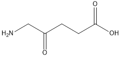
| 分子式 |
C5H9NO3
|
|---|---|
| 分子量 |
131.12986
|
| 精确质量 |
131.058
|
| 元素分析 |
C, 45.80; H, 6.92; N, 10.68; O, 36.60
|
| CAS号 |
106-60-5
|
| 相关CAS号 |
5-Aminolevulinic acid hydrochloride;5451-09-2;5-Aminolevulinic acid-13C;123253-93-0
|
| PubChem CID |
137
|
| 外观&性状 |
Typically exists as solid at room temperature
|
| 密度 |
1.2±0.1 g/cm3
|
| 沸点 |
298.4±20.0 °C at 760 mmHg
|
| 熔点 |
156-158 °C
156 - 158 °C |
| 闪点 |
134.3±21.8 °C
|
| 蒸汽压 |
0.0±1.3 mmHg at 25°C
|
| 折射率 |
1.482
|
| LogP |
-0.93
|
| tPSA |
80.39
|
| 氢键供体(HBD)数目 |
2
|
| 氢键受体(HBA)数目 |
4
|
| 可旋转键数目(RBC) |
4
|
| 重原子数目 |
9
|
| 分子复杂度/Complexity |
121
|
| 定义原子立体中心数目 |
0
|
| SMILES |
NCC(=O)CCC(O)=O
|
| InChi Key |
ZGXJTSGNIOSYLO-UHFFFAOYSA-N
|
| InChi Code |
InChI=1S/C5H9NO3/c6-3-4(7)1-2-5(8)9/h1-3,6H2,(H,8,9)
|
| 化学名 |
5-amino-4-oxopentanoic acid
|
| 别名 |
5-Aminolevulinic acid; Aminolevulinic acid; 106-60-5; 5-Amino-4-oxopentanoic acid; 5-Aminolevulinate; Pentanoic acid, 5-amino-4-oxo-; delta-aminolevulinic acid; Aladerm; 5451-09-2 (HCl); 106-60-5 (free); 868074-65-1 (phosphate)
|
| HS Tariff Code |
2934.99.9001
|
| 存储方式 |
Powder -20°C 3 years 4°C 2 years In solvent -80°C 6 months -20°C 1 month |
| 运输条件 |
Room temperature (This product is stable at ambient temperature for a few days during ordinary shipping and time spent in Customs)
|
| 溶解度 (体外实验) |
DMSO : ~100 mg/mL (~762.60 mM)
|
|---|---|
| 溶解度 (体内实验) |
配方 1 中的溶解度: ≥ 2.5 mg/mL (19.07 mM) (饱和度未知) in 10% DMSO + 40% PEG300 + 5% Tween80 + 45% Saline (这些助溶剂从左到右依次添加,逐一添加), 澄清溶液。
例如,若需制备1 mL的工作液,可将100 μL 25.0 mg/mL澄清DMSO储备液加入到400 μL PEG300中,混匀;然后向上述溶液中加入50 μL Tween-80,混匀;加入450 μL生理盐水定容至1 mL。 *生理盐水的制备:将 0.9 g 氯化钠溶解在 100 mL ddH₂O中,得到澄清溶液。 配方 2 中的溶解度: ≥ 2.5 mg/mL (19.07 mM) (饱和度未知) in 10% DMSO + 90% (20% SBE-β-CD in Saline) (这些助溶剂从左到右依次添加,逐一添加), 澄清溶液。 例如,若需制备1 mL的工作液,可将 100 μL 25.0 mg/mL澄清DMSO储备液加入900 μL 20% SBE-β-CD生理盐水溶液中,混匀。 *20% SBE-β-CD 生理盐水溶液的制备(4°C,1 周):将 2 g SBE-β-CD 溶解于 10 mL 生理盐水中,得到澄清溶液。 View More
配方 3 中的溶解度: ≥ 2.5 mg/mL (19.07 mM) (饱和度未知) in 10% DMSO + 90% Corn Oil (这些助溶剂从左到右依次添加,逐一添加), 澄清溶液。 1、请先配制澄清的储备液(如:用DMSO配置50 或 100 mg/mL母液(储备液)); 2、取适量母液,按从左到右的顺序依次添加助溶剂,澄清后再加入下一助溶剂。以 下列配方为例说明 (注意此配方只用于说明,并不一定代表此产品 的实际溶解配方): 10% DMSO → 40% PEG300 → 5% Tween-80 → 45% ddH2O (或 saline); 假设最终工作液的体积为 1 mL, 浓度为5 mg/mL: 取 100 μL 50 mg/mL 的澄清 DMSO 储备液加到 400 μL PEG300 中,混合均匀/澄清;向上述体系中加入50 μL Tween-80,混合均匀/澄清;然后继续加入450 μL ddH2O (或 saline)定容至 1 mL; 3、溶剂前显示的百分比是指该溶剂在最终溶液/工作液中的体积所占比例; 4、 如产品在配制过程中出现沉淀/析出,可通过加热(≤50℃)或超声的方式助溶; 5、为保证最佳实验结果,工作液请现配现用! 6、如不确定怎么将母液配置成体内动物实验的工作液,请查看说明书或联系我们; 7、 以上所有助溶剂都可在 Invivochem.cn网站购买。 |
| 制备储备液 | 1 mg | 5 mg | 10 mg | |
| 1 mM | 7.6260 mL | 38.1301 mL | 76.2602 mL | |
| 5 mM | 1.5252 mL | 7.6260 mL | 15.2520 mL | |
| 10 mM | 0.7626 mL | 3.8130 mL | 7.6260 mL |
1、根据实验需要选择合适的溶剂配制储备液 (母液):对于大多数产品,InvivoChem推荐用DMSO配置母液 (比如:5、10、20mM或者10、20、50 mg/mL浓度),个别水溶性高的产品可直接溶于水。产品在DMSO 、水或其他溶剂中的具体溶解度详见上”溶解度 (体外)”部分;
2、如果您找不到您想要的溶解度信息,或者很难将产品溶解在溶液中,请联系我们;
3、建议使用下列计算器进行相关计算(摩尔浓度计算器、稀释计算器、分子量计算器、重组计算器等);
4、母液配好之后,将其分装到常规用量,并储存在-20°C或-80°C,尽量减少反复冻融循环。
计算结果:
工作液浓度: mg/mL;
DMSO母液配制方法: mg 药物溶于 μL DMSO溶液(母液浓度 mg/mL)。如该浓度超过该批次药物DMSO溶解度,请首先与我们联系。
体内配方配制方法:取 μL DMSO母液,加入 μL PEG300,混匀澄清后加入μL Tween 80,混匀澄清后加入 μL ddH2O,混匀澄清。
(1) 请确保溶液澄清之后,再加入下一种溶剂 (助溶剂) 。可利用涡旋、超声或水浴加热等方法助溶;
(2) 一定要按顺序加入溶剂 (助溶剂) 。
 L-Histidinol-d3
 D-myo-Inositol-3-phosphate sodium
 (±)19(20)-DiHDPA
 4-Hydroxy-2-butanone
InvivoChem的所有产品仅用于作科学研究,不面向患者销售
Copyright 2020 InvivoChem LLC | All Rights Reserved 粤ICP备20063088号-1


 463611831
