| 规格 | 价格 | 库存 | 数量 |
|---|---|---|---|
| 10 mM * 1 mL in DMSO |
|
||
| 5mg |
|
||
| 10mg |
|
||
| 25mg |
|
||
| 50mg |
|
||
| 100mg |
|
||
| 250mg |
|
||
| 500mg |
|
||
| Other Sizes |
|

| 靶点 |
p110α (IC50 = 32 nM); p110α E545K (IC50 = 30 nM); p110α H1047R (IC50 = 43 nM); p110γ (IC50 = 3480 nM); PI3K-C2β (IC50 = 462 nM); PI4Kβ (IC50 = 236 nM)
|
|---|---|
| 体外研究 (In Vitro) |
p110α 的致癌形式,如 p110α E545K 和 p110α H1047R,也被 A66 有效抑制,IC50 值分别为 30 nM 和 43 nM。与其他 I 类 PI3K 同工型相比,A66 对 p110α 的选择性大于 100 倍,而 PIK-75 则相反。在 II 类 PI3K、III 类 PI3K 和 PI4K 中,A66 仅与 II 类 PI3K PI3KC2β 和 PI4K 的 PI4Kβ 亚型表现出有限的交叉反应性,IC50 分别为 462 nM 和 236 nM。相关激酶 DNA-PK 和 mTOR 以及其他脂质激酶不受 A66 抑制。与 PIK-75 相比,在 10 μM 浓度下针对两个相当大的组(110 种蛋白激酶和 318 种激酶)进行测试时,A66 具有更高水平的特异性。在一些 PIK3CA 具有 H1047R 突变且 p110 和 Ia 类 PI3K 活性水平较高的细胞系中,通过 A66 治疗抑制 p110α 足以阻断向 Akt/PKB 发出的胰岛素信号传导。 [1]高度转化的 p85 iSH2 突变体 KS459delN、DKRMN-S560del 和 K379E 在接受 0.7 μM A66 后,病灶形成减少 75–80%,并且所有 p85α iSH2 突变体都减少了 T308 上的 Akt 磷酸化。 [2]
抑制剂特异性[1] 首先对A66进行了表征,并证实它是野生型和致癌型p110α的强效抑制剂,但不是其他I类PI3K亚型(表1)。发现A66对p110α的选择性比PIK-75高得多。鉴于II类PI3Ks、III类PI3K[38]和PI4Ks(磷酸肌醇4-激酶)在生长因子信号传导中的重要作用,我们还评估了A66对这些的活性,发现与II类PI3K PI3K-C2β和PI4K的PI4Kβ亚型存在一些有限的交叉反应(表2)。对其他脂质激酶或相关激酶DNA-PK和mTOR没有抑制作用(表2)。我们还测试了10μM A66效应对两组110种蛋白激酶的抑制作用(补充图S1http://www.BiochemJ.org/bj/438/bj4380053add.htm)318种激酶(补充图S2http://www.BiochemJ.org/bj/438/bj4380053add.htm). 这些表明A66是p110α的一种非常特异的抑制剂,而PIK-75,即之前描述为p110α选择性抑制剂的化合物,在该浓度下抑制了大量蛋白激酶(补充图S1)。我们使用HTRF测定法生成的TGX-221和IC87114数据与之前使用其他测定方法的研究一致,并证实这些分别是p110β和p110δ的高选择性抑制剂(表1),尽管TGX-221在较高浓度下会与p110δ发生交叉反应。我们进一步报告说,这些抑制剂对110种蛋白激酶没有任何重大影响(补充图1)。 特异性抑制p110α对细胞功能的影响[1] 为了研究p110α在调节PI3K依赖性信号通路近端元件中的作用,我们通过Ser473和Thr308的磷酸化评估了不同浓度的A66S形式在一系列细胞系中急性阻断Akt/PKB激活的能力(图3)。通过对整个PKB进行重新编程来控制负载(见补充图S3http://www.BiochemJ.org/bj/438/bj4380053add.htm). 我们发现,在所有测试的细胞系中,Ser473和Thr308的磷酸化对LY294002都很敏感,这意味着激活Akt/PKB需要I类PI3K活性。然而,我们发现抑制Ser473和Thr308磷酸化所需的A66S形式的量遵循两种不同的模式,要么对A66 S形式在与通过p110α起作用的浓度一致的浓度下的抑制敏感,要么具有抗性。敏感细胞系最明显的特征是它们在PIK3CA中携带H1047R突变,而所有其他细胞系都是抗性的。作为对照,我们测试了A66 R形式的作用,发现它不能抑制Akt/PKB的磷酸化(见补充图S4http://www.BiochemJ.org/bj/438/bj4380053add.htm). |
| 体内研究 (In Vivo) |
在体内,给药后 1 小时和 6 小时,单剂量 100 mg/kg 的 A66 导致 Akt/PKB 和 p70 S6 激酶的磷酸化显着降低,但 ERK 的磷酸化没有显着降低。与众所周知的泛 PI3K 抑制剂 BEZ-235 相比,A66 导致 SK-OV-3 异种移植肿瘤的生长速度显着减慢,每日一次 100 mg/kg 剂量给药时,平均 TGI 分别为 45.9% 和 29.9% (QD) 持续 21 天或 75 mg/kg 每天两次 (BID) 持续 16 天。 A66 的 QD 给药虽然导致 U87MG 异种移植模型中的肿瘤体积没有显着减少,但也导致 HCT-116 异种移植模型中的肿瘤体积显着减少,TGI 为 77.2%。 [1]在雄性 CD1 小鼠中,给予 10 mg/kg A66 会导致 ITT(胰岛素耐量试验)和 GTT(葡萄糖耐量试验)显着受损,并且在 PTT(丙酮酸耐量试验)期间葡萄糖产生增加,几乎与泛 PI3K 抑制剂引起的症状一样严重。 [3]
根据药代动力学和药效学研究结果,在肿瘤疗效研究中,每天一次以100mg/kg体重给药A66S,持续21天,或每天两次以75mg/kg体重给药,持续16天。两种给药策略都显著延迟了SK-OV-3异种移植物肿瘤的生长,甚至超过了公认的泛PI3K抑制剂BEZ-235诱导的生长(图7A)。在给药的最后一天,A66 S型的平均TGI为对照组的45.9%(QD;P<0.05)和对照组的29.9%(BID;P<0.01)(表4)。QDA66S在该异种移植物模型中耐受良好,体重减轻最小;然而,BID治疗与中度体重减轻和两例死亡有关,尽管尚不清楚死亡是由于药物毒性还是其他原因,因为这些小鼠没有表现出明显的体重减轻(图7B)。相比之下,BEZ-235诱导的肿瘤生长没有显著减少,甚至耐受性更低,体重适度减轻,4人死亡。HCT-116异种移植物模型中每天一次给予A66S也诱导了肿瘤体积的显著减少,给药结束时TGI为对照组的77.2%(P<0.01),但在U87MG异种移植模型中,肿瘤体积没有显著减少(表4和补充图S5http://www.BiochemJ.org/bj/438/bj4380053add.htm). 相比之下,BEZ-235显著降低了U87MG肿瘤的生长(TGI=对照组的61.1%;P<0.05),但对HCT-116肿瘤没有影响。尽管SK-OV-3研究中BEZ-235的剂量水平相同,但U87MG模型和HCT-116模型中的药物耐受性良好,在HCT-116模式中,由于对照组治疗的小鼠体重中度减轻,使用了较低剂量(10mg/kg体重)的BEZ-235。[1] 本研究首次研究了选择性p110α抑制剂(A66)对体内葡萄糖代谢的影响。我们发现A66会损害体内胰岛素作用的所有指标,几乎与pan-PI3K抑制剂的水平相同。这提供了强有力的药理学证据,表明p110α是急性调节葡萄糖代谢途径中最重要的亚型,PI3K亚型之间的功能冗余不太可能是体内调节葡萄糖代谢主要途径的主要特征。A66对葡萄糖代谢的影响是p110α激酶死亡形式全局表达杂合小鼠的表型。然而,尽管A66在全球范围内抑制p110α,但本研究的结果也与仅在肝脏中急性或慢性缺失Pik3ca基因的小鼠的结果非常相似。结合我们的PTT结果,这表明p110α在调节胰岛素对葡萄糖代谢的影响方面的主要作用位点在肝脏[3]。 |
| 酶活实验 |
PI3K(人类)HTRF 检测用于计算 IC50 值。 Invitrogen 提供 p85α/p110δ 。所有其他亚型都是通过将全长人 p85α 与适当的全长人催化亚基结合而现场制备的,该亚基在 N 末端标有组氨酸标签以方便纯化。 PI3K 的使用浓度在其 EC65 和 EC80 值之间进行滴定。使用 p85α N-SH2(N-Src 同源 2)结构域的抗体,测量免疫沉淀物中的 PI3K 活性。国家蛋白激酶分析中心和 Invitrogen 药物发现服务中心对其他脂质激酶和蛋白激酶进行检测[1]。
|
| 细胞实验 |
细胞培养和转染。[2]
受精鸡蛋(白色Leghorn)购自Charles River育种实验室。如前所述制备和培养原代CEF。对于转染,细胞在含有5.8%补铁FCS和1%L-谷氨酰胺-青霉素-链霉素溶液的F-10中以80%的融合率铺板。第二天,使用二甲亚砜/聚异戊二烯法用RCAS载体转染CEF。在血清存在下传代两次后,收获细胞进行进一步分析。 HEK 293-T细胞在添加了10%FCS和1%L-谷氨酰胺-青霉素-链霉素溶液的DMEM中培养。根据制造商的方案,使用脂质体PLUS进行输血。将70%融合的MP6板中的HEK293T细胞用Opti-MEM培养基洗涤一次,并在0.8mL Opti-MEM中孵育。在室温下将总共1μg质粒DNA与0.1mL Opti-MEM和2μL Lipofectamine PLUS混合15分钟。将含有6μL Lipofectamine的Opti-MEM(0.1 mL)加入DNA-PLUS混合物中,并在室温下孵育15分钟。将DNA、PLUS和Lipofectamine的混合物加入细胞中并孵育过夜。第二天,将培养基换成含有10%FCS和1%L-谷氨酰胺-青霉素-链霉素溶液的DMEM。转染40小时后,收集细胞,裂解并分析特定蛋白质。 焦点分析。[2] 如前所述,使用感染性逆转录病毒载体进行了重点检测。使用二甲亚砜/聚异戊二烯法用适当的RCAS构建体转染CEF,每隔一天用营养琼脂覆盖2至3周,直至观察到焦点形成。用结晶紫对平板进行染色,并计数转化细胞的焦点。为了检测不同化合物对焦点形成的抑制作用,将10μM LY294002、100 nM NVP-BEZ-235、2 nM雷帕霉素、5μM ZK-93、250 nM TGX221、5μM IC87114或5μM AS604850添加到每个覆盖层的营养琼脂中。 细胞增殖。[2] 转染后,将CEF分成含有F-10的增殖试验培养基,其中添加了2%的FCS和1%的鸡血清。在第二次分裂时,将细胞以每孔4000个细胞的速度接种到96孔板中。在接种后第1至5天,CEF在37°C的增殖试验培养基中与10μg/mL的Resazurin Na一起孵育4小时。在560nm的激发波长和590nm的发射下测定荧光。 Western Blot和免疫沉淀。[2] 如前所述进行蛋白质印迹,但稍作修改。细胞在改良的Nonidet P-40裂解缓冲液中裂解(20 mM Tris-Cl、150 mM NaCl、1 mM MgCl2、1%Nonident P-40和10%甘油,含1 mM PMSF、1 mM DTT、50 mM NaF、1 mmol Na3VO4、50 mMβ-甘油磷酸盐和蛋白酶抑制剂混合物)。在4°C下以18000×g离心10分钟后,测定上清液的蛋白质浓度。为了检查抑制剂信号传导,在收集之前,在含血清的条件下用250 nM TGX-221或5μM IC87114处理细胞2小时。为了进行免疫沉淀,将含有40μg蛋白质的细胞裂解物与抗FLAG M2琼脂糖在4°C下孵育过夜。琼脂糖珠用裂解缓冲液洗涤四次,加热至95°C,然后在SDS-PAGE凝胶上分离。转移到Immobilon P膜后,这些膜在室温下用含0.1%吐温-20(TBS-T)的Tris缓冲盐水中的5%BSA封闭2小时,然后用1:2000的抗FLAG或1:1000的抗p110α或抗p110β一抗稀释液孵育过夜。将膜在TBS-T中洗涤三次,并在室温下与过氧化物酶偶联的山羊抗小鼠或山羊抗兔抗体在5%BSA/TBS-T溶液中孵育1小时。反应带通过SuperSignal West Pico化学发光底物可视化。 对于蛋白质印迹,在SDS-PAGE凝胶上分离含有10μg总蛋白的细胞裂解物,并将其转移到Immobilon P膜上。将膜与1:1000稀释的针对p85、pAkt(T308)、Akt、p4E-BP、4E-BP和β-actin的一抗一起孵育。如上所述,开发了蛋白质印迹。抗FLAG抗体(FLAG-M2 F3165)购自xxx。从xyz获得抗p110α抗体(#4255)、抗p110β抗体(#3011S)、抗p85抗体(#4292)、抗Akt(#2967)、抗磷酸化Akt(Thr-308)(#9275S)、抗4E-BP1(#9452)和抗磷酸化4E-BPl(Ser-65)(#9451S)抗体。 |
| 动物实验 |
小鼠:将 5×10⁶ 个 U87MG、SK-OV-3 或 HCT-116 细胞悬浮于 PBS 中,皮下接种于年龄匹配的无特定病原体 Rag1-/- 或 NIH-III 小鼠的右侧腹部。根据公式 (L×w²)×π/6(其中 L 为肿瘤最长直径,w 为垂直直径),使用电子游标卡尺测量肿瘤直径,计算肿瘤体积 (mm³)。BEZ-235 溶于 10% 乙醇溶液中,A66 溶于 20% 2-羟丙基-β-环糊精水溶液中。对照组小鼠仅给予 A66 给药溶剂。药物以游离碱当量 10 mL/kg 体重进行腹腔注射。当肿瘤直径生长至约 8 至 9 毫米时,小鼠接受单次注射 A66 或对照物质,用于肿瘤药效学研究。取出肿瘤,进行生物粉碎,并在末次给药后 1 小时或 6 小时处死动物前测量蛋白质浓度。
异种移植方法 [1] 将年龄匹配的无特定病原体 Rag1−/− 或 NIH-III 小鼠右侧腹部皮下接种 5×10⁶ 个 U87MG、SK-OV-3 或 HCT-116 细胞(溶于 PBS)。使用电子游标卡尺测量肿瘤直径,并根据公式 (L×w²)×π/6 计算肿瘤体积 (mm³)(其中 L=肿瘤最长直径,w=垂直直径)。 A66溶于20% 2-羟丙基-β-环糊精水溶液中给药,而BEZ-235溶于10%乙醇溶液中给药。对照组小鼠仅给予A66给药溶剂。药物以游离碱当量进行腹腔注射,给药体积为10 ml/kg体重。在肿瘤药效学研究中,当肿瘤直径达到约8-9 mm时,小鼠单次注射A66或对照溶剂。给药后1小时或6小时处死动物,取出肿瘤,进行生物粉碎,并测定蛋白质浓度。在抗肿瘤疗效研究中,当肿瘤已充分形成,平均直径约为7 mm时开始给药。给药方案为每日一次(QD)或每日两次(BID),两次注射间隔至少约8小时。根据肿瘤生长速度和对照小鼠的体重耐受情况,三种异种移植模型采用了不同的给药方案。动物每日给药一次,持续21天;或每日两次给药,持续16天(SK-OV-3);每日给药一次,持续14天(U87MG);每日给药一次,持续7天(HCT-116)。每日监测动物的毒性反应,并记录体重。若小鼠出现中度毒性反应或体重下降超过初始体重的20%,则处死小鼠。在给药的最后一天,通过计算药物治疗组小鼠的相对肿瘤大小占对照组小鼠平均相对肿瘤大小的百分比来评估肿瘤生长抑制率(TGI)。使用GraphPad Prism 5.02软件,通过单因素方差分析(ANOVA)和Bonferroni多重比较检验来确定TGI值的统计学意义。 GTT、ITT 和 PTT [3] GTT、ITT 和 PTT 以及胰岛素水平测定均按先前所述方法进行,不同之处在于使用雄性 CD1 小鼠代替大鼠。进行 GTT 和 PTT 时,小鼠需禁食过夜;进行 ITT 时,小鼠需在实验开始前 2 小时停止进食。药物在黑暗周期结束后1小时腹腔注射,并在腹腔注射葡萄糖或丙酮酸(2 g/kg体重)或胰岛素(0.75单位/kg体重)前1小时注射。 代谢笼研究[3] 使用Oxymax/CLAMS(Columbus Instruments)定量氧耗量(V̇O2)、二氧化碳生成量(V̇CO2)、基础代谢率(BMR)、食物摄入量、饮水摄入量和动物活动量,具体方法如前所述。BMR根据先前研究的建议,以瘦体重为自变量表示。所有数据均使用EchoMRI-100定量磁共振系统进行标准化,以达到瘦体重,具体方法如前所述。动物在笼中适应24小时后,收集接下来24小时的数据。 药物浓度分析[3] 在喂食的CD1雄性小鼠(体重30克)中进行药代动力学研究。通过灌胃或腹腔注射给予动物指定的PI3K抑制剂,并在给药后15分钟、1小时、2小时、4小时、6小时和24小时用EDTA抗凝管采集终末血样。所有药物均溶于DMSO。血液经离心(2000 g,10分钟,4℃)后分离血浆用于药物定量。药物定量采用液相色谱串联质谱法(LC-MS/MS)。简而言之,将300 μl 100%甲醇加入100 μl血浆中。将样品轻轻混合后离心(2000 g,10 分钟,4°C)。取上清液 50 μl 加入小瓶中进行 LC-MS/MS 分析。离子源类型为电喷雾电离(ESI),条件如下:喷雾电压5500 V,鞘气压力50单位,离子扫描气压力0.0单位,辅助气压力2单位,毛细管温度370°C,毛细管偏置电压35 V。色谱柱为HPLC Kinetex柱(100 mm × 3 mm,2.6 μm C18(2)-HST;Phenomenex)。流动相为10%(0.1%甲酸水溶液)和90%甲醇的等度洗脱。流速为0.2 mL/min。保留时间分别为2.64 min(PI-103)、2.76 min(TGX221)和2.35 min(IC87114)。未知浓度根据标准曲线和内标确定。 |
| 药代性质 (ADME/PK) |
首先,我们通过在CD-1小鼠腹腔注射10 mg/kg体重的药物后,研究其药代动力学,确定了异种移植研究的最佳给药策略。尽管A66S的半衰期仅为0.42小时,但其在给药后30分钟即达到的Cmax值高达8247 nM,确保了其AUC0-inf(曲线下面积,从零时间到无穷大)(6809 nM·h)与半衰期为2.73小时的BEZ-235(7333 nM·h)相似(表3)。此外,我们采用单次100 mg/kg体重的剂量,在体内测试了A66S对SK-OV-3肿瘤组织的影响,以确定该药物是否能在靶组织中产生持久作用(图6)。研究表明,给药后1小时和6小时,A66 S均能显著降低Akt/PKB和p70 S6激酶的磷酸化水平,但对ERK(细胞外信号调节激酶)的磷酸化水平没有影响(图6)。这与A66 S在此期间对肿瘤中PI3K信号通路具有完全抑制作用的结果相一致。在本研究中,给药后1小时和6小时,血浆中A66 S的浓度分别为21.1±1.2 μM和9.1±1.1 μM,而肿瘤组织中A66 S的浓度在相同时间点分别为22.7±2.1 μM和16.0±1.3 μM。因此,药物在肿瘤中的滞留可能是抑制作用持续存在的原因。 [1]
药代动力学方法[1] 将年龄匹配的特定病原体清除(SPF)雄性CD-1小鼠单次注射A66(10 mg/kg体重),溶于20% 2-羟丙基-β-环糊精水溶液中;或注射BEZ-235,溶于15% (v/v) DMSO、20% (v/v) 0.1 M HCl、0.7% Tween 20和64.3% (v/v)生理盐水中。给药后分别在5或6个时间点处死小鼠(每个时间点n=3),通过心脏穿刺采集血液至EDTA抗凝管中。血液样本在20℃下以6000转/分钟离心10分钟,保留血浆上清液。向血浆中加入甲醇进行蛋白质提取。采用安捷伦 6460 三重四极杆液相色谱-串联质谱仪 (LC-MS/MS) 进行定量分析,使用多反应监测 (MRM) 和电喷雾电离 (ESI) 模式。色谱分离采用安捷伦 Zorbax SB-C18 色谱柱 (2.1 mm×50 mm; 5 μm),流动相梯度为 20%~100% 甲醇(含 0.1% 甲酸和 5 mM 甲酸铵),流速为 0.4 ml/min。血浆药物浓度根据已知浓度范围为 10~10000 nM 的校准曲线进行定量,并设置了 65、650 和 6500 nM 的质控样品。为防止先前样品的污染,在每个血浆样品之间运行甲醇冲洗。药代动力学参数采用 WinNonlin 5.3 软件进行非房室模型分析。 |
| 参考文献 | |
| 其他信息 |
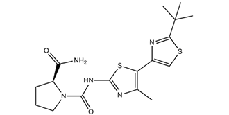
(2S)-N1-[5-(2-叔丁基-4-噻唑基)-4-甲基-2-噻唑基]吡咯烷-1,2-二甲酰胺是一种脯氨酸衍生物。
PI3K(磷脂酰肌醇3-激酶)信号通路的基因改变在癌症中很常见,包括PTEN(10号染色体上缺失的磷酸酶和张力蛋白同源物)的缺失、PIK3CA的扩增以及PIK3CA基因两个不同区域的突变。这提示靶向PI3K(尤其是p110α)的药物可能对癌症治疗有效。广谱抑制PI3K可有效抑制生长因子信号传导和肿瘤生长,但目前尚无合适的p110α抑制剂来研究单独抑制该亚型的效果。在本研究中,我们对一种新型小分子化合物A66进行了表征,结果表明其S-对映体是一种高度特异性和选择性的p110α抑制剂。我们利用分子建模和生化研究解释了这种选择性的基础。使用一系列亚型选择性抑制剂,我们发现,在所有测试的细胞系中,抑制p110α/p110β/p110δ的叠加效应均可减弱胰岛素向Akt/PKB(蛋白激酶B)的信号传导。然而,在某些细胞系中,单独抑制p110α就足以阻断胰岛素向Akt/PKB的信号传导。这些响应性细胞系均携带PIK3CA的H1047R突变,并且具有高水平的p110α和Ia类PI3K活性。这可能解释了这些细胞对p110α抑制剂的更高敏感性。我们评估了异种移植模型中 Akt/PKB 的激活和肿瘤生长情况,发现源自两种反应性细胞系的肿瘤在体内也对 A66 有反应。这些结果表明,单独抑制 p110α 有可能阻断生长因子信号传导,并减少部分肿瘤的生长。[1] 磷脂酰肌醇 3-激酶 (PI3K) 的调节亚基 p85α 的 iSH2(SH2 间区)和 nSH2(N 端 SH2)结构域中的癌症特异性突变表现出功能获得性。它们诱导致癌细胞转化,刺激细胞增殖,并增强 PI3K 信号传导。致癌活性的定量测定揭示了 p85α 各个突变体之间存在显著差异。突变蛋白仍然能够与催化亚基 p110α 和 p110β 结合。使用p110亚型特异性抑制剂的研究表明,成纤维细胞中p85突变体的表达仅导致p110α的激活,并且p110α是p85突变体诱导致癌转化的唯一介质。p85突变体的特征与以下假设相符:这些突变削弱了p85α和p110α之间的抑制性相互作用,同时保留了p85α iSH2与p110α衔接结合域之间的稳定相互作用。[2]体外研究表明,I类PI3K(磷脂酰肌醇3-激酶)、II类PI3K和mTOR(雷帕霉素靶蛋白)均参与葡萄糖代谢的调控。然而,它们各自在体内正常葡萄糖代谢信号通路中的相对作用尚不明确。基因敲除和基因敲入小鼠模型已提供了一些证据,表明I类PI3K亚型p110α、p110β以及在较小程度上p110γ对于调节葡萄糖代谢和食欲的过程是必需的。然而,在这些模型中,PI3K活性长期降低。因此,我们分析了单独急性抑制PI3K亚型或同时急性抑制PI3K和mTOR对葡萄糖代谢和食物摄入的影响。在本研究中,我们观察到,使用泛PI3K/mTOR抑制剂PI-103和NVP-BEZ235治疗的小鼠出现葡萄糖耐量受损、胰岛素耐量降低以及肝脏葡萄糖输出增加。ZSTK474具有类似作用,表明这些作用是由PI3K抑制而非mTOR抑制引起的。 p110α选择性抑制剂PIK75和A66也诱导了这些表型,但p110β、p110δ或p110γ抑制剂仅产生轻微影响。这些药物对基础代谢率(BMR)、耗氧量或饮水量没有显著影响,但BEZ235、PI-103和PIK75确实导致食物摄入量略有下降。令人惊讶的是,泛PI3K抑制剂或p110α抑制剂导致动物活动量减少,尽管其原因尚不清楚。综上所述,这些研究提供了药理学证据,支持PI3K的p110α亚型在急性调节葡萄糖代谢的通路中发挥着重要作用。[3] |
| 分子式 |
C17H23N5O2S2
|
|---|---|
| 分子量 |
393.5268
|
| 精确质量 |
393.129
|
| 元素分析 |
C, 51.89; H, 5.89; N, 17.80; O, 8.13; S, 16.29
|
| CAS号 |
1166227-08-2
|
| 相关CAS号 |
1166227-08-2
|
| PubChem CID |
42636535
|
| 外观&性状 |
Off-white to light yellow solid powder
|
| 密度 |
1.4±0.1 g/cm3
|
| 折射率 |
1.640
|
| LogP |
0.63
|
| tPSA |
162.17
|
| 氢键供体(HBD)数目 |
2
|
| 氢键受体(HBA)数目 |
6
|
| 可旋转键数目(RBC) |
4
|
| 重原子数目 |
26
|
| 分子复杂度/Complexity |
556
|
| 定义原子立体中心数目 |
1
|
| SMILES |
S1C([H])=C(C2=C(C([H])([H])[H])N=C(N([H])C(N3C([H])([H])C([H])([H])C([H])([H])[C@@]3([H])C(N([H])[H])=O)=O)S2)N=C1C(C([H])([H])[H])(C([H])([H])[H])C([H])([H])[H]
|
| InChi Key |
HBPXWEPKNBHKAX-NSHDSACASA-N
|
| InChi Code |
InChI=1S/C17H23N5O2S2/c1-9-12(10-8-25-14(20-10)17(2,3)4)26-15(19-9)21-16(24)22-7-5-6-11(22)13(18)23/h8,11H,5-7H2,1-4H3,(H2,18,23)(H,19,21,24)/t11-/m0/s1
|
| 化学名 |
(2S)-1-N-[5-(2-tert-butyl-1,3-thiazol-4-yl)-4-methyl-1,3-thiazol-2-yl]pyrrolidine-1,2-dicarboxamide
|
| 别名 |
A-66; A66; 1166227-08-2; A66; (S)-N1-(2-(tert-butyl)-4'-methyl-[4,5'-bithiazol]-2'-yl)pyrrolidine-1,2-dicarboxamide; A 66; CHEMBL3218581; (2S)-1-N-[5-(2-tert-butyl-1,3-thiazol-4-yl)-4-methyl-1,3-thiazol-2-yl]pyrrolidine-1,2-dicarboxamide; 1,2-Pyrrolidinedicarboxamide, N1-[2-(1,1-dimethylethyl)-4'-methyl[4,5'-bithiazol]-2'-yl]-, (2S)-; A 66
|
| HS Tariff Code |
2934.99.9001
|
| 存储方式 |
Powder -20°C 3 years 4°C 2 years In solvent -80°C 6 months -20°C 1 month |
| 运输条件 |
Room temperature (This product is stable at ambient temperature for a few days during ordinary shipping and time spent in Customs)
|
| 溶解度 (体外实验) |
DMSO: ~79 mg/mL (~200.7 mM)
Water: ~1 mg/mL (~2.5 mM) Ethanol: ~4 mg/mL (~26.8 mM) |
|---|---|
| 溶解度 (体内实验) |
配方 1 中的溶解度: ≥ 2.5 mg/mL (6.35 mM) (饱和度未知) in 10% DMSO + 40% PEG300 + 5% Tween80 + 45% Saline (这些助溶剂从左到右依次添加,逐一添加), 澄清溶液。
例如,若需制备1 mL的工作液,可将100 μL 25.0 mg/mL澄清DMSO储备液加入到400 μL PEG300中,混匀;然后向上述溶液中加入50 μL Tween-80,混匀;加入450 μL生理盐水定容至1 mL。 *生理盐水的制备:将 0.9 g 氯化钠溶解在 100 mL ddH₂O中,得到澄清溶液。 配方 2 中的溶解度: ≥ 2.5 mg/mL (6.35 mM) (饱和度未知) in 10% DMSO + 90% (20% SBE-β-CD in Saline) (这些助溶剂从左到右依次添加,逐一添加), 澄清溶液。 例如,若需制备1 mL的工作液,可将 100 μL 25.0 mg/mL澄清DMSO储备液加入900 μL 20% SBE-β-CD生理盐水溶液中,混匀。 *20% SBE-β-CD 生理盐水溶液的制备(4°C,1 周):将 2 g SBE-β-CD 溶解于 10 mL 生理盐水中,得到澄清溶液。 View More
配方 3 中的溶解度: ≥ 2.5 mg/mL (6.35 mM) (饱和度未知) in 10% DMSO + 90% Corn Oil (这些助溶剂从左到右依次添加,逐一添加), 澄清溶液。 配方 4 中的溶解度: 15% Captisol: 8mg/mL 1、请先配制澄清的储备液(如:用DMSO配置50 或 100 mg/mL母液(储备液)); 2、取适量母液,按从左到右的顺序依次添加助溶剂,澄清后再加入下一助溶剂。以 下列配方为例说明 (注意此配方只用于说明,并不一定代表此产品 的实际溶解配方): 10% DMSO → 40% PEG300 → 5% Tween-80 → 45% ddH2O (或 saline); 假设最终工作液的体积为 1 mL, 浓度为5 mg/mL: 取 100 μL 50 mg/mL 的澄清 DMSO 储备液加到 400 μL PEG300 中,混合均匀/澄清;向上述体系中加入50 μL Tween-80,混合均匀/澄清;然后继续加入450 μL ddH2O (或 saline)定容至 1 mL; 3、溶剂前显示的百分比是指该溶剂在最终溶液/工作液中的体积所占比例; 4、 如产品在配制过程中出现沉淀/析出,可通过加热(≤50℃)或超声的方式助溶; 5、为保证最佳实验结果,工作液请现配现用! 6、如不确定怎么将母液配置成体内动物实验的工作液,请查看说明书或联系我们; 7、 以上所有助溶剂都可在 Invivochem.cn网站购买。 |
| 制备储备液 | 1 mg | 5 mg | 10 mg | |
| 1 mM | 2.5411 mL | 12.7055 mL | 25.4110 mL | |
| 5 mM | 0.5082 mL | 2.5411 mL | 5.0822 mL | |
| 10 mM | 0.2541 mL | 1.2706 mL | 2.5411 mL |
1、根据实验需要选择合适的溶剂配制储备液 (母液):对于大多数产品,InvivoChem推荐用DMSO配置母液 (比如:5、10、20mM或者10、20、50 mg/mL浓度),个别水溶性高的产品可直接溶于水。产品在DMSO 、水或其他溶剂中的具体溶解度详见上”溶解度 (体外)”部分;
2、如果您找不到您想要的溶解度信息,或者很难将产品溶解在溶液中,请联系我们;
3、建议使用下列计算器进行相关计算(摩尔浓度计算器、稀释计算器、分子量计算器、重组计算器等);
4、母液配好之后,将其分装到常规用量,并储存在-20°C或-80°C,尽量减少反复冻融循环。
计算结果:
工作液浓度: mg/mL;
DMSO母液配制方法: mg 药物溶于 μL DMSO溶液(母液浓度 mg/mL)。如该浓度超过该批次药物DMSO溶解度,请首先与我们联系。
体内配方配制方法:取 μL DMSO母液,加入 μL PEG300,混匀澄清后加入μL Tween 80,混匀澄清后加入 μL ddH2O,混匀澄清。
(1) 请确保溶液澄清之后,再加入下一种溶剂 (助溶剂) 。可利用涡旋、超声或水浴加热等方法助溶;
(2) 一定要按顺序加入溶剂 (助溶剂) 。
|
|
|
|
 NF-κB-IN-19
 PI3K-IN-57
 PI3K-IN-52
 SR-3-65
InvivoChem的所有产品仅用于作科学研究,不面向患者销售
Copyright 2020 InvivoChem LLC | All Rights Reserved 粤ICP备20063088号-1


 COA
COA


 463611831
