| 规格 | 价格 | 库存 | 数量 |
|---|---|---|---|
| 10 mM * 1 mL in DMSO |
|
||
| 1mg |
|
||
| 5mg |
|
||
| 10mg |
|
||
| 25mg |
|
||
| 50mg |
|
||
| 100mg |
|
||
| 250mg |
|
||
| 500mg |
|
||
| 1g |
|
||
| Other Sizes |
|

| 靶点 |
Cysteine protease
Cysteine proteases: - Cathepsin B (human recombinant): Ki ≈ 0.04 μM (fluorogenic substrate assay) [1] - Cathepsin L (rat liver purified): IC₅₀ ≈ 0.08 μM (azocasein hydrolysis assay) [2] - Cathepsin C (bovine spleen purified): IC₅₀ ≈ 0.2 μM (glycyl-phenylalanyl-4-methoxy-β-naphthylamide cleavage assay) [1] - Selectivity over serine proteases: No inhibition of trypsin or chymotrypsin (10 μM Aloxistatin, casein hydrolysis assay) [1] |
|---|---|
| 体外研究 (In Vitro) |
Aloxistatin(也称为 E-64d、E-64c 乙酯;Loxistatin;NSC 694281)是一种有效的、选择性的、不可逆的、广谱的、细胞膜可渗透的半胱氨酸蛋白酶抑制剂。 Aloxistatin 可防止体外雨蛙蛋白诱导的胰蛋白酶原激活。阿洛司他汀可以进入完整细胞并抑制钙蛋白酶。 E-64d 已被证明对于治疗人类阿尔茨海默病是安全的。 Aloxistatin 通过调节异常的锌信号转导以及通过海马中的 ApoE/clusterin 途径调节脂质代谢改变,可潜在用于治疗发育性癫痫诱发的脑损伤。阿洛司他汀可以进入完整的血小板,通过抑制钙蛋白酶来抑制蛋白水解。 Aloxistatin 在体外可抑制甲状旁腺激素 (PTH) 诱导的细胞增殖并抑制成骨细胞的分化。激酶测定:在 24 孔板中,用抑制剂 L1 (10-20 μM) 或阿洛司他丁 (20-30 μM) 在 37°C 下处理 CTL 和 NK 细胞 (0.8×10⁶/mL) 24 小时。然后将细胞用于 51 Cr 释放测定或裂解以在蛋白质印迹中检查穿孔素。如所示,在一些 51 Cr 释放测定中,在 4 小时反应期间也以相同浓度添加抑制剂。使用 NP-40 裂解缓冲液(25 mM HEPES、250 mM NaCl、2.5 mM 乙二胺四乙酸、0.1% 体积/体积 Nonidet P-40)制备细胞裂解物,并使用 Bradford 测定法测定总蛋白浓度。等量的蛋白质上样并在 8% SDS-PAGE 凝胶上解析。使用指定的适当抗体检测人或小鼠穿孔素。抗肌动蛋白抗体用作上样对照。细胞测定:用 A23187 加钙激活血小板,已知这种情况会导致钙蛋白酶催化 ABP 和 talin 蛋白水解。在没有抑制剂的情况下,A23187 导致 ABP 和talin 完全降解。 E 64d,渗透抑制剂,确实抑制细胞内蛋白水解。在最低测试浓度 20 μg/ml 下观察到了一些抑制作用,而在 50 μg/ml 下则获得了基本上完全的抑制作用。
半胱氨酸蛋白酶抑制活性(文献[1]、[2]): 1. 组织蛋白酶B抑制:Aloxistatin(0.01–1 μM)浓度依赖性抑制人重组组织蛋白酶B。0.1 μM时抑制率达~90%(Z-Arg-Arg-AMC荧光底物实验)[1] 2. 组织蛋白酶L抑制:0.1 μM Aloxistatin抑制大鼠肝脏组织蛋白酶L介导的偶氮酪蛋白降解~85%,IC₅₀≈0.08 μM[2] - 癌细胞自噬调控: 1. 人乳腺癌MCF-7细胞:10 μM Aloxistatin处理24小时,LC3-II/LC3-I比值上调~3.2倍(Western blot),提示自噬诱导;自噬底物p62水平较对照组降低~60%[6] 2. 小鼠黑色素瘤B16细胞:5 μM Aloxistatin增强自噬流:mRFP-GFP-LC3斑点(自噬体)荧光强度上调~2.8倍(共聚焦显微镜)[6] - 阿尔茨海默病(AD)相关活性: 1. 大鼠原代皮质神经元:2 μM Aloxistatin处理48小时,Aβ₄₂分泌减少~45%(ELISA)。Western blot显示β-分泌酶(BACE1)蛋白水平无变化,而Aβ降解酶组织蛋白酶B活性上调~30%[5] |
| 体内研究 (In Vivo) |
E-64d 治疗后,用青霉素治疗小鼠以诱导复发性癫痫发作。结果表明,E-64d显着减少了齿状回颗粒上区和海马CA3亚区的异常苔藓纤维萌芽。在未经E-64d治疗的大鼠中,在齿状回和CA3亚区的锥体层中存在明显的苔藓纤维末端聚集。在用 E-64d 治疗的大鼠中,齿状回颗粒上区域和 CA3 亚区的苔藓纤维末端聚集显着减少。
小鼠AD模型(APP/PS1转基因小鼠,文献[5]): 1. 分组:6月龄小鼠(n=8/组)随机分为2组:(1)溶剂对照组(腹腔注射10% DMSO+90%生理盐水);(2)Aloxistatin 1 mg/kg组[5] 2. 给药方案:腹腔注射,每日1次,持续4周[5] 3. 疗效: - 脑内Aβ₄₂水平:海马区较对照组降低~40%,皮质区降低~35%(ELISA); - 认知功能:Morris水迷宫实验显示逃避潜伏期较对照组缩短~30%; - 小胶质细胞活化:海马区Iba1⁺小胶质细胞数量减少~25%(免疫组织化学)[5] - 小鼠黑色素瘤异种移植模型: 1. 给药方案:B16肿瘤体积达~100 mm³时开始,Aloxistatin 5 mg/kg(腹腔注射,每2天1次),持续14天[6] 2. 疗效: - 肿瘤体积:较溶剂对照组减少~55%; - 肿瘤自噬:肿瘤组织中LC3-II水平上调~2.5倍(Western blot); - 对小鼠体重无显著影响(较基线变化<5%)[6] |
| 酶活实验 |
将抑制剂 L1 (10–20 μM) 或 Aloxistatin (20–30 μM) 应用于 CTL 和 NK 细胞 (0.8×10⁶/mL),在 37°C 下在 24 孔板中持续 24 小时。之后,将细胞用于 51 Cr 释放实验或裂解细胞以在蛋白质印迹分析中观察穿孔素。一些 51Cr 释放测定还在 4 小时反应期间添加相同浓度的抑制剂,如图所示。使用 NP-40 裂解缓冲液(25 mM HEPES、250 mM NaCl、2.5 mM 乙二胺四乙酸、0.1% 体积/体积 Nonidet P-40)制备细胞裂解液,并使用 Bradford 法测定总蛋白浓度。将等量的蛋白质上样并解析到 8% SDS-PAGE 凝胶上。按照指示使用正确的抗体来检测人或小鼠穿孔素。使用抗肌动蛋白抗体作为上样对照。
组织蛋白酶B活性抑制实验: 1. 蛋白制备:人重组组织蛋白酶B在大肠杆菌中表达,镍螯合层析纯化,在50 mM醋酸钠缓冲液(pH5.5)中用10 mM DTT激活[1] 2. 反应体系:100 μL混合物含激活的组织蛋白酶B(0.5 μg)、荧光底物Z-Arg-Arg-AMC(20 μM)、Aloxistatin(0.01–1 μM)及50 mM醋酸钠缓冲液(pH5.5),溶剂(DMSO)作为对照[1] 3. 孵育与检测:37℃孵育60分钟,每10分钟测定荧光强度(激发光360 nm,发射光460 nm)。抑制率=(1–药物组荧光强度/对照组荧光强度)×100%[1] 4. 数据分析:Lineweaver-Burk双倒数作图(竞争性抑制模型)计算Ki值[1] - 组织蛋白酶L活性抑制实验: 1. 蛋白制备:大鼠肝脏中通过离子交换层析纯化组织蛋白酶L,在0.1 M Tris-HCl缓冲液(pH7.5)中用5 mM DTT激活[2] 2. 反应体系:200 μL混合物含组织蛋白酶L(1 μg)、底物偶氮酪蛋白(1%,w/v)、Aloxistatin(0.02–0.5 μM)及0.1 M Tris-HCl缓冲液(pH7.5)[2] 3. 检测:37℃孵育4小时,5%三氯乙酸终止反应,测定上清液366 nm处吸光度,计算抑制率[2] |
| 细胞实验 |
增殖标记物 Ki67 或凋亡标记物 cleaved caspase 3 染色可用于评估细胞增殖和凋亡。极性标记的过程与之前相同。四天后,MCF10 变体在 3D rBM 覆盖培养物中生长,并用 5 μM CA074Me、5 μM Aloxistatin 或 0.1% DMSO 处理。通过使用 Zeiss Axiophot 落射荧光显微镜对两个不同盖玻片上总共 100 个结构进行计数,可以确定 Ki67 或裂解的 caspase 3 呈阳性的结构的百分比。如果一个结构至少有一个 Ki67 染色细胞,则被认为是 Ki67 阳性。当某个结构具有一个或多个裂解 caspase 3 阳性的细胞,并且这些细胞不位于发育管腔的中心时,该结构被称为 caspase 3 阳性[3]。
MCF-7细胞自噬实验: 1. 细胞接种:MCF-7细胞以2×10⁵个细胞/孔接种于6孔板,使用含10% FBS的RPMI 1640培养基[6] 2. 药物处理:加入Aloxistatin(1–20 μM),37℃、5% CO₂孵育24小时。检测自噬流时,最后4小时共孵育100 nM巴弗洛霉素A1[6] 3. 检测: - Western blot:含蛋白酶抑制剂的RIPA缓冲液裂解细胞,30 μg蛋白进行免疫印迹(一抗:抗LC3、抗p62、抗β-actin); - 免疫荧光:药物处理前24小时转染mRFP-GFP-LC3质粒,共聚焦显微镜计数荧光斑点[6] - 原代皮质神经元Aβ分泌实验: 1. 细胞培养:大鼠胚胎(E18)皮质神经元以1×10⁵个细胞/孔接种于24孔板,使用含2% B27添加剂的神经基础培养基[5] 2. 药物处理:加入Aloxistatin(0.5–5 μM),孵育48小时[5] 3. 检测:收集上清液,夹心ELISA量化Aβ₄₂水平;细胞裂解后进行Western blot(一抗:抗BACE1、抗组织蛋白酶B)[5] |
| 动物实验 |
小鼠和猪:使用平均体重 400 克(约 6 周龄)的雄性 Hartley 品系豚鼠。在雄性转基因小鼠中表达伦敦突变型 β-分泌酶位点序列和含有野生型 β-分泌酶位点的 AβPP。虽然灌胃给药可以实现精确的剂量,但这种方法创伤性较大,仅适用于短时间给药(最长约一周)。在豚鼠研究中,采用灌胃给药。将推荐剂量的阿洛昔他汀(0.1、1.0、5 和 10 mg/kg)悬浮于二亚硫酸甲酯 (Me2SO) 中,并通过饲管每日一次口服给药。对照组动物仅灌胃给予 Me2SO。
大鼠:使用近交系雄性 DS 大鼠。断奶后的大鼠在 7 周龄之前饲喂含 0.3% NaCl 的实验室饲料。 DS大鼠喂食8% NaCl饮食7周后,在12周时出现向心性左心室肥厚代偿的迹象,这与高血压有关。19周时,大鼠出现致命性左心室衰竭的明显迹象,并伴有肺充血。为此,DS大鼠从7周龄开始喂食8% NaCl饮食。从12周龄到19周龄,将它们随机分为三组:高脂饮食组(HF)、阿洛昔他汀组(10 mg/kg/天,隔日腹腔注射)和RNH-6270组(3 mg/kg/天混入饲料中)(每组n=10)。阿洛昔他汀和RNH-6270(一种血管紧张素受体阻滞剂)的剂量由初步实验和既往研究确定。年龄匹配的对照组(对照组,n=10)为喂食0.3% NaCl饮食的DS大鼠。所有大鼠均在19周龄时通过腹腔注射过量(50 mg/kg)的NSC 10816处死,并取出心脏进行组织学和生物学检查。为测定肾素活性,从腹主动脉抽取动脉血。从7周龄开始,每周对清醒的大鼠进行无创尾套法测量心率和收缩压。在另一项独立研究中,每组n=5只12周龄的DS大鼠(从7周龄开始喂食低盐饮食)分别以与先前研究相同的方式给予赋形剂、RNH-6270或Aloxistatin。用于检测靶向mRNA和蛋白水平的LV组织随后立即用液氮冷冻,并保存在-80°C。 APP/PS1转基因小鼠AD模型方案: 1. 动物饲养:6月龄APP/PS1转基因小鼠(雄性,25-30 g)饲养于SPF级动物房(22-25°C,12小时光照/黑暗循环),自由摄食饮水[5] 2. 分组和治疗:小鼠随机分为载体对照组和阿洛司他汀组。阿洛司他汀溶于10% DMSO + 90%生理盐水中,以1 mg/kg的剂量,每日一次,腹腔注射(10 μL/g体重),持续4周。对照组仅接受溶剂处理[5] 3. 监测和分析: - 认知功能:每周进行Morris水迷宫测试(逃避潜伏期、平台穿越次数); - 脑组织采集:小鼠经CO₂吸入安乐死;解剖海马和皮层,用于Aβ ELISA和免疫组织化学(Iba1染色)[5] - 小鼠B16黑色素瘤异种移植方案: 1. 肿瘤植入:将B16细胞(5×10⁶个细胞/只小鼠)重悬于100 μL PBS中,皮下注射到C57BL/6小鼠(6-8周龄,雌性)右侧腹部[6] 2. 治疗:肿瘤体积达到约100 mm³(第0天)时,随机分组。将阿洛昔他汀溶于 5% DMSO + 95% 生理盐水中,以 5 mg/kg 的剂量腹腔注射,每 2 天一次,连续 14 天 [6] 3. 分析:每 3 天测量一次肿瘤体积(体积 = 长 × 宽² / 2);处死动物后切除肿瘤进行蛋白质印迹分析(LC3、p62)[6] |
| 药代性质 (ADME/PK) |
大鼠腹腔注射药代动力学:
1. 药代动力学参数(5 mg/kg 腹腔注射剂量): - Cmax:~12 μM(Tmax = 0.5 小时); - AUC₀-24h:~35 μM·h; - 末端半衰期 (t₁/₂):~2.8 小时; - 清除率 (CL):~140 mL/h/kg [3] 2. 组织分布(5 mg/kg 腹腔注射,给药后 1 小时): - 肝脏:~25 μM; - 肾脏:~18 μM; - 脑组织浓度:~2.5 μM(中枢神经系统穿透性低)[3] - 口服药代动力学: 1. 口服生物利用度:~15%(大鼠,10 mg/kg 口服剂量与腹腔注射剂量相比);肝脏首过代谢显著[1] 2. 排泄:约 60% 的给药剂量在 72 小时内经尿液(以代谢物形式)排出;约 20% 经粪便排出(原药:~5%)[3] |
| 毒性/毒理 (Toxicokinetics/TK) |
体外毒性(文献[1]、[6]):
1. 正常人成纤维细胞 (MRC-5):20 μM 阿洛昔他汀(72 小时处理)使细胞活力降低 <10%(MTT 法)[1] 2. 原代大鼠皮层神经元:5 μM 阿洛昔他汀 未显示明显的细胞毒性;神经元存活率 >90%(NeuN 染色)[5] - 体内毒性(文献[3]、[6]): 1. 急性毒性(小鼠): - 单次腹腔注射 LD₅₀ ≈ 80 mg/kg; - 过量症状:短暂性共济失调和活动减少,24 小时内缓解 [3] 2. 亚慢性毒性(大鼠,5 mg/kg 腹腔注射,每日一次,持续 4 周): - 无死亡;体重变化 <5%(与基线相比); - 血清生化指标(ALT、AST、肌酐)在正常范围内 [3] - 血浆蛋白结合率:约 85%(人血浆,37°C 平衡透析)[1] |
| 参考文献 | |
| 其他信息 |
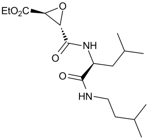
阿洛昔他汀是一种L-亮氨酸衍生物,它是(2S,3S)-3-(乙氧羰基)环氧乙烷-2-羧酸的羧基与N-(3-甲基丁基)-L-亮氨酰胺的氨基缩合而成的酰胺。它是一种组织蛋白酶B抑制剂和抗冠状病毒药物。它是一种L-亮氨酸衍生物、单羧酸酰胺、环氧化物和乙酯。
阿洛昔他汀是一种半胱氨酸蛋白酶抑制剂,具有抑制血小板聚集的活性。阿洛昔他汀是一种不可逆的、可透过细胞膜的溶酶体和胞质半胱氨酸蛋白酶抑制剂,能够抑制完整血小板中的钙蛋白酶活性。 从日本曲霉培养物中分离得到的E-64是一种特异性半胱氨酸蛋白酶抑制剂。 E-64-c是E-64的合成类似物,仅在腹腔注射并通过渗透泵给药时,才能在肌营养不良动物模型中发挥疗效。口服给药时,由于其肠道吸收率低,因此无效。EST是E-64-c的乙酯,由于其亲脂性高于E-64-c,预计更容易被肠膜吸收。EST和E-64-c对半胱氨酸蛋白酶的特异性均与E-64相似,但体外组织蛋白酶抑制实验中,E-64-c的活性比EST强100至1000倍。然而,口服给药时,EST的组织蛋白酶抑制活性强于E-64-c。仓鼠口服100 mg/kg体重的雌二醇(EST)后,骨骼肌、心脏和肝脏中的组织蛋白酶B和L活性(组织蛋白酶B和L的总活性)迅速受到显著抑制。这种抑制作用至少持续3小时,然后逐渐消失。在EST处理的仓鼠血浆中检测到了E-64-c,但未检测到未代谢的EST。这些结果表明,EST在穿过肠膜的过程中转化为活性更高的E-64-c。采用原位袢吸收法进行的吸收实验也证实了EST向E-64-c的转化。因此,EST被证实可作为口服药物,并有望在动物模型治疗试验中发挥疗效。[1] E-64d是E-64c的膜渗透性衍生物,E-64c是一种硫醇蛋白酶抑制剂(Tamai等人,1986,《药理生物动力学杂志》9,672-677),我们测试了E-64d抑制完整血小板中钙蛋白酶活性的能力。钙蛋白酶活性通过肌动蛋白结合蛋白和肌动蛋白(钙蛋白酶的两种已知底物)的蛋白水解来测定。在裂解前用E-64c(非膜渗透性)或E-64d孵育血小板可阻止裂解后的蛋白水解。当先用E-64c或E-64d孵育血小板,然后在裂解前洗涤以去除药物时,只有E-64d抑制了蛋白水解。当血小板与 E-64c 或 E-64d 孵育,然后用 A23187 和钙激活(该处理可激活血小板内钙蛋白酶)时,只有 E-64d 抑制了蛋白水解。这些结果表明 E-64d 可以进入完整的细胞并抑制钙蛋白酶。[2]甲状旁腺激素 (PTH) 激活钙蛋白酶 I 和 II(钙激活的木瓜蛋白酶样蛋白酶),并刺激成骨细胞中组织蛋白酶 B(一种溶酶体半胱氨酸蛋白酶)的合成和分泌。合成代谢剂量的 PTH 还刺激骨祖细胞增殖和分化为成熟的、功能齐全的成骨细胞,这些成骨细胞能够生成骨基质;而分解代谢剂量的 PTH 则刺激钙动员和基质周转。先前在其他细胞类型的研究表明,钙激活的钙蛋白酶通过催化核蛋白、转录因子和酶的有限调控性蛋白水解,在调节细胞增殖和分化中发挥重要作用。我们检验了以下假设:抑制细胞内半胱氨酸蛋白酶(如钙蛋白酶)会消除PTH介导的成骨细胞增殖和分化,而增殖和分化是骨合成代谢的两个基本指标。在短期PTH处理前,用膜通透性不可逆半胱氨酸蛋白酶抑制剂E64d(10微克/毫升)进行短暂预孵育,可减弱亚融合培养物中PTH诱导的细胞增殖,并减弱长期融合培养物中的增殖和分化。这证实了以下假设:半胱氨酸蛋白酶(如钙蛋白酶)在介导PTH对培养的MC3T3-E1细胞的增殖、促分化或合成代谢作用中发挥重要作用。免疫荧光定位显示,钙蛋白酶 I、钙蛋白酶 II 和钙蛋白酶抑制剂(内源性钙蛋白酶抑制剂)在活跃增殖的 MC3T3-E1 前成骨细胞中含量丰富且分布广泛。由于钙蛋白酶在成骨细胞中中性细胞内pH值下具有活性和稳定性,而组织蛋白酶则不具备这种活性和稳定性,我们的研究结果支持这些钙激活的调节性蛋白酶在介导PTH对骨骼的合成代谢作用中发挥作用。[3] 背景:阿洛司他汀(Loxistatin;E64d,NSC-694281)是一种细胞渗透性、不可逆的半胱氨酸蛋白酶(例如组织蛋白酶B/L/C)抑制剂,最初是为研究蛋白酶相关疾病(阿尔茨海默病、癌症、炎症)而开发的。[1][5][6] - 作用机制:与半胱氨酸蛋白酶活性位点的半胱氨酸残基共价结合,不可逆地抑制其活性。在阿尔茨海默病中,它增强了组织蛋白酶B介导的Aβ降解;在癌症中,它根据细胞类型诱导保护性自噬或细胞毒性自噬[5][6] - 治疗潜力:在阿尔茨海默病转基因小鼠(减少Aβ,改善认知)和癌症模型(抑制肿瘤生长)中的临床前疗效支持其作为蛋白酶靶向疗法的潜力[5][6] |
| 分子式 |
C17H30N2O5
|
|
|---|---|---|
| 分子量 |
342.43
|
|
| 精确质量 |
342.215
|
|
| 元素分析 |
C, 59.63; H, 8.83; N, 8.18; O, 23.36
|
|
| CAS号 |
88321-09-9
|
|
| 相关CAS号 |
|
|
| PubChem CID |
65663
|
|
| 外观&性状 |
White to off-white solid powder
|
|
| 密度 |
1.2±0.1 g/cm3
|
|
| 沸点 |
470.5±55.0 °C at 760 mmHg
|
|
| 熔点 |
126.2°C
|
|
| 闪点 |
238.4±31.5 °C
|
|
| 蒸汽压 |
0.0±2.6 mmHg at 25°C
|
|
| 折射率 |
1.530
|
|
| LogP |
3.64
|
|
| tPSA |
97.03
|
|
| 氢键供体(HBD)数目 |
2
|
|
| 氢键受体(HBA)数目 |
5
|
|
| 可旋转键数目(RBC) |
11
|
|
| 重原子数目 |
24
|
|
| 分子复杂度/Complexity |
450
|
|
| 定义原子立体中心数目 |
3
|
|
| SMILES |
C([C@H]1O[C@@H]1C(=O)OCC)(=O)N[C@@H](CC(C)C)C(=O)NCCC(C)C
|
|
| InChi Key |
SRVFFFJZQVENJC-IHRRRGAJSA-N
|
|
| InChi Code |
InChI=1S/C17H30N2O5/c1-6-23-17(22)14-13(24-14)16(21)19-12(9-11(4)5)15(20)18-8-7-10(2)3/h10-14H,6-9H2,1-5H3,(H,18,20)(H,19,21)/t12-,13-,14-/m0/s1
|
|
| 化学名 |
ethyl (2S,3S)-3-[[(2S)-4-methyl-1-(3-methylbutylamino)-1-oxopentan-2-yl]carbamoyl]oxirane-2-carboxylate
|
|
| 别名 |
|
|
| HS Tariff Code |
2934.99.9001
|
|
| 存储方式 |
Powder -20°C 3 years 4°C 2 years In solvent -80°C 6 months -20°C 1 month |
|
| 运输条件 |
Room temperature (This product is stable at ambient temperature for a few days during ordinary shipping and time spent in Customs)
|
| 溶解度 (体外实验) |
|
|||
|---|---|---|---|---|
| 溶解度 (体内实验) |
配方 1 中的溶解度: ≥ 2.5 mg/mL (7.30 mM) (饱和度未知) in 10% EtOH + 40% PEG300 + 5% Tween80 + 45% Saline (这些助溶剂从左到右依次添加,逐一添加), 澄清溶液。
例如,若需制备1 mL的工作液,可将100 μL 25.0 mg/mL 澄清 EtOH 储备液加入到400 μL PEG300中,混匀;再向上述溶液中加入50 μL Tween-80,混匀;然后加入450 μL 生理盐水定容至1 mL。 *生理盐水的制备:将 0.9 g 氯化钠溶解在 100 mL ddH₂O中,得到澄清溶液。 配方 2 中的溶解度: ≥ 2.5 mg/mL (7.30 mM) (饱和度未知) in 10% EtOH + 90% (20% SBE-β-CD in Saline) (这些助溶剂从左到右依次添加,逐一添加), 澄清溶液。 例如,若需制备1 mL的工作液,可将 100 μL 25.0 mg/mL 澄清乙醇储备液加入 900 μL 20% SBE-β-CD 生理盐水溶液中,混匀。 *20% SBE-β-CD 生理盐水溶液的制备(4°C,1 周):将 2 g SBE-β-CD 溶解于 10 mL 生理盐水中,得到澄清溶液。 View More
配方 3 中的溶解度: ≥ 2.5 mg/mL (7.30 mM) (饱和度未知) in 10% EtOH + 90% Corn Oil (这些助溶剂从左到右依次添加,逐一添加), 澄清溶液。 配方 4 中的溶解度: 2.08 mg/mL (6.07 mM) in 10% DMSO + 40% PEG300 + 5% Tween80 + 45% Saline (这些助溶剂从左到右依次添加,逐一添加), 悬浊液; 超声助溶。 例如,若需制备1 mL的工作液,可将100 μL 20.8 mg/mL澄清的DMSO储备液加入400 μL PEG300中,混匀;再向上述溶液中加入50 μL Tween-80,混匀;然后加入450 μL生理盐水定容至1 mL。 *生理盐水的制备:将 0.9 g 氯化钠溶解在 100 mL ddH₂O中,得到澄清溶液。 配方 5 中的溶解度: ≥ 2.08 mg/mL (6.07 mM) (饱和度未知) in 10% DMSO + 90% Corn Oil (这些助溶剂从左到右依次添加,逐一添加), 澄清溶液。 例如,若需制备1 mL的工作液,可将100 μL 20.8 mg/mL 澄清 DMSO 储备液加入900 μL 玉米油中,混合均匀。 配方 6 中的溶解度: 2% DMSO+corn oil: 5mg/mL 1、请先配制澄清的储备液(如:用DMSO配置50 或 100 mg/mL母液(储备液)); 2、取适量母液,按从左到右的顺序依次添加助溶剂,澄清后再加入下一助溶剂。以 下列配方为例说明 (注意此配方只用于说明,并不一定代表此产品 的实际溶解配方): 10% DMSO → 40% PEG300 → 5% Tween-80 → 45% ddH2O (或 saline); 假设最终工作液的体积为 1 mL, 浓度为5 mg/mL: 取 100 μL 50 mg/mL 的澄清 DMSO 储备液加到 400 μL PEG300 中,混合均匀/澄清;向上述体系中加入50 μL Tween-80,混合均匀/澄清;然后继续加入450 μL ddH2O (或 saline)定容至 1 mL; 3、溶剂前显示的百分比是指该溶剂在最终溶液/工作液中的体积所占比例; 4、 如产品在配制过程中出现沉淀/析出,可通过加热(≤50℃)或超声的方式助溶; 5、为保证最佳实验结果,工作液请现配现用! 6、如不确定怎么将母液配置成体内动物实验的工作液,请查看说明书或联系我们; 7、 以上所有助溶剂都可在 Invivochem.cn网站购买。 |
| 制备储备液 | 1 mg | 5 mg | 10 mg | |
| 1 mM | 2.9203 mL | 14.6015 mL | 29.2030 mL | |
| 5 mM | 0.5841 mL | 2.9203 mL | 5.8406 mL | |
| 10 mM | 0.2920 mL | 1.4602 mL | 2.9203 mL |
1、根据实验需要选择合适的溶剂配制储备液 (母液):对于大多数产品,InvivoChem推荐用DMSO配置母液 (比如:5、10、20mM或者10、20、50 mg/mL浓度),个别水溶性高的产品可直接溶于水。产品在DMSO 、水或其他溶剂中的具体溶解度详见上”溶解度 (体外)”部分;
2、如果您找不到您想要的溶解度信息,或者很难将产品溶解在溶液中,请联系我们;
3、建议使用下列计算器进行相关计算(摩尔浓度计算器、稀释计算器、分子量计算器、重组计算器等);
4、母液配好之后,将其分装到常规用量,并储存在-20°C或-80°C,尽量减少反复冻融循环。
计算结果:
工作液浓度: mg/mL;
DMSO母液配制方法: mg 药物溶于 μL DMSO溶液(母液浓度 mg/mL)。如该浓度超过该批次药物DMSO溶解度,请首先与我们联系。
体内配方配制方法:取 μL DMSO母液,加入 μL PEG300,混匀澄清后加入μL Tween 80,混匀澄清后加入 μL ddH2O,混匀澄清。
(1) 请确保溶液澄清之后,再加入下一种溶剂 (助溶剂) 。可利用涡旋、超声或水浴加热等方法助溶;
(2) 一定要按顺序加入溶剂 (助溶剂) 。
|
|---|
|
InvivoChem的所有产品仅用于作科学研究,不面向患者销售
Copyright 2020 InvivoChem LLC | All Rights Reserved 粤ICP备20063088号-1


 COA
COA


 463611831
