| 规格 | 价格 | 库存 | 数量 |
|---|---|---|---|
| 10 mM * 1 mL in DMSO |
|
||
| 1mg |
|
||
| 5mg |
|
||
| 10mg |
|
||
| 25mg |
|
||
| 50mg |
|
||
| 100mg |
|
||
| 250mg |
|
||
| Other Sizes |
|
| 靶点 |
Antifungal; Glycosylphosphatidylinositol (GPI) biosynthesis pathway (no specific IC50 or Ki values provided) [1]
|
|---|---|
| 体外研究 (In Vitro) |
即使浓度高达 100 μM,Manogepix 对人 Pig-Wp 也没有抑制作用,但它确实降低了白色念珠菌 Gwt1p 和烟曲霉 Gwt1p 的肌醇酰化活性,IC50 为 0.3 至 0.6 μM。对用 Manogepix 处理的白色念珠菌细胞表面 GPI 锚定蛋白 ALS1 的表达进行了检查,结果显示其表达远低于未处理的细胞,证实了真菌糖基磷脂酰肌醇 (GPI) 生物合成的抑制。当浓度高于其最低抑菌浓度 (MIC) 时,manogepix 会抑制白色念珠菌的芽管产生、聚苯乙烯表面粘附以及生物膜形成 [1]。
- 抗白色念珠菌活性:E1210抑制白色念珠菌生长的最低抑菌浓度(MIC)为0.125–0.5 μg/mL。它以剂量依赖性方式抑制菌丝生长和芽管形成,在浓度≥0.5 μg/mL时观察到显著抑制。显微镜分析显示细胞壁形态异常且表面粘附能力降低[1]。 - 机制相关效应:E1210阻断白色念珠菌的GPI锚生物合成,表现为细胞表面GPI锚定蛋白(如ALS1)表达减少。Western blot分析证实,处理后的细胞中这些蛋白水平较未处理组降低[1]。 |
| 体内研究 (In Vivo) |
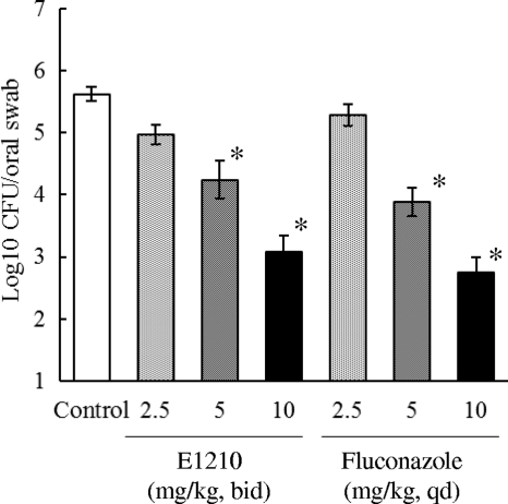
通过 manogepix 治疗(口服给药;每天两次;持续三天;无特定病原体雌性 ICR 小鼠;2.5 mg/kg、5 mg/kg 和 10 mg/kg),活 C 的数量减少。口腔白色念珠菌细胞呈剂量依赖性[2]。
- 小鼠念珠菌病模型疗效:口服E1210(5–20 mg/kg)显著降低播散性念珠菌病小鼠的真菌负荷,肾脏菌落形成单位(CFU)较对照组减少1.5–2.5 log。在口腔念珠菌病中,2.5–10 mg/kg剂量使口腔真菌负荷减少2–3 log CFU [2]。 - 小鼠曲霉病和镰刀菌病模型疗效:E1210(10–20 mg/kg,口服)提高肺曲霉病小鼠的存活率(50–60% vs 对照组20%)并降低肺部真菌负荷;在播散性镰刀菌病中,存活率提高至40–50%(对照组10%),且器官真菌播散减少[2]。 |
| 酶活实验 |
体外药敏试验。[2]
E1210和参比化合物的mic采用临床与实验室标准协会(CLSI) M27-A3和M38-A2文件中详细说明的肉汤微量稀释法测定。使用RPMI 1640培养基,用0.165 m3 -(N-morpholino)-丙磺酸(MOPS)缓冲至pH 7.0。结果表示为三个独立实验得出的每种化合物的中位数MIC。 对通过抑制糖基磷脂酰肌醇(GPI)生物合成而起作用的新型抗真菌药物的持续研究导致了E1210的设计。本研究通过评价E1210对白色念珠菌GWT1 (Orf19.6884)蛋白、烟曲霉GWT1 (AFUA_1G14870)蛋白和人猪- w蛋白抑制活性的选择性,在GPI生物合成途径早期催化GPI肌醇酰化,进而评价E1210对白色念珠菌关键毒力因子的影响。E1210在0.3 ~ 0.6 μM的50%抑制浓度(IC(50)s)范围内对白色念珠菌Gwt1p和烟曲霉Gwt1p肌醇酰化活性有抑制作用,但在100 μM的浓度下对人猪- wp无抑制作用。为了证实真菌GPI生物合成的抑制作用,研究了E1210处理后白色念珠菌细胞表面上GPI锚定蛋白ALS1蛋白的表达,结果表明E1210处理后白色念珠菌细胞表面上GPI锚定蛋白ALS1蛋白的表达明显低于未经处理的细胞。然而,粗提物中的ALS1蛋白水平与细胞表面的RHO1蛋白水平几乎相同。此外,当E1210浓度高于其MIC时,E1210可抑制白色念珠菌芽管的形成、对聚苯乙烯表面的粘附以及生物膜的形成。这些结果表明,E1210选择性地抑制了Gwt1p催化的真菌特异性GPI的肌醇酰化,从而抑制GPI锚定蛋白的成熟,并且E1210通过抑制GPI的生物合成抑制了一些重要毒力因子的表达。[1] |
| 细胞实验 |
同时测定了白念珠菌细胞粗提物中Als1p的含量。细胞在E1210存在下,35℃孵育1 h。孵育后,将培养物在4℃下1000 × g离心10分钟,将微球悬浮在含有真菌蛋白酶抑制剂鸡尾酒的50 mM磷酸钾缓冲液(pH 7.4)中。将细胞悬浮液与等重量的玻璃微珠混合,并用细胞干扰剂均匀化。然后将未破碎的细胞和玻璃珠离心取出。所得上清液为粗提物。用ELISA法测定粗提物中Als1蛋白的含量。捕获抗体为抗als1抗体,捕获抗体为抗c。白念珠菌用酶标兔抗体(ViroStat Inc., Portland, ME)作为二抗。每个浓度的酶联免疫吸附试验一式两份,最终值以三次测定的平均值表示。估计蛋白浓度是为了标准化Als1p的量。[1]
- 菌丝生长抑制实验:白色念珠菌细胞在含E1210(0.125–2 μg/mL)的RPMI 1640培养基中37°C培养3–6小时。显微镜定量菌丝长度和芽管形成,结果显示0.5 μg/mL的E1210可减少>50%的菌丝生长[1]。 - GPI锚定蛋白检测:白色念珠菌细胞经E1210(0.5–1 μg/mL)处理16小时后裂解,通过Western blot分析GPI锚定蛋白(如ALS1)。密度分析显示,与未处理细胞相比,蛋白水平降低40–60% [1]。 |
| 动物实验 |
动物/疾病模型: 感染白色念珠菌的无特定病原体雌性ICR小鼠(5周龄;约25克)[2]
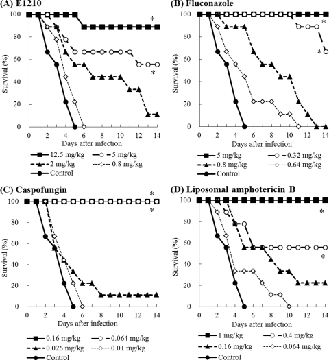
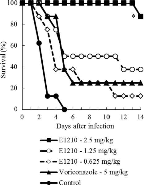
剂量: 2.5 mg/kg、5 mg/kg 和 10 mg/kg 给药途径: 口服;每日两次;持续3天 实验结果: 以剂量依赖的方式降低了口腔内活白色念珠菌的数量。 口咽念珠菌病模型。[2] 用白色念珠菌感染经皮质酮免疫抑制的小鼠,并在药物治疗后测量每只小鼠口腔内白色念珠菌的数量。 ICR小鼠在感染前1天和感染后3天皮下注射4 mg醋酸可的松进行免疫抑制。为预防细菌感染,小鼠饮用水中添加1 mg/ml盐酸四环素,从注射醋酸可的松当天开始,持续整个实验过程。白色念珠菌IFM49971在35℃的沙氏葡萄糖琼脂(SDA)培养基上培养2天。将菌体悬浮于无菌生理盐水中。使用血细胞计数板计数菌体,并用无菌生理盐水调整至所需浓度。随后,小鼠皮下注射0.5 mg盐酸氯丙嗪进行麻醉。使用微量移液器将10 μl白色念珠菌IFM49971菌悬液接种到麻醉小鼠的口腔内。随后,每只小鼠接种4 × 10⁵ CFU的白色念珠菌(CFU)。感染后3天开始,连续3天,分别给予E1210(每日两次,BID)或氟康唑(每日一次,QD)口服。对照组给予等体积的5%葡萄糖溶液(每日两次,BID)。在最后一次给药后的第二天,用盐酸氯丙嗪(0.5 mg/只,皮下注射)麻醉小鼠。通过测定每只小鼠口腔内白色念珠菌的数量来评估药物疗效。使用细尖棉签彻底擦拭小鼠口腔(即颊部、舌部和软腭)。擦拭后,将棉签末端放入装有1 ml无菌生理盐水的试管中。回收的细胞用涡旋混合器混匀后悬浮于无菌生理盐水中,然后进行10倍系列稀释,接种于添加了氨苄青霉素(0.1 mg/ml)的SDA培养基平板上进行培养。SDA平板于35℃培养过夜,然后计数活细胞,以菌落形成单位(CFU)表示。细胞数量以log10 CFU/拭子表示。口腔中可检测到的最低细胞数为10 CFU(1 log10 CFU)。活细胞计数重复两次。[2] 播散性念珠菌病模型。[2] ICR小鼠在感染前6天皮下注射5-氟尿嘧啶(5-FU),剂量为200 mg/kg体重,进行免疫抑制。为了预防内源性细菌感染,这些小鼠在感染前2~3天至感染后5~7天,通过饮用水口服给予0.1 mg/ml的环丙沙星。白色念珠菌IFM49971、白色念珠菌IFM49738和热带念珠菌E83037分别在35℃的SDA平板上培养2天。将琼脂平板表面的菌体悬浮于无菌生理盐水中,并用血细胞计数器进行计数。最后用无菌生理盐水将菌液浓度调整至所需浓度。通过尾静脉注射0.2 ml白色念珠菌细胞悬液(0.8至1.4 × 10⁴ CFU/只小鼠,或IFM49971菌株为5.3 × 10⁴ CFU/只小鼠)或热带念珠菌细胞悬液(3.0 × 10⁵ CFU/只小鼠)诱导中性粒细胞减少小鼠发生感染。感染后1小时或24小时开始抗真菌治疗,并持续3天(第0至2天或第1至3天)。E1210或伏立康唑每日口服2至3次,氟康唑每日口服1次,卡泊芬净或脂质体两性霉素B每日静脉注射1次。对照组每日口服等体积的赋形剂(5%葡萄糖溶液,10 ml/kg)2至3次。在我们的初步研究中,口服载体对照组小鼠的生存曲线与静脉注射载体对照组小鼠的生存曲线相似。因此,我们没有设置静脉注射载体的对照组。生存率和生存期在14天内测定。[2] - 播散性念珠菌病模型:小鼠经静脉注射感染白色念珠菌(1×10⁵ CFU)。E1210溶于0.5%甲基纤维素溶液中,于感染后2小时开始,每日一次口服5、10或20 mg/kg,连续5天。通过在琼脂平板上接种匀浆来评估肾脏真菌负荷,并监测7天的生存情况[2]。 - 肺曲霉病模型:免疫缺陷小鼠经鼻内感染烟曲霉(1×10⁶ 分生孢子)。 E1210(10 或 20 mg/kg)每日口服一次,连续 7 天,从感染后 2 小时开始。评估肺部菌落形成单位 (CFU) 计数和存活率 [2]。 - 口咽念珠菌病模型:小鼠经免疫抑制后,经口感染白色念珠菌。E1210(2.5、5 或 10 mg/kg)每日口服两次,连续 3 天。采集口腔拭子,通过菌落形成单位 (CFU) 计数确定真菌载量 [2]。 查看更多肺曲霉病模型。[2] 播散性镰刀菌病模型。[2] DBA/2N小鼠在感染前6天皮下注射200 mg/kg的5-氟尿嘧啶(5-FU)进行免疫抑制。为预防细菌感染,小鼠在感染前3天至感染后7天期间,饮用水中添加0.1 mg/ml的环丙沙星进行口服。将茄病镰刀菌IFM50956菌株接种于PDA琼脂平板上,30℃培养7天。将琼脂平板表面的菌体悬浮于含0.05% Tween 80的无菌生理盐水中,并使用血细胞计数板进行计数。使用含0.05% Tween 80的无菌生理盐水将最终接种物调整至所需密度。通过尾静脉注射0.2 ml F. solani细胞悬液(5.0 × 10³ 个细胞/只小鼠)诱导中性粒细胞减少小鼠发生感染。感染后1小时开始抗真菌治疗,并持续5天(第0至4天)。E1210每日三次口服给药。对照组每日三次口服等体积的5%葡萄糖溶液。在14天内测定存活率和存活期。[2] 药代动力学研究。[2] E1210静脉注射或口服给药于雄性ICR小鼠。给予E1210后,在指定时间点(0.08、0.25、0.5、1、2、4、6、8 小时)从每只小鼠的下腔静脉抽取血样。通过离心血液获得血浆样本。用甲醇脱蛋白后,提取的样本采用液相色谱-串联质谱法(LC/MS/MS)进行分析。使用 MassLynx 软件,通过内标法测定血浆中E1210的浓度。E1210的药代动力学参数采用模型非依赖性分析法计算。[2] 毒理学研究。 [2] E1210以灌胃法连续7天,每日一次,分别以100、300或1000 mg/kg的剂量灌胃给予雄性和雌性Sprague-Dawley大鼠(每组每性别3只)。对照组给予等体积(10 ml/kg)的溶剂(0.4 mol/L盐酸)。所有死亡或濒死的大鼠均立即进行尸检,所有存活的大鼠在给药7天后进行尸检。评估指标包括:死亡率、临床症状、体重、食物消耗量、血液学、血液化学、毒代动力学、肝脏药物代谢酶以及宏观和微观病理学。[2] |
| 参考文献 |
|
| 其他信息 |
E1210 是一种首创的广谱抗真菌药物,其作用机制新颖——抑制真菌糖基磷脂酰肌醇的生物合成。本研究在小鼠口咽念珠菌病、播散性念珠菌病、肺曲霉病和播散性镰刀菌病模型中评估了 E1210 和参考抗真菌药物的疗效。口服 E1210 对念珠菌属、曲霉属和茄病镰刀菌引起的感染表现出剂量依赖性疗效。在口咽念珠菌病的治疗中,E1210 和氟康唑均能显著降低口腔菌落形成单位(CFU)的数量,优于对照组(P < 0.05)。在播散性念珠菌病模型中,接受 E1210、氟康唑、卡泊芬净或脂质体两性霉素 B 治疗的小鼠的存活率显著高于对照组小鼠(P < 0.05)。E1210 对由唑类耐药的白色念珠菌或热带念珠菌引起的播散性念珠菌病也具有显著疗效。治疗开始时间延迟 24 小时对 E1210 治疗播散性念珠菌病的疗效影响甚微。在黄曲霉肺曲霉病模型中,接受 E1210、伏立康唑或卡泊芬净治疗的小鼠的存活率显著高于对照组小鼠(P < 0.05)。E1210 对烟曲霉肺曲霉病也具有疗效。与许多抗真菌药物不同,E1210 对由茄病镰刀菌(F. solani)引起的播散性镰刀菌病也有效。总之,E1210 在小鼠口咽念珠菌病、播散性念珠菌病、肺曲霉病和播散性镰刀菌病模型中均表现出稳定的疗效。这些数据表明,有必要开展进一步研究,以确定 E1210 治疗播散性真菌感染的潜力。[2] - 作用机制:E1210 通过抑制 GPI 生物合成途径发挥其抗真菌作用,该途径对于蛋白质正确锚定到真菌细胞壁至关重要。这种破坏会导致细胞壁完整性受损和毒力降低[1]。
- 广谱活性:E1210在体外和体内均显示出对多种真菌的活性,包括念珠菌属、烟曲霉和茄病镰刀菌,在某些模型中其疗效与现有抗真菌药物相当或更优[2]。 |
| 分子式 |
C21H18N4O2
|
|---|---|
| 分子量 |
358.393224239349
|
| 精确质量 |
358.142
|
| 元素分析 |
C, 70.38; H, 5.06; N, 15.63; O, 8.93
|
| CAS号 |
936339-60-5
|
| 相关CAS号 |
Fosmanogepix;2091769-17-2
|
| PubChem CID |
16719049
|
| 外观&性状 |
White to light yellow solid powder
|
| 密度 |
1.3±0.1 g/cm3
|
| 沸点 |
569.4±45.0 °C at 760 mmHg
|
| 闪点 |
298.1±28.7 °C
|
| 蒸汽压 |
0.0±1.6 mmHg at 25°C
|
| 折射率 |
1.640
|
| LogP |
3.75
|
| tPSA |
87.1
|
| 氢键供体(HBD)数目 |
1
|
| 氢键受体(HBA)数目 |
6
|
| 可旋转键数目(RBC) |
6
|
| 重原子数目 |
27
|
| 分子复杂度/Complexity |
444
|
| 定义原子立体中心数目 |
0
|
| SMILES |
N1=C(CC2C=CC(COC3C=CC=CN=3)=CC=2)C=C(C2C(N)=NC=CC=2)O1
|
| InChi Key |
WSEKTEUGRLFBSE-UHFFFAOYSA-N
|
| InChi Code |
InChI=1S/C21H18N4O2/c22-21-18(4-3-11-24-21)19-13-17(25-27-19)12-15-6-8-16(9-7-15)14-26-20-5-1-2-10-23-20/h1-11,13H,12,14H2,(H2,22,24)
|
| 化学名 |
(3-(3-(4-(((Pyridin-2-yl)oxy)methyl)benzyl)isoxazol-5-yl)pyridin-2-)amine
|
| 别名 |
E1210; APX001A; E-1210; APX 001 A; 936339-60-5; E1210; Manogepix; 7B1P18ID9L; APX-001A; (pharmaceutical); Manogepix; E-1210; 3-(3-(4-((Pyridin-2-yloxy)methyl)benzyl)isoxazol-5-yl)pyridin-2-amine; (3-(3-(4-(((Pyridin-2-yl)oxy)methyl)benzyl)isoxazol-5-yl)pyridin-2-)amine; 7B1P18ID9L; E 1210; APX-001-A; APX001-A; Manogepix
|
| HS Tariff Code |
2934.99.9001
|
| 存储方式 |
Powder -20°C 3 years 4°C 2 years In solvent -80°C 6 months -20°C 1 month |
| 运输条件 |
Room temperature (This product is stable at ambient temperature for a few days during ordinary shipping and time spent in Customs)
|
| 溶解度 (体外实验) |
DMSO : ~100 mg/mL (~279.03 mM)
|
|---|---|
| 溶解度 (体内实验) |
配方 1 中的溶解度: ≥ 2.08 mg/mL (5.80 mM) (饱和度未知) in 10% DMSO + 40% PEG300 + 5% Tween80 + 45% Saline (这些助溶剂从左到右依次添加,逐一添加), 澄清溶液。
例如,若需制备1 mL的工作液,可将100 μL 20.8 mg/mL澄清DMSO储备液加入400 μL PEG300中,混匀;然后向上述溶液中加入50 μL Tween-80,混匀;加入450 μL生理盐水定容至1 mL。 *生理盐水的制备:将 0.9 g 氯化钠溶解在 100 mL ddH₂O中,得到澄清溶液。 配方 2 中的溶解度: ≥ 2.08 mg/mL (5.80 mM) (饱和度未知) in 10% DMSO + 90% (20% SBE-β-CD in Saline) (这些助溶剂从左到右依次添加,逐一添加), 澄清溶液。 例如,若需制备1 mL的工作液,可将 100 μL 20.8 mg/mL澄清DMSO储备液加入900 μL 20% SBE-β-CD生理盐水溶液中,混匀。 *20% SBE-β-CD 生理盐水溶液的制备(4°C,1 周):将 2 g SBE-β-CD 溶解于 10 mL 生理盐水中,得到澄清溶液。 View More
配方 3 中的溶解度: ≥ 2.08 mg/mL (5.80 mM) (饱和度未知) in 10% DMSO + 90% Corn Oil (这些助溶剂从左到右依次添加,逐一添加), 澄清溶液。 1、请先配制澄清的储备液(如:用DMSO配置50 或 100 mg/mL母液(储备液)); 2、取适量母液,按从左到右的顺序依次添加助溶剂,澄清后再加入下一助溶剂。以 下列配方为例说明 (注意此配方只用于说明,并不一定代表此产品 的实际溶解配方): 10% DMSO → 40% PEG300 → 5% Tween-80 → 45% ddH2O (或 saline); 假设最终工作液的体积为 1 mL, 浓度为5 mg/mL: 取 100 μL 50 mg/mL 的澄清 DMSO 储备液加到 400 μL PEG300 中,混合均匀/澄清;向上述体系中加入50 μL Tween-80,混合均匀/澄清;然后继续加入450 μL ddH2O (或 saline)定容至 1 mL; 3、溶剂前显示的百分比是指该溶剂在最终溶液/工作液中的体积所占比例; 4、 如产品在配制过程中出现沉淀/析出,可通过加热(≤50℃)或超声的方式助溶; 5、为保证最佳实验结果,工作液请现配现用! 6、如不确定怎么将母液配置成体内动物实验的工作液,请查看说明书或联系我们; 7、 以上所有助溶剂都可在 Invivochem.cn网站购买。 |
| 制备储备液 | 1 mg | 5 mg | 10 mg | |
| 1 mM | 2.7903 mL | 13.9513 mL | 27.9026 mL | |
| 5 mM | 0.5581 mL | 2.7903 mL | 5.5805 mL | |
| 10 mM | 0.2790 mL | 1.3951 mL | 2.7903 mL |
1、根据实验需要选择合适的溶剂配制储备液 (母液):对于大多数产品,InvivoChem推荐用DMSO配置母液 (比如:5、10、20mM或者10、20、50 mg/mL浓度),个别水溶性高的产品可直接溶于水。产品在DMSO 、水或其他溶剂中的具体溶解度详见上”溶解度 (体外)”部分;
2、如果您找不到您想要的溶解度信息,或者很难将产品溶解在溶液中,请联系我们;
3、建议使用下列计算器进行相关计算(摩尔浓度计算器、稀释计算器、分子量计算器、重组计算器等);
4、母液配好之后,将其分装到常规用量,并储存在-20°C或-80°C,尽量减少反复冻融循环。
计算结果:
工作液浓度: mg/mL;
DMSO母液配制方法: mg 药物溶于 μL DMSO溶液(母液浓度 mg/mL)。如该浓度超过该批次药物DMSO溶解度,请首先与我们联系。
体内配方配制方法:取 μL DMSO母液,加入 μL PEG300,混匀澄清后加入μL Tween 80,混匀澄清后加入 μL ddH2O,混匀澄清。
(1) 请确保溶液澄清之后,再加入下一种溶剂 (助溶剂) 。可利用涡旋、超声或水浴加热等方法助溶;
(2) 一定要按顺序加入溶剂 (助溶剂) 。
| NCT Number | Recruitment | interventions | Conditions | Sponsor/Collaborators | Start Date | Phases |
| NCT05491733 | Completed | Drug: APX001 Drug: APX001A |
Invasive Fungal Infections | Basilea Pharmaceutica | 2021-03-02 | Phase 1 |
| NCT04166669 | Completed | Drug: APX001 Drug: Itraconazole Drug: Rifampin |
Fungal Infection | Basilea Pharmaceutica | 2019-11-12 | Phase 1 |
| NCT04240886 | Terminated | Drug: fosmanogepix | Invasive Fungal Infections | Basilea Pharmaceutica | 2020-01-04 | Phase 2 |
| NCT05582187 | Recruiting | Drug: Fosmanogepix | Hepatic Impairment | Basilea Pharmaceutica | 2022-10-31 | Phase 1 |
|
|
|
 Geranic acid
 Bromomonilicin
 Econazole-d6
 Succinate dehydrogenase-IN-11
InvivoChem的所有产品仅用于作科学研究,不面向患者销售
Copyright 2020 InvivoChem LLC | All Rights Reserved 粤ICP备20063088号-1


 COA
COA
 463611831
