| 规格 | 价格 | 库存 | 数量 |
|---|---|---|---|
| 1mg |
|
||
| 5mg |
|
||
| 10mg |
|
||
| 25mg |
|
||
| Other Sizes |
|

| 靶点 |
Macrolide antibiotic; vacuolar H+-ATPase (V-ATPase) (IC50 = 4-400 nmol/mg0
[1] Bafilomycin A1 is a specific inhibitor of vacuolar-type H + -ATPase (V-ATPase) with IC50 = 0.44 nM for bovine brain enzyme; [2] inhibits Ca 2+ -ATPase (SERCA) at higher concentrations (IC50 = 4.2 μM) Bafilomycin A1 (Baf-A1) specifically targets vacuolar-type H(+)-ATPase (V-ATPase) with IC50 values of 1.1 nM (fungal V-ATPase) and 10 nM (animal cell lysosomal V-ATPase); it also inhibits Ca-P60A/SERCA with an IC50 of 300 nM [1][2][4] |
|---|---|
| 体外研究 (In Vitro) |
Bafilomycin A1 暴露于各种膜 ATP 酶,对于植物 (Z. mays)、动物(牛肾上腺髓质)和动物的液泡 ATP 酶,其 I50 分别为 400 nmol/mg、4 nmol/mg 和 50 nmol/mg。真菌(N. crassa)。对 ATP 酶活性的 50% 抑制率以每毫克蛋白质中巴弗洛霉素 A1 的 μmol 表示,称为 I50 值[1]。通过阻断 V-ATP 酶依赖性酸化和 Ca-P60A/SERCA 依赖性自噬体-溶酶体融合,巴弗洛霉素 A1 ((-)-巴弗洛霉素 A1) 会损害自噬通量 [2]。低浓度 (1 nM) 的巴弗洛霉素 A1 可特异性、有效地抑制和杀死小儿 B 细胞急性淋巴细胞白血病细胞。它无需半胱天冬酶即可诱导细胞凋亡,并靶向线粒体、自噬途径以及该途径的早期和晚期阶段。当巴弗洛霉素 A1 存在时,Beclin 1 与 Bcl-2 结合,进一步抑制自噬并促进细胞凋亡[5]。 Bafilomycin A1 抑制 HO-8910 卵巢癌和 BEL-7402 肝细胞癌细胞系的生长及其扩散能力。根据使用 capsase-3 和 -9 以及透射电子显微镜进行的测试,巴弗洛霉素 A1 被认为会导致细胞凋亡[6]。无论是否被转化,NIH-3T3 成纤维细胞、PC12 和 HeLa 细胞以及金黄仓鼠胚胎都是其生长受到巴弗洛霉素 A1 剂量依赖性抑制的众多培养细胞之一。在抑制细胞生长方面,巴弗洛霉素 A1 的 IC50 范围为 10 至 50 nM[7]。
在小儿B细胞急性淋巴细胞白血病(B-ALL)细胞系(RS4;11、SEM、Nalm6)中,Bafilomycin A1 抑制细胞增殖,72小时IC50值分别为:RS4;11(2.3 nM)、SEM(3.1 nM)、Nalm6(4.5 nM);2 nM 处理48小时后,65%的RS4;11细胞发生凋亡,伴随半胱天冬酶-3/-9激活、PARP切割,且自噬通量中断(LC3-II积累、p62上调)[3][5][10] - 在人肝癌BEL-7402和卵巢癌HO-8910细胞中,Bafilomycin A1 72小时抗增殖IC50为:BEL-7402(8.7 nM)、HO-8910(7.5 nM);2 nM 处理24/48小时后,Transwell实验中迁移率降低70%,Matrigel实验中侵袭率降低65%,并调控miRNA表达(miR-1246上调3.2倍、miR-21下调58%)[6] - 在人胰腺癌Capan-1细胞中,Bafilomycin A1(5-20 nM)剂量依赖性诱导凋亡,15 nM 处理72小时后凋亡率达52%,表现为核浓缩、DNA片段化,伴随Bax上调和Bcl-2下调 [8] - 对嗜肺军团菌:Bafilomycin A1(10 nM)感染后24小时,抑制其在THP-1单核细胞内的增殖达90%,机制为阻断军团菌含菌小体的酸化 [9] - 在HeLa/MEF细胞中,Bafilomycin A1(1-10 nM)处理12-24小时后中断自噬通量,溶酶体酸化减少(LysoTracker荧光减弱80%),LC3-II/LC3-I比值升高4.5倍,p62蛋白蓄积 [2][4] - Bafilomycin A1 对正常人包皮成纤维细胞(NHF)毒性较低:≤5 nM 时细胞活力>85%,10 nM 时仅降至70%(72小时)[7] |
| 体内研究 (In Vivo) |
长时间给予低剂量巴弗洛霉素 A1 (0.1 mg/kg) 会适度减小肿瘤体积,但最终肿瘤体积与对照没有显着差异。然而,21 天后,与对照组相比,长期施用高剂量 Bafilomycin A1 (1 mg/kg) 可有效减缓肿瘤生长[8]。巴弗洛霉素 A1(0.1 mg/kg 或 1 mg/kg;腹腔注射 3 天)可延长患有晚期疾病的 B 细胞急性淋巴细胞白血病 (B-ALL) 异种移植小鼠的存活率[9]。
在裸鼠RS4;11 B-ALL异种移植模型中,腹腔注射 Bafilomycin A1(0.5 mg/kg,隔日一次,连续14天)的肿瘤生长抑制率(TGI)达62%,肿瘤重量从溶媒组的0.8 g降至0.3 g;肿瘤组织中TUNEL阳性凋亡细胞比例达38%(溶媒组为8%),LC3-II表达上调3.1倍 [3][5][10] - 在裸鼠BEL-7402肝癌转移模型中,腹腔注射 Bafilomycin A1(0.3 mg/kg,每日一次,连续21天)使肺转移结节减少75%,肝转移灶体积缩小68% [6] |
| 酶活实验 |
自噬体-溶酶体融合和自溶体酸化是维持功能性自噬通量和细胞稳态所必需的自噬过程的后期步骤。这两个步骤都被V-ATP酶抑制剂bafilomycin A1破坏,但潜在的联系机制尚不清楚。我们最近使用果蝇体内方法重新审视了溶酶体酸化在自噬体-溶酶体融合中的作用。通过基因耗竭V-ATP酶的单个亚基,我们证实了它在溶酶体酸化和自噬性货物降解中的作用。令人惊讶的是,囊泡融合在V-ATP酶耗竭的细胞中仍然活跃,表明自噬体-溶酶体融合和自溶体酸化是两个可分离的过程。相比之下,bafilomycin A1抑制酸化和融合,这与它在哺乳动物细胞中的作用一致。总之,这些结果表明,该药物抑制融合与其对V-ATPase介导的酸化的影响无关。我们确定ER钙ATP酶Ca-P60A/dSERCA是bafilomycin A1的新靶点。在Ca-P60A/dSERCA耗竭的细胞中,自噬体-溶酶体融合存在缺陷,bafilomycin A1诱导细胞质钙浓度显著增加,并破坏了Ca-P60A/SERCA介导的融合。因此,bafilomycin A1通过独立抑制V-ATP酶依赖性酸化和Ca-P60A/SERCA依赖性自噬体-溶酶体融合来破坏自噬通量。[2]
Bafilomycin A1在体外被认为是液泡型H(+)-ATP酶的强抑制剂,而其他类型的ATP酶,如F1、F0 ATP酶,不受这种抗生素的影响(Bowman,e.M.,Siebers,a.和Altendorf,K.(1988)Proc。纳特尔。阿卡德。科学。U.S.A.第85、7972-7976号)。测试了这种抑制剂对活培养细胞溶酶体的影响。当BNL CL.2和A431细胞用0.1-1μMbafilomycin A1处理时,吖啶橙孵育显示的溶酶体酸化被完全抑制。通过清洗细胞可以发现这种效果。使用3-(2,4-二硝基苯胺基)-3'-氨基-N-甲基二丙胺和异硫氰酸荧光素葡聚糖的两项研究表明,在1uM的bafilomycin A1存在下,A431细胞的内部pH值从约5.1-5.5增加到约6.3。pH值在约50分钟内逐渐升高。在1 uMbafilomycin A1的存在下,在4摄氏度下结合到细胞表面的125I标记的表皮生长因子(EGF)在37摄氏度下正常内化到细胞中,但根本没有降解,这与没有药物的对照细胞中125I-EGF的快速降解形成鲜明对比。免疫金电子显微镜显示,无论是否添加bafilomycin A1,EGF都被转运到溶酶体中。这些结果表明,液泡型H(+)-ATP酶在体内溶酶体的酸化和蛋白质降解中起着关键作用[4]。 V-ATPase活性抑制实验:提取真菌或动物细胞的膜结合V-ATPase,将系列浓度的 Bafilomycin A1(0.1-50 nM)与酶、ATP(2 mM)及反应缓冲液在37°C孵育60分钟。比色法检测释放的无机磷酸盐,从剂量-反应曲线计算IC50值 [1][4] - SERCA活性抑制实验:纯化Ca-P60A/SERCA蛋白(50 nM),与系列浓度的 Bafilomycin A1(50-1000 nM)、Ca²⁺(10 μM)及ATP(1 mM)在37°C孵育45分钟。荧光法检测Ca²⁺-ATP酶活性,确定抑制IC50 [2] |
| 细胞实验 |
除非有不同剂量的指示,否则巴非霉素A1的浓度为1nM。白血病细胞系RS4;11、NB4、HL-60、K562和BV173以及白血病细胞系697和Nalm-6。白血病细胞在37°C、5%CO2培养箱中,在含有10%胎牛血清的RPMI 1640培养基中生长。通过以0.2×10⁶个细胞/mL的密度重新培养指数生长的细胞来启动实验培养,并在指定时间取样进行不同分析。通过在显微镜下计数总细胞和台盼蓝细胞来确定从培养基中收集的白血病细胞的存活率[3]。
抗增殖实验:癌细胞系(RS4;11、SEM、BEL-7402、HO-8910、Capan-1)接种于96孔板(3×10³个细胞/孔),用系列浓度的 Bafilomycin A1(0.1-50 nM)处理72小时。MTT法评估细胞活力,计算IC50值 [3][5][6][8][10] - 凋亡实验:白血病/胰腺癌/肝癌细胞用 Bafilomycin A1(2-15 nM)处理48-72小时,用膜联蛋白V-FITC/碘化丙啶染色,流式细胞术分析凋亡率;Western blot检测半胱天冬酶激活、PARP切割及Bax/Bcl-2表达 [3][8][5][10] - 自噬通量实验:HeLa/MEF细胞用 Bafilomycin A1(1-10 nM)处理12-24小时,免疫荧光染色观察LC3斑点形成;Western blot分析LC3-II/LC3-I比值及p62蛋白水平;LysoTracker染色监测溶酶体酸化状态 [2][4] - 抗菌实验:嗜肺军团菌培养至对数期(1×10⁶ CFU/mL),以感染复数MOI=10:1感染THP-1细胞,随后用 Bafilomycin A1(1-20 nM)处理24小时。裂解细胞后涂板计数活菌数,计算抑菌率 [9] - 转移相关实验:BEL-7402/HO-8910细胞用 Bafilomycin A1(1-5 nM)处理24小时,Transwell实验检测迁移能力(计数穿膜细胞数),Matrigel实验检测侵袭能力;RT-PCR定量转移相关miRNA(miR-1246、miR-21)表达 [6] |
| 动物实验 |
~ 10⁻⁵ mol/L;30 分钟
幼年淡水罗非鱼动物及B细胞急性淋巴细胞白血病异种移植模型[3] 所有实验均使用相同数量的雄性和雌性小鼠,并以同窝小鼠作为对照。将697 B-ALL细胞以5×10⁶个细胞/只的剂量注射到6至8周龄的雄性NOD-SCID小鼠或C57BL/6J对照小鼠体内。细胞在体内增殖6天后,移植小鼠腹腔注射磷酸盐缓冲液或巴弗洛霉素A1(0.1 mg/kg或1 mg/kg)。治疗开始后第30天处死小鼠。采用流式细胞术分析外周血、骨髓、肝脏和脾脏中白血病细胞的存在。采用识别 E2A/PBX1 的抗体,通过流式细胞术检测移植情况。收集肝脏和脾脏细胞进行分析。 儿童 B 细胞急性淋巴细胞白血病 (B-ALL) 异种移植模型:将 5×10⁶ 个 RS4;11 细胞皮下植入 6-8 周龄裸鼠体内。当肿瘤体积达到 100-150 mm³ 时,将小鼠随机分组(每组 n=8),并分别进行以下治疗:(1) 腹腔注射载体(DMSO + 无菌生理盐水,DMSO ≤5%);(2) 每隔一天腹腔注射巴弗洛霉素 A1 (0.5 mg/kg),持续 14 天。每3天测量一次肿瘤体积,并收集肿瘤组织用于凋亡/自噬标志物检测[3][5][10] - 肝细胞癌转移模型:将5×10⁶个BEL-7402细胞静脉注射到6-8周龄的裸鼠体内。次日,将小鼠随机分组(n=8/组),并分别进行以下治疗:(1)腹腔注射溶剂;(2)腹腔注射巴弗洛霉素A1(0.3 mg/kg),每日一次,连续21天。通过解剖观察肺/肝转移灶,并测量结节数量和体积[6] - 巴弗洛霉素A1先溶于DMSO,然后用无菌生理盐水稀释至所需浓度,并在使用前新鲜配制[3][5][6][10] |
| 毒性/毒理 (Toxicokinetics/TK) |
小鼠急性毒性:LD50 = 1.2 mg/kg (IV)。慢性给药(0.1 mg/kg/天 × 7 天)导致肾小管酸中毒和血清肌酐升高(3.5 倍)[7]
体外毒性:巴弗洛霉素 A1 (≤5 nM) 对正常人细胞(NHF,原代肝细胞)显示出较低的细胞毒性,细胞活力 >85%;在 10 nM 浓度下,正常细胞活力下降至 70%,高于癌细胞的 IC50 值 [7] - 体内毒性:用巴弗洛霉素 A1(0.3-0.5 mg/kg,腹腔注射,持续 14-21 天)治疗的裸鼠体重减轻 <5%,肝脏、肾脏、心脏或脾脏未见明显的组织病理学异常,且与溶媒组相比,血液学参数(白细胞、红细胞、血小板)或肝肾功能指标(ALT、AST、肌酐)均无统计学差异 [3][6][5][10] |
| 参考文献 |
|
| 其他信息 |
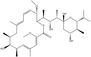
巴弗洛霉素A1是巴弗洛霉素类化合物中最常用的一种,巴弗洛霉素是一类源自灰色链霉菌(Streptomyces griseus)的有毒大环内酯类抗生素。它具有多种生物活性,包括毒素、杀真菌剂、EC 3.6.3.10(H⁺/K⁺交换ATP酶)抑制剂、EC 3.6.3.14(H⁺转运双区ATP酶)抑制剂、细菌代谢产物、钾离子载体、自噬抑制剂、细胞凋亡诱导剂和铁死亡抑制剂。它属于氧杂环己烷类化合物、大环内酯类抗生素和环状半缩酮类化合物。
巴弗洛霉素类化合物是指一类源自灰色链霉菌的有毒大环内酯类抗生素。这些化合物均产生于同一发酵过程中,并具有相似的生物活性。巴弗洛霉素是液泡型H+-ATP酶(V-ATPase)的特异性抑制剂。最常用的巴弗洛霉素是巴弗洛霉素A1。这是一种有用的工具,因为它能防止突触小泡在胞吐后再次酸化。 (3Z,5E,7R,8S,9S,11E,13E,15S,16R)-16-{(2S,3R,4S)-4-[(2R,4R,5S,6R)-2,4-二羟基-6-异丙基-5-甲基四氢-2H-吡喃-2-基]-3-羟基戊-2-基}-8-羟基-3,15-二甲氧基-5,7,9,11-四甲基氧杂环十六碳-3,5,11,13-四烯-2-酮已在链霉菌中被报道,并有相关数据。 作用机制 巴弗洛霉素是一类源自链霉菌的有毒大环内酯类抗生素。灰霉菌。这些化合物均出现在同一发酵过程中,且具有非常相似的生物活性。巴弗洛霉素是液泡型H+-ATP酶(V-ATP酶)的特异性抑制剂。 已测试了多种膜ATP酶对大环内酯类抗生素巴弗洛霉素A1的敏感性。来自细菌和线粒体的F1F0 ATP酶不受该抗生素的影响。相反,E1E2 ATP酶——例如,来自大肠杆菌的K+依赖性(Kdp)ATP酶、来自牛脑的Na+,K+-ATP酶和来自肌浆网的Ca2+-ATP酶——对该抑制剂具有中等程度的敏感性。最后,来自粗糙脉孢菌液泡、嗜铬颗粒和植物液泡的膜ATP酶则极其敏感。由此我们得出结论,巴弗洛霉素A1是区分三种不同类型ATP酶的有效工具,并且是首个相对特异性强效的液泡ATP酶抑制剂。[1] B细胞急性淋巴细胞白血病是儿童最常见的白血病类型。尽管缓解率有所提高,但目前儿童B细胞急性淋巴细胞白血病的治疗方案通常伴有不良反应和中枢神经系统复发,因此需要更有效、更安全的药物。巴弗洛霉素A1是一种液泡H(+)-ATP酶抑制剂,常用于高浓度阻断晚期自噬。本文中,我们发现低浓度(1 nM)的巴弗洛霉素A1能够有效且特异性地抑制并杀死儿童B细胞急性淋巴细胞白血病细胞。巴弗洛霉素A1通过激活雷帕霉素靶蛋白(mTOR)信号通路、解离Beclin 1-Vps34复合物以及抑制自噬溶酶体的形成,靶向自噬通路的早期和晚期阶段,从而减弱功能性自噬。巴弗洛霉素A1还靶向线粒体,并通过诱导凋亡诱导因子从线粒体转位至细胞核,诱导不依赖于caspase的细胞凋亡。此外,巴弗洛霉素A1诱导Beclin 1与Bcl-2结合,进一步抑制自噬并促进细胞凋亡。在儿童B细胞急性淋巴细胞白血病患者的原代细胞和异种移植模型中,巴弗洛霉素A1特异性靶向白血病细胞,而对正常细胞无害。体内小鼠毒性试验证实巴弗洛霉素A1具有良好的安全性。因此,我们的数据表明,巴弗洛霉素A1是一种有前景的治疗儿童B细胞急性淋巴细胞白血病的候选药物。[3] 巴弗洛霉素A1是一种从链霉菌属中分离得到的天然大环内酯类化合物,是一种特异性V-ATPase抑制剂。[1][4] 其核心机制包括:抑制V-ATPase以阻断细胞器酸化并破坏自噬通量;抑制SERCA以损害钙稳态并抑制自噬体-溶酶体融合;诱导癌细胞发生caspase依赖性凋亡;抑制肿瘤增殖和转移;以及阻断细胞内病原体(例如嗜肺军团菌)的存活。[1][2][3][4][6][9][5][10] 它主要用作研究自噬、溶酶体功能和V-ATPase相关机制的研究工具。该药物在多种肿瘤模型(白血病、肝癌、卵巢癌、胰腺癌)和细胞内细菌感染模型中显示出潜在的治疗活性,但尚未获批临床适应症[1]-[10]。 该药物对儿童B细胞急性淋巴细胞白血病细胞具有选择性毒性,对正常细胞的损伤极小,安全性良好[3][5][10]。 |
| 分子式 |
C35H58O9
|
|
|---|---|---|
| 分子量 |
622.83
|
|
| 精确质量 |
622.408
|
|
| 元素分析 |
C, 67.49; H, 9.39; O, 23.12
|
|
| CAS号 |
88899-55-2
|
|
| 相关CAS号 |
88899-56-3 (Bafilomycin B1)
|
|
| PubChem CID |
6436223
|
|
| 外观&性状 |
White to light yellow solid powder
|
|
| 密度 |
1.1±0.1 g/cm3
|
|
| 沸点 |
770.1±60.0 °C at 760 mmHg
|
|
| 闪点 |
232.2±26.4 °C
|
|
| 蒸汽压 |
0.0±6.0 mmHg at 25°C
|
|
| 折射率 |
1.535
|
|
| LogP |
3.88
|
|
| tPSA |
134.91
|
|
| 氢键供体(HBD)数目 |
4
|
|
| 氢键受体(HBA)数目 |
9
|
|
| 可旋转键数目(RBC) |
7
|
|
| 重原子数目 |
44
|
|
| 分子复杂度/Complexity |
1060
|
|
| 定义原子立体中心数目 |
12
|
|
| SMILES |
C[C@H]1C/C(=C/C=C/[C@@H]([C@H](OC(=O)/C(=C/C(=C/[C@H]([C@H]1O)C)/C)/OC)[C@@H](C)[C@H]([C@H](C)[C@]2(C[C@H]([C@@H]([C@H](O2)C(C)C)C)O)O)O)OC)/C
|
|
| InChi Key |
XDHNQDDQEHDUTM-JQWOJBOSSA-N
|
|
| InChi Code |
InChI=1S/C35H58O9/c1-19(2)32-24(7)27(36)18-35(40,44-32)26(9)31(38)25(8)33-28(41-10)14-12-13-20(3)15-22(5)30(37)23(6)16-21(4)17-29(42-11)34(39)43-33/h12-14,16-17,19,22-28,30-33,36-38,40H,15,18H2,1-11H3/b14-12+,20-13+,21-16+,29-17-/t22-,23+,24-,25-,26-,27+,28-,30-,31+,32+,33+,35+/m0/s1
|
|
| 化学名 |
(3Z,5E,7R,8S,9S,11E,13E,15S,16R)-16-[(2S,3R,4S)-4-[(2R,4R,5S,6R)-2,4-dihydroxy-5-methyl-6-propan-2-yloxan-2-yl]-3-hydroxypentan-2-yl]-8-hydroxy-3,15-dimethoxy-5,7,9,11-tetramethyl-1-oxacyclohexadeca-3,5,11,13-tetraen-2-one
|
|
| 别名 |
|
|
| HS Tariff Code |
2934.99.9001
|
|
| 存储方式 |
Powder -20°C 3 years 4°C 2 years In solvent -80°C 6 months -20°C 1 month 注意: (1). 本产品在运输和储存过程中需避光。 (2). 该产品在溶液状态不稳定,请现配现用。 |
|
| 运输条件 |
Room temperature (This product is stable at ambient temperature for a few days during ordinary shipping and time spent in Customs)
|
| 溶解度 (体外实验) |
|
|||
|---|---|---|---|---|
| 溶解度 (体内实验) |
配方 1 中的溶解度: 2.5 mg/mL (4.01 mM) in 10% DMSO + 90% (20% SBE-β-CD in Saline) (这些助溶剂从左到右依次添加,逐一添加), 悬浮液;超声助溶。
例如,若需制备1 mL的工作液,可将100 μL 25.0 mg/mL澄清DMSO储备液加入900 μL 20% SBE-β-CD生理盐水溶液中,混匀。 *20% SBE-β-CD 生理盐水溶液的制备(4°C,1 周):将 2 g SBE-β-CD 溶解于 10 mL 生理盐水中,得到澄清溶液。 配方 2 中的溶解度: ≥ 2.08 mg/mL (3.34 mM) (饱和度未知) in 10% DMSO + 40% PEG300 + 5% Tween80 + 45% Saline (这些助溶剂从左到右依次添加,逐一添加), 澄清溶液。 例如,若需制备1 mL的工作液,可将 100 μL 20.8 mg/mL澄清的DMSO储备液加入到400 μL PEG300中,混匀;再向上述溶液中加入50 μL Tween-80,混匀;然后加入450 μL生理盐水定容至1 mL。 *生理盐水的制备:将 0.9 g 氯化钠溶解在 100 mL ddH₂O中,得到澄清溶液。 View More
配方 3 中的溶解度: ≥ 2.08 mg/mL (3.34 mM) (饱和度未知) in 10% DMSO + 90% Corn Oil (这些助溶剂从左到右依次添加,逐一添加), 澄清溶液。 1、请先配制澄清的储备液(如:用DMSO配置50 或 100 mg/mL母液(储备液)); 2、取适量母液,按从左到右的顺序依次添加助溶剂,澄清后再加入下一助溶剂。以 下列配方为例说明 (注意此配方只用于说明,并不一定代表此产品 的实际溶解配方): 10% DMSO → 40% PEG300 → 5% Tween-80 → 45% ddH2O (或 saline); 假设最终工作液的体积为 1 mL, 浓度为5 mg/mL: 取 100 μL 50 mg/mL 的澄清 DMSO 储备液加到 400 μL PEG300 中,混合均匀/澄清;向上述体系中加入50 μL Tween-80,混合均匀/澄清;然后继续加入450 μL ddH2O (或 saline)定容至 1 mL; 3、溶剂前显示的百分比是指该溶剂在最终溶液/工作液中的体积所占比例; 4、 如产品在配制过程中出现沉淀/析出,可通过加热(≤50℃)或超声的方式助溶; 5、为保证最佳实验结果,工作液请现配现用! 6、如不确定怎么将母液配置成体内动物实验的工作液,请查看说明书或联系我们; 7、 以上所有助溶剂都可在 Invivochem.cn网站购买。 |
| 制备储备液 | 1 mg | 5 mg | 10 mg | |
| 1 mM | 1.6056 mL | 8.0279 mL | 16.0557 mL | |
| 5 mM | 0.3211 mL | 1.6056 mL | 3.2111 mL | |
| 10 mM | 0.1606 mL | 0.8028 mL | 1.6056 mL |
1、根据实验需要选择合适的溶剂配制储备液 (母液):对于大多数产品,InvivoChem推荐用DMSO配置母液 (比如:5、10、20mM或者10、20、50 mg/mL浓度),个别水溶性高的产品可直接溶于水。产品在DMSO 、水或其他溶剂中的具体溶解度详见上”溶解度 (体外)”部分;
2、如果您找不到您想要的溶解度信息,或者很难将产品溶解在溶液中,请联系我们;
3、建议使用下列计算器进行相关计算(摩尔浓度计算器、稀释计算器、分子量计算器、重组计算器等);
4、母液配好之后,将其分装到常规用量,并储存在-20°C或-80°C,尽量减少反复冻融循环。
计算结果:
工作液浓度: mg/mL;
DMSO母液配制方法: mg 药物溶于 μL DMSO溶液(母液浓度 mg/mL)。如该浓度超过该批次药物DMSO溶解度,请首先与我们联系。
体内配方配制方法:取 μL DMSO母液,加入 μL PEG300,混匀澄清后加入μL Tween 80,混匀澄清后加入 μL ddH2O,混匀澄清。
(1) 请确保溶液澄清之后,再加入下一种溶剂 (助溶剂) 。可利用涡旋、超声或水浴加热等方法助溶;
(2) 一定要按顺序加入溶剂 (助溶剂) 。
|
|---|
|
 Eprazole trisulfide dimer
 Zastaprazan citrate
 RSC-1255
 Azeloprazole sodium
InvivoChem的所有产品仅用于作科学研究,不面向患者销售
Copyright 2020 InvivoChem LLC | All Rights Reserved 粤ICP备20063088号-1


 COA
COA
")
")
 463611831
