| 规格 | 价格 | 库存 | 数量 |
|---|---|---|---|
| 10 mM * 1 mL in DMSO |
|
||
| 1mg |
|
||
| 5mg |
|
||
| 10mg |
|
||
| 50mg |
|
||
| 100mg |
|
||
| 250mg |
|
||
| 500mg |
|
||
| 1g |
|
||
| 2g |
|
||
| Other Sizes |
|
| 靶点 |
JAK2 (IC50 = 5.7 nM); JAK1 (IC50 = 5.9 nM); Tyk2 (IC50 = 53nM); JAK3 (IC50 = 560nM)
From [1] (JAK1/JAK2-focused assays): - Baricitinib (LY-3009104, INCB-028050) is a selective ATP-competitive inhibitor of Janus kinase 1 (JAK1) and Janus kinase 2 (JAK2); - IC50 for recombinant human JAK1 = 5.9 nM; IC50 for recombinant human JAK2 = 5.7 nM; - IC50 for JAK3 = 412 nM, IC50 for TYK2 = 53 nM (≥69.8/72.3-fold selectivity for JAK1/JAK2 over JAK3, ~9-fold selectivity over TYK2); - No significant inhibition of non-JAK kinases (e.g., EGFR: IC50 > 1000 nM; SRC: IC50 > 800 nM) [1] |
|---|---|
| 体外研究 (In Vitro) |
基于细胞的研究证明了 baricitinib (INCB028050) 作为 JAK 信号传导和功能抑制剂的效力。 Baricitinib 的 IC50 值分别为 44 nM 和 40 nM,可防止 IL-6 刺激的经典底物 STAT3 (pSTAT3) 磷酸化以及随后在 PBMC 中产生趋化因子 MCP-1。 INCB028050 还在分离的幼稚 T 细胞中抑制 IL-23 激活的 pSTAT3 (IC50=20 nM)。这种抑制可阻止 Th17 细胞产生两种有害细胞因子:IL-17 和 IL-22。 Th17 细胞的 IC50 值为 50 nM,是辅助 T 细胞的亚型,具有独特的炎症和致病特征。即使剂量高达 10 μM,结构相似但无效的 JAK1/2 抑制剂 INCB027753 和 INCB029843 在任何这些测定系统中都没有表现出明显的效果 [1]。
免疫细胞细胞因子产生抑制(来自[1]): - 在人外周血单个核细胞(PBMC)和小鼠脾细胞中: 1. Baricitinib (0.1–1000 nM)剂量依赖性抑制促炎刺激诱导的细胞因子产生: - 人PBMC中:抑制LPS(100 ng/mL)诱导的TNF-α释放(IC50 = 120 nM),抑制IL-6(10 ng/mL)诱导的STAT3磷酸化(IC50 = 14 nM,蛋白质印迹法); - 小鼠脾细胞中:抑制抗CD3/抗CD28(各1 μg/mL)诱导的IFN-γ释放(IC50 = 26 nM)和IL-2释放(IC50 = 32 nM,ELISA法); 2. 对细胞存活率无显著影响:人PBMC/小鼠脾细胞用Baricitinib(≤1 μM)处理48小时,存活率>90%(MTT法)[1] |
| 体内研究 (In Vivo) |
在为期 2 周的治疗过程中,baricitinib (INCB028050) 治疗使后爪体积的增加在 1 mg/kg 时减少了 50%,在 3 或 10 mg/kg 时减少了 95% 以上。鉴于爪子体积的基线测量是在治疗第 0 天表现出明显疾病迹象的动物中获得的,那些肿胀明显改善的动物可能表现出 >100% 的抑制作用 [1]。给予巴瑞克替尼(0.7 mg/kg/天)的小鼠表现出 I 类和 II 类 MHC 表达显着降低,CD8 浸润减少,炎症显着减少(通过 H&E 染色测量)。与载体对照动物相比,用巴瑞克替尼治疗的小鼠中 CD8+NKG2D+ 细胞的数量明显较低,CD8+NKG2D+ 细胞是人和小鼠斑秃 (AA) 疾病的重要效应细胞。
啮齿动物关节炎模型疗效(来自[1]): 1. DBA/1小鼠胶原诱导关节炎(CIA,雄性,6–8周龄): - 小鼠通过胶原免疫(第0、21天)诱导关节炎,第28天(关节炎发作:关节肿胀评分≥2)开始治疗; - 分组(n=8/组):溶剂组(0.5%甲基纤维素,口服每日1次)、Baricitinib 1 mg/kg、3 mg/kg、10 mg/kg(口服每日1次); - 疗效(第42天):10 mg/kg使关节肿胀评分降低75%(从8.2降至2.1,0–16分制),血清IL-6降低80%、TNF-α降低70%(ELISA);组织病理学:10 mg/kg使滑膜增生和炎症细胞浸润减少80%(HE染色); 2. Lewis大鼠佐剂诱导关节炎(AIA,雄性,8–10周龄): - 大鼠通过完全弗氏佐剂免疫(第0天)诱导关节炎,第14天(关节炎发作)开始治疗; - Baricitinib 3 mg/kg(口服每日1次)在第28天使爪体积减少65%,步态评分改善70%(0–4分制)[1] - 斑秃患者疗效(来自[2]): - 2例患者(1例全秃,1例普秃),对既往治疗(皮质类固醇、外用米诺地尔)无效: - 治疗方案:Baricitinib 4 mg/天,口服,持续治疗; - 患者1(全秃):第12周开始毛发生长,第24周头皮毛发覆盖率达90%; - 患者2(普秃):第8周开始毛发生长,第20周头皮毛发覆盖率达85%; - 无严重不良事件,1例患者出现轻微上呼吸道感染(自行缓解)[2] |
| 酶活实验 |
生化测定[1]
使用具有重组表位标记的激酶结构域(JAK1837-1142;JAK2828-1132;JAK3718-1124;Tyk2873-1187)或全长酶(cMET和Chk2)和肽底物的均匀时间分辨荧光测定法进行酶测定。在测定缓冲液中使用或不使用测试化合物(11点稀释)、JAK、cMET或Chk2酶、500nM(对于Chk2为100nM)肽、ATP(在每种激酶特异性的Km或1mM)和2.0%DMSO进行每种酶反应。计算出的IC50值是抑制50%荧光信号所需的化合物浓度。使用200 nM的标准条件在Cerep进行额外的激酶测定。测试的酶包括:Abl、Akt1、AurA、AurB、CDC2、CDK2、CDK4、CHK2、c-kit、EGFR、EphB4、ERK1、ERK2、FLT-1、HER2、IGF1R、IKKα、IKKβ、JNK1、Lck、MEK1、p38α、p70S6K、PKA、PKCα、Src和ZAP70。 JAK1/JAK2激酶活性实验(基于HTRF,来自[1]): 1. JAK1/JAK2检测:将纯化人JAK1(0.2 μg/mL)或JAK2(0.1 μg/mL)与生物素化STAT底物(JAK1用STAT3,JAK2用STAT5,1 μg/mL)、ATP(10 μM)在实验缓冲液(50 mM Tris-HCl pH 7.5、10 mM MgCl₂、1 mM DTT)中37°C孵育15分钟。 2. 加入系列浓度Baricitinib(0.01–1000 nM),继续孵育30分钟。 3. 用20 mM EDTA终止反应,加入抗磷酸化STAT穴状化合物抗体(JAK1用抗p-STAT3,JAK2用抗p-STAT5)和链霉亲和素-铕偶联物。 4. 检测时间分辨荧光(激发光340 nm,发射光665 nm/620 nm比值),通过四参数逻辑回归计算IC50[1] |
| 细胞实验 |
细胞测定[1]
通过leukapetheresis和Ficoll-Hypaque离心分离人PBMC。为了测定IL-6诱导的MCP-1的产生,在存在或不存在各种浓度的INCB028050的情况下,将PBMC以3.3×10⁵个细胞/孔的速度接种在RPMI 1640+10%FCS中。在室温下与化合物预孵育10分钟后,通过向每个孔中加入10ng/ml人重组IL-6来刺激细胞。将细胞在37°C、5%CO2下孵育48小时。采集上清液并通过ELISA分析人MCP-1的水平。INCB028050抑制IL-6诱导的MCP-1分泌的能力被报道为50%抑制所需的浓度(IC50)。使用Cell Titer Glo在标准测定条件下在3天内进行Ba/F3-TEL-JAK3细胞的增殖。 为了测定IL-23诱导的IL-17和IL-22,将PBMC维持在补充有10%FBS、2mM l-谷氨酰胺、100μg/ml链霉素和100U/ml青霉素的RPMI 1640培养基中。通过用抗CD3和抗CD28 Abs培养来活化T细胞。2天后,洗涤细胞并用IL-23(100ng/ml)、IL-2(10ng/ml)和各种浓度的INCB028050再培养。将细胞在37°C下再孵育4天,然后收集上清液,并通过ELISA测量IL-17和IL-22的分泌。INCB028050抑制IL-23诱导的IL-17和IL-22分泌的能力被报道为50%抑制所需的浓度(IC50)。 磷酸-STAT3分析[1] 分离的细胞。[1] 为了分析人PBMC或PHA刺激的T细胞中的磷酸化-STAT3,在用不同浓度的INCB028050预孵育10−15分钟并用IL-6(100 ng/ml)、IL-12(20 ng/ml)或IL-23(100 ng/ml)刺激细胞15分钟后制备细胞提取物。然后通过使用磷酸化-STAT3特异性ELISA分析提取物的磷酸化STAT3。 全血。[1] 将从大鼠抽取的血液收集到肝素化管中,然后等分到微量离心管中(每个样品0.3ml)。在刺激实验中,在37°C下用人IL-6(100 ng/ml)刺激15分钟之前,加入不同浓度的INCB028050 10分钟。使用低渗条件裂解RBCs。然后将WBC快速成丸并裂解以制备总的细胞提取物。使用磷酸化STAT3特异性ELISA分析提取物中的磷酸化STAT3。在INCB028050给药后的不同时间从给药INCB02805的动物中抽取血液,并如上所述进行处理。 人PBMC细胞因子抑制实验(ELISA法,来自[1]): 1. 从健康供体分离人PBMC,用含10% FBS的RPMI 1640培养基调整浓度至2×10⁶细胞/mL,接种于96孔板(100 μL/孔)。 2. 加入系列浓度Baricitinib(0.1/1/10/100/1000 nM)或溶剂,37°C(5% CO₂)预孵育1小时。 3. 加入刺激物:LPS(100 ng/mL)诱导TNF-α/IL-6,或IL-6(10 ng/mL)诱导STAT3磷酸化;继续培养24小时。 4. 收集上清用ELISA检测TNF-α/IL-6;裂解细胞用蛋白质印迹法检测p-STAT3和总STAT3[1] - 小鼠脾细胞IFN-γ/IL-2抑制实验(来自[1]): 1. 从C57BL/6小鼠分离脾细胞,调整浓度至1×10⁶细胞/mL,接种于96孔板。 2. 加入Baricitinib(0.1–1000 nM)+抗CD3/抗CD28(各1 μg/mL),孵育48小时。 3. 收集上清用ELISA检测IFN-γ/IL-2[1] |
| 动物实验 |
溶于 5% 二甲基乙酰胺、0.5% 甲基纤维素;180 mg/kg/天;灌胃给药;JAK2V617F 驱动的小鼠模型体内实验[1]
动物饲养于经国际实验动物评估和认证协会 (AAALAC-I) 认证的屏障设施中。所有操作均按照美国公共卫生署关于人道对待和使用实验动物的政策以及 Incyte 动物护理和使用委员会的指导方针进行。动物饲喂标准啮齿动物饲料,并可自由饮水。 药代动力学[1] 雌性大鼠(每组每性别 n = 6)以 10 ml/kg 的剂量灌胃给予溶于 0.5% 甲基纤维素的 10 mg/kg INCB028050。前三只大鼠分别于给药前(0 小时)、给药后 2 小时、8 小时和 24 小时采血,后三只大鼠分别于给药后 1 小时、4 小时和 12 小时采血。采用 EDTA 作为抗凝剂,离心后获得血浆。我们开发了一种用于定量分析 INCB028050 的分析方法,并将其用于毒理学研究样本的分析。该方法结合了 10% 甲醇乙腈溶液的蛋白质沉淀提取和 LC/MS/MS 分析。使用 0.1 ml 研究样本,该方法的线性检测范围为 1–5000 nM。数据处理采用 Analyst 1.3.1 软件。使用加权线性回归 (1/x2) 根据峰面积比值与浓度确定标准曲线。 查看更多大鼠佐剂诱导关节炎。[1]根据既定方法在大鼠中诱导佐剂诱导关节炎。将100 μl CFA乳剂(10 mg/ml丁酸分枝杆菌溶于弗氏不完全佐剂)注射到Lewis大鼠(150–200 g,雌性)尾根部。注射CFA后2周内,大鼠出现炎症症状。通过目测观察,对每只大鼠的爪子进行评分,评分范围为0–3分(0 = 正常;1 = 指趾轻微肿胀和发红;2 = 指趾和/或爪子中度肿胀;3 = 指趾和/或爪子严重肿胀)。将每只动物的爪子评分相加,记录为四只爪子评分的总和,并报告各组总和的平均值。临床评分/严重程度的抑制百分比使用以下公式计算:此外,使用体积描记器测量基线和研究结束时的爪子体积。实验结束后,对安乐死的实验鼠取其爪子进行组织学分析。当观察到明显的疾病症状时开始治疗,并将动物分组,使平均评分相等——通常在佐剂注射后2周进行。图表仅反映治疗开始前后(治疗第0天)收集的终点数据。每组包含6只动物,治疗组和载体对照组之间的统计学差异采用双尾Student t检验或ANOVA方差分析(必要时进行Dunnett检验)进行评估。 胶原诱导性关节炎。[1] DBA/1j小鼠(4-5周龄雄性)购自杰克逊实验室(缅因州巴港)。该模型的建立方法如前所述,并稍作修改。将100 μl牛II型胶原蛋白CFA溶液皮内注射至小鼠尾根部进行免疫。 21天后,小鼠再次接受免疫接种,注射50 μl溶于IFA的胶原蛋白溶液。观察小鼠爪子和踝关节的临床症状,并根据0-3分进行评分(0 = 正常;1 = 轻微发红;2 = 中度发红和肿胀;3 = 中度/重度发红和肿胀)。本研究的实验中,当所有动物至少有一只爪子受累时开始治疗,并将各组随机分组,以确保平均评分相近。每组包含6只动物。使用Rheumera ELISA平台,按照制造商的说明测定抗II型胶原蛋白抗体滴度(每组n = 4)。血清样本稀释1:100,000后冷冻保存,以备分析。采用双尾学生t检验比较各治疗组与对照组。 抗胶原抗体诱导的关节炎。[1] BALB/c小鼠(7-8周龄,雌性)购自Charles River Laboratories。模型建立方法参照文献所述,并稍作修改。小鼠腹腔注射200 μl致关节的抗胶原抗体。两天后,小鼠腹腔注射LPS(大肠杆菌来源,25 μg),次日开始治疗(每组n = 5)。小鼠评分方法与上述胶原诱导关节炎模型中的评分方法类似。采用双尾学生t检验分析研究结束时(最后一天)临床评分差异的显著性。血液学参数采用Bayer Advia120进行测定。采用双尾学生t检验比较各治疗组与对照组。 CIA小鼠模型方案(引自[1]): 1. 动物:雄性DBA/1小鼠(6-8周龄,20-22克),每组n=8。 2. 关节炎诱导:第0天:在尾根部皮下注射200 μg II型胶原蛋白(用弗氏完全佐剂(CFA)乳化);第21天:加强注射100 μg胶原蛋白(用弗氏不完全佐剂(IFA)乳化)。。 3. 开始治疗:第28天(关节炎确诊:每个关节肿胀评分≥2/4,共4个关节)。 4. 分组: - 载体组:0.5% 甲基纤维素 PBS 溶液,灌胃,每日一次; - 巴瑞替尼 1 mg/kg 组:溶于 0.5% 甲基纤维素溶液,灌胃,每日一次; - 巴瑞替尼 3 mg/kg 组:溶剂/给药途径与 1 mg/kg 组相同; - 巴瑞替尼 10 mg/kg 组:溶剂/给药途径与 1 mg/kg 组相同。5. 监测:每日关节肿胀评分(0-4 分/关节),每周称重;第 42 天:处死动物,采集血清(细胞因子 ELISA),取后肢关节(HE 染色,组织病理学检查)[1] - AIA 大鼠模型方案(引自 [1]): 1. 动物:雄性 Lewis 大鼠(8-10 周龄,180-200 g),每组 n=6。 2. 关节炎诱导:第0天:在左后爪皮下注射0.1 mL完全弗氏佐剂(含1 mg/mL热灭活结核分枝杆菌)。3. 开始治疗:第14天(关节炎确诊:爪体积较基线增加≥50%)。4. 分组:赋形剂组(每日口服)和巴瑞替尼3 mg/kg组(每日口服)。5. 监测:每周测量爪体积(使用体积描记器),步态评分(0-4分制);第28天:安乐死,采集爪组织进行组织学检查[1] |
| 药代性质 (ADME/PK) |
吸收、分布和排泄
巴瑞替尼的绝对生物利用度约为 80%。口服给药后 1 小时达到血药峰浓度 (Cmax)。高脂饮食使巴瑞替尼的平均 AUC 和 Cmax 分别降低约 11% 和 18%,并使达峰时间 (Tmax) 延迟 0.5 小时。 巴瑞替尼主要经肾脏排泄。它通过肾小球滤过和主动分泌清除。约 75% 的给药剂量经尿液排出,其中 20% 为原药。约20%的剂量经粪便排出,其中15%为原药。 静脉给药后,分布容积为76升,表明药物分布于组织中。 在类风湿性关节炎患者中,巴瑞替尼的总清除率为8.9升/小时。在接受鼻胃管(NG)或口胃管(OG)给药的COVID-19插管患者中,巴瑞替尼的总清除率和半衰期为14.2升/小时。 代谢/代谢物 巴瑞替尼由CYP3A4代谢。口服给药后,约6%的剂量在尿液和粪便中被鉴定为代谢物;然而,在血浆中未检测到巴瑞替尼的代谢物。 生物半衰期 类风湿性关节炎患者的消除半衰期约为 12 小时。经鼻胃管 (NG) 或口胃管 (OG) 给予巴瑞替尼的 COVID-19 插管患者的消除半衰期为 10.8 小时。 大鼠/小鼠的口服生物利用度(来自 [1]):- 大鼠(雄性 Sprague-Dawley,250–300 g,每组 n=4):- 口服 10 mg/kg:Cmax=3.8 μg/mL,Tmax=1.5 h,t1/2=4.6 h,AUC0-24h=22.3 μg·h/mL; - 静脉注射 2 mg/kg:Cmax=9.2 μg/mL,t1/2=4.2 h,AUC0-∞=5.7 μg·h/mL;- 口服生物利用度=79%;- 小鼠(雄性 C57BL/6,20–22 g,每组 n=3):- 口服 10 mg/kg:Cmax=5.1 μg/mL,Tmax=1.0 h,t1/2=3.8 h,AUC0-24h=18.7 μg·h/mL [1] - 血浆蛋白结合率(来自 [1]):- 人血浆:97%(平衡透析,37°C,4 h);- 大鼠血浆:96%;小鼠血浆:95% [1] - CIA 小鼠的组织分布(引自 [1]): - 口服 10 mg/kg,给药后 2 小时: - 滑膜组织浓度=3.5 μg/g(血浆浓度 3.2 μg/mL 的 1.1 倍); - 肝脏浓度=4.8 μg/g,脾脏浓度=4.2 μg/g [1] |
| 毒性/毒理 (Toxicokinetics/TK) |
肝毒性
在类风湿性关节炎的大型上市前临床试验中,接受巴瑞替尼治疗的受试者中,高达 17% 出现血清转氨酶升高,而安慰剂组为 11%。这些升高通常较轻且短暂,1% 至 2% 的患者出现高于正常值上限 (ULN) 3 倍以上的升高。这些升高有时会导致提前停药,但更多时候即使不调整剂量也能自行恢复。在类风湿性关节炎、斑秃和其他风湿性和免疫介导性疾病的上市前研究中,未发现巴瑞替尼引起的临床明显肝损伤病例。自巴瑞替尼获批上市并广泛应用以来,尚未有关于其使用相关的肝毒性的已发表报告。 已有报道称巴瑞替尼联合瑞德西韦用于治疗重症 COVID-19 肺炎,但关于其潜在肝损伤的信息很少。重症SARS-CoV-2感染患者常出现血清转氨酶水平升高,偶见黄疸。此外,瑞德西韦治疗期间也曾出现血清转氨酶升高,但通常程度较轻至中度,停药后迅速恢复正常。巴瑞替尼是否会增加COVID-19期间肝损伤的风险尚待证实,但在早期针对重症COVID-19患者的研究中,肝毒性并非主要特征。最后,巴瑞替尼是一种免疫调节剂,可能导致包括乙型肝炎在内的病毒感染复发。在临床试验中,血清HBsAg阳性的患者被排除在外,但抗-HBc阳性但HBsAg阴性的患者则被允许入组。虽然并非所有患者都接受了常规的病毒再激活监测,但在接受巴瑞替尼治疗的类风湿性关节炎抗-HBc阳性患者中,至少有15%出现了病毒学上的再激活证据,表现为血清中新出现的低水平HBV DNA。所有病例的病毒血症持续时间都很短,且未伴有血清转氨酶升高或黄疸。因此,巴瑞替尼似乎能够引起HBV再激活,但通常为亚临床型。此外,用于治疗重症 COVID-19 的短期巴瑞替尼疗程尚未发现与乙型肝炎病毒 (HBV) 再激活相关。 可能性评分:E(不太可能引起特异性临床表现明显的肝损伤,但有可能导致乙型肝炎病毒再激活)。 妊娠和哺乳期影响 ◉ 哺乳期用药概述 目前尚无关于哺乳期使用巴瑞替尼的信息。大多数资料建议母亲在服用巴瑞替尼期间不要哺乳。尤其是在哺乳新生儿或早产儿时,最好选择其他药物。制造商建议女性在治疗期间以及末次给药后4天内避免哺乳。 ◉ 对母乳喂养婴儿的影响 截至修订日期,未找到相关的已发表信息。 ◉ 对哺乳和母乳的影响 截至修订日期,未找到相关的已发表信息。 蛋白结合 巴瑞替尼与血浆蛋白的结合率约为50%,与血清蛋白的结合率约为45%。 大鼠28天重复给药毒性(引自[1]):- 雄性/雌性Sprague-Dawley大鼠(每性别每组n=4),口服剂量:每日1 mg/kg、10 mg/kg、30 mg/kg。- 无死亡或明显毒性(嗜睡、腹泻);NOAEL=30 mg/kg。 - 30 mg/kg 组:轻度可逆性血小板减少症(较对照组减少 15%),肝脏/肾脏/滑膜无组织病理学改变;血清 ALT/AST/肌酐/BUN 正常 [1] - 小鼠急性毒性(引自 [1]):- 雄性 C57BL/6 小鼠,单次口服剂量高达 200 mg/kg:无死亡,体重变化 ≤5% [1] - 临床安全性(引自 [2]):- 2 例患者,巴瑞替尼 4 mg/天口服,持续 24/20 周:无严重不良事件;1 例患者出现轻度上呼吸道感染(自行痊愈);肝肾功能(ALT/AST/肌酐)正常 [2] |
| 参考文献 |
|
| 其他信息 |
药效学
巴瑞替尼是一种改善病情抗风湿药(DMARD),用于缓解类风湿关节炎的症状并延缓其进展。在炎症性关节炎动物模型中,巴瑞替尼显示出显著的抗炎作用,同时还能保护软骨和骨骼,且未检测到体液免疫抑制或不良血液学反应。巴瑞替尼可降低类风湿关节炎患者的免疫球蛋白和血清C反应蛋白水平。 作用机制(引自[1,2]):1. 在关节炎中:巴瑞替尼抑制JAK1/JAK2,通过抑制STAT磷酸化阻断细胞因子(IL-6、TNF-α、IFN-γ)信号传导,从而减轻滑膜炎症和关节损伤[1]; 2. 在斑秃治疗中:抑制JAK1/JAK2,减少毛囊Th1/Th22细胞产生的IFN-γ/IL-15/IL-22,减轻毛囊周围炎症,恢复毛囊生长[2] - 治疗潜力(引自[1,2]): - 临床前数据支持其用于治疗类风湿性关节炎(RA)[1]; - 临床病例数据支持其治疗斑秃(难治性病例)的潜力[2] - 药物类别(引自[1]):巴瑞替尼属于吡咯并[2,3-d]嘧啶类JAK抑制剂,针对JAK1/JAK2选择性和口服生物利用度进行了优化[1] |
| 分子式 |
C16H17N7O2S
|
|
|---|---|---|
| 分子量 |
371.42
|
|
| 精确质量 |
371.116
|
|
| 元素分析 |
C, 51.74; H, 4.61; N, 26.40; O, 8.62; S, 8.63
|
|
| CAS号 |
1187594-09-7
|
|
| 相关CAS号 |
Baricitinib phosphate;1187595-84-1;Baricitinib-d5;1564241-79-7;Baricitinib-d3;1564242-30-3
|
|
| PubChem CID |
44205240
|
|
| 外观&性状 |
Typically exists as white to gray solids at room temperature
|
|
| 密度 |
1.6±0.1 g/cm3
|
|
| 沸点 |
707.2±70.0 °C at 760 mmHg
|
|
| 闪点 |
381.5±35.7 °C
|
|
| 蒸汽压 |
0.0±2.3 mmHg at 25°C
|
|
| 折射率 |
1.763
|
|
| LogP |
-0.06
|
|
| tPSA |
128.94
|
|
| 氢键供体(HBD)数目 |
1
|
|
| 氢键受体(HBA)数目 |
7
|
|
| 可旋转键数目(RBC) |
5
|
|
| 重原子数目 |
26
|
|
| 分子复杂度/Complexity |
678
|
|
| 定义原子立体中心数目 |
0
|
|
| SMILES |
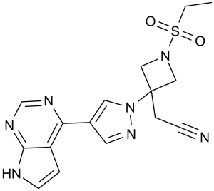
S(C([H])([H])C([H])([H])[H])(N1C([H])([H])C(C([H])([H])C#N)(C1([H])[H])N1C([H])=C(C2=C3C([H])=C([H])N([H])C3=NC([H])=N2)C([H])=N1)(=O)=O
|
|
| InChi Key |
XUZMWHLSFXCVMG-UHFFFAOYSA-N
|
|
| InChi Code |
InChI=1S/C16H17N7O2S/c1-2-26(24,25)22-9-16(10-22,4-5-17)23-8-12(7-21-23)14-13-3-6-18-15(13)20-11-19-14/h3,6-8,11H,2,4,9-10H2,1H3,(H,18,19,20)
|
|
| 化学名 |
|
|
| 别名 |
|
|
| HS Tariff Code |
2934.99.9001
|
|
| 存储方式 |
Powder -20°C 3 years 4°C 2 years In solvent -80°C 6 months -20°C 1 month |
|
| 运输条件 |
Room temperature (This product is stable at ambient temperature for a few days during ordinary shipping and time spent in Customs)
|
| 溶解度 (体外实验) |
|
|||
|---|---|---|---|---|
| 溶解度 (体内实验) |
配方 1 中的溶解度: ≥ 2.5 mg/mL (6.73 mM) (饱和度未知) in 10% DMSO + 40% PEG300 + 5% Tween80 + 45% Saline (这些助溶剂从左到右依次添加,逐一添加), 澄清溶液。
例如,若需制备1 mL的工作液,可将100 μL 25.0 mg/mL澄清DMSO储备液加入到400 μL PEG300中,混匀;然后向上述溶液中加入50 μL Tween-80,混匀;加入450 μL生理盐水定容至1 mL。 *生理盐水的制备:将 0.9 g 氯化钠溶解在 100 mL ddH₂O中,得到澄清溶液。 配方 2 中的溶解度: ≥ 2.5 mg/mL (6.73 mM) (饱和度未知) in 10% DMSO + 90% (20% SBE-β-CD in Saline) (这些助溶剂从左到右依次添加,逐一添加), 澄清溶液。 例如,若需制备1 mL的工作液,可将 100 μL 25.0 mg/mL澄清DMSO储备液加入900 μL 20% SBE-β-CD生理盐水溶液中,混匀。 *20% SBE-β-CD 生理盐水溶液的制备(4°C,1 周):将 2 g SBE-β-CD 溶解于 10 mL 生理盐水中,得到澄清溶液。 View More
配方 3 中的溶解度: ≥ 2.5 mg/mL (6.73 mM) (饱和度未知) in 10% DMSO + 90% Corn Oil (这些助溶剂从左到右依次添加,逐一添加), 澄清溶液。 配方 4 中的溶解度: 0.5% CMC+0.25% Tween 80:30mg/mL 配方 5 中的溶解度: 2.5 mg/mL (6.73 mM) in 0.5% Methylcellulose/saline water (这些助溶剂从左到右依次添加,逐一添加), 悬浊液; 超声助溶。 *生理盐水的制备:将 0.9 g 氯化钠溶解在 100 mL ddH₂O中,得到澄清溶液。 1、请先配制澄清的储备液(如:用DMSO配置50 或 100 mg/mL母液(储备液)); 2、取适量母液,按从左到右的顺序依次添加助溶剂,澄清后再加入下一助溶剂。以 下列配方为例说明 (注意此配方只用于说明,并不一定代表此产品 的实际溶解配方): 10% DMSO → 40% PEG300 → 5% Tween-80 → 45% ddH2O (或 saline); 假设最终工作液的体积为 1 mL, 浓度为5 mg/mL: 取 100 μL 50 mg/mL 的澄清 DMSO 储备液加到 400 μL PEG300 中,混合均匀/澄清;向上述体系中加入50 μL Tween-80,混合均匀/澄清;然后继续加入450 μL ddH2O (或 saline)定容至 1 mL; 3、溶剂前显示的百分比是指该溶剂在最终溶液/工作液中的体积所占比例; 4、 如产品在配制过程中出现沉淀/析出,可通过加热(≤50℃)或超声的方式助溶; 5、为保证最佳实验结果,工作液请现配现用! 6、如不确定怎么将母液配置成体内动物实验的工作液,请查看说明书或联系我们; 7、 以上所有助溶剂都可在 Invivochem.cn网站购买。 |
| 制备储备液 | 1 mg | 5 mg | 10 mg | |
| 1 mM | 2.6924 mL | 13.4618 mL | 26.9237 mL | |
| 5 mM | 0.5385 mL | 2.6924 mL | 5.3847 mL | |
| 10 mM | 0.2692 mL | 1.3462 mL | 2.6924 mL |
1、根据实验需要选择合适的溶剂配制储备液 (母液):对于大多数产品,InvivoChem推荐用DMSO配置母液 (比如:5、10、20mM或者10、20、50 mg/mL浓度),个别水溶性高的产品可直接溶于水。产品在DMSO 、水或其他溶剂中的具体溶解度详见上”溶解度 (体外)”部分;
2、如果您找不到您想要的溶解度信息,或者很难将产品溶解在溶液中,请联系我们;
3、建议使用下列计算器进行相关计算(摩尔浓度计算器、稀释计算器、分子量计算器、重组计算器等);
4、母液配好之后,将其分装到常规用量,并储存在-20°C或-80°C,尽量减少反复冻融循环。
计算结果:
工作液浓度: mg/mL;
DMSO母液配制方法: mg 药物溶于 μL DMSO溶液(母液浓度 mg/mL)。如该浓度超过该批次药物DMSO溶解度,请首先与我们联系。
体内配方配制方法:取 μL DMSO母液,加入 μL PEG300,混匀澄清后加入μL Tween 80,混匀澄清后加入 μL ddH2O,混匀澄清。
(1) 请确保溶液澄清之后,再加入下一种溶剂 (助溶剂) 。可利用涡旋、超声或水浴加热等方法助溶;
(2) 一定要按顺序加入溶剂 (助溶剂) 。
| NCT Number | Recruitment | interventions | Conditions | Sponsor/Collaborators | Start Date | Phases |
| NCT04901325 | Recruiting | Drug: Baricitinib | RPyoderma Gangrenosum Skin Diseases |
Oregon Health and Science University | October 2023 | Phase 2 |
| NCT05852171 | Recruiting | Drug: Baricitinib | Mastitis Chronic Idiopathic Granulomatous Mastitis |
First Affiliated Hospital of Zhejiang University |
January 1, 2023 | Phase 2 |
| NCT05074420 | Recruiting | Drug: Baricitinib | Covid19 Corona Virus Infection |
Eli Lilly and Company | December 21, 2021 | Phase 3 |
| NCT06240351 | Not yet recruiting | Drug: Baricitinib 4 MG Oral Tablet | Frontal Fibrosing Alopecia | University of Alabama at Birmingham |
June 1, 2024 | Phase 4 |
Cellular activity of INCB028050.J Immunol.2010 May 1;184(9):5298-307. |
Anti-inflammatory and DMARD activity of once daily INCB028050 in rats with established disease in the adjuvant arthritis model.J Immunol.2010 May 1;184(9):5298-307. |
Suppression of delayed-type hypersensitivity by INCB028050.J Immunol.2010 May 1;184(9):5298-307. |
INCB028050 is efficacious and well tolerated independently of effects on humoral immunity.J Immunol.2010 May 1;184(9):5298-307. |
INCB028050 improves clinical and histologic signs of disease in the murine CIA model. |
 JAK2-IN-14
 JAK1/TYK2-IN-5
 Gusacitinib-d4
 Povorcitinib-d6
InvivoChem的所有产品仅用于作科学研究,不面向患者销售
Copyright 2020 InvivoChem LLC | All Rights Reserved 粤ICP备20063088号-1


 COA
COA
")
")
")
")
")
 463611831
