| 规格 | 价格 | 库存 | 数量 |
|---|---|---|---|
| 5mg |
|
||
| 10mg |
|
||
| 25mg |
|
||
| 50mg |
|
||
| 100mg |
|
||
| 250mg |
|
||
| 500mg |
|
||
| Other Sizes |
|

| 靶点 |
Guanylate cyclase
|
||
|---|---|---|---|
| 体外研究 (In Vitro) |
体外活性:在体外,BAY 41-2272导致人和兔海绵体的浓度依赖性松弛,EC50 分别为 489.1 nM 和 406.3 nM。激酶测定:BAY 41-2272是一氧化氮敏感鸟苷酸环化酶(NO 敏感 GC)的激活剂,在存在和不存在 100 nmol/L DEA-NO 的情况下,EC50 值为 0.3 μmol/L 和 3 μmol/L,分别。细胞测定:在血小板中,使用 3 μmol/L(次最大有效浓度)的 GSNO 来评估 BAY 41-2272 对 NO 敏感的 GC 可能的致敏作用。在不存在 BAY 41-2272 的情况下,应用该浓度的 NO 产生的 cGMP 响应仅是微不足道的。在 100 μmol/L BAY 41-2272 存在的情况下,用 3 μmol/L GSNO 处理导致 cGMP 快速增加至 1000 pmol/109 血小板。
背景:一氧化氮最重要的受体是可溶性鸟苷酸环化酶(sGC),一种含血红素的异二聚体。最近,一种与YC-1结构相关的吡唑并吡啶衍生物BAY 41-2272被鉴定为以NO非依赖的方式刺激可溶性鸟苷酸环化酶,从而产生血管舒张和抗血小板活性。这里描述的研究涉及可溶性鸟苷酸环化酶上NO非依赖性位点的鉴定。 结果:我们通过在氚标记化合物中引入叠氮基团,为直接和NO非依赖性可溶性鸟苷酸环化酶(sGC)刺激剂BAY 41-2272开发了一种光亲和标记物(3H-meta-PAL)。合成的光亲和标记直接刺激纯化的sGC,并与NO结合显示出对sGC活性的协同作用。3H-meta-PAL与高纯度sGC一起用紫外光照射,导致与酶的α1亚基共价结合。这种结合被未标记的间PAL、YC-1和BAY 41-2272阻断。为了进一步鉴定NO非依赖性调控位点,3H-meta-PAL标记的sGC被溴化氰消化片段化。3H-meta-PAL与由α1亚基的氨基酸236-290组成的溴化氰片段结合。通过测序该溴化氰片段来确定单个PTH循环的放射性,检测到半胱氨酸238和243是3H-meta-PAL的结合残基。 结论:我们的数据表明,sGCα1亚基中半胱氨酸238和243周围的区域可能在sGC活性的调节中发挥重要作用,并可能成为这种新型sGC刺激剂的靶点。[1] 通过与sGC中该结合位点的相互作用,BAY 41-2272具有独特的体外和体内药理学特征。苯肾上腺素预收缩(3× 10⁻⁸g ml-1)兔主动脉环,BAY 41-2272 诱导浓度依赖性舒张,半最大抑制浓度(IC50)为304 ± 63 nM(n=6),而用作对照的三硝酸甘油和YC-1的IC50为1300 ± 385 nM(n=6)和10035 ± 632 nM(n=26)。 我们和其他人已经证明,YC-1在体外、人血小板、sGC过表达细胞系和平滑肌细胞中激活纯化的sGC并使酶对NO敏感。与BAY 41-2272相比,需要大约100倍的YC-1浓度才能在sGC上产生类似的刺激。BAY 41-2272和YC-1之间的结构相似性可能表明这两种化合物的作用机制相同。然而,与YC-1(参考文献21)相比,BAY 41-2272高达10-5 M、 不具有任何PDE-5抑制活性。[2] BAY 41-2272导致人和兔海绵体的浓度依赖性舒张(平均EC50+/-SEM分别为489.1+/-22.5和406.3+/-21.5nM)。该化合物的效力是YC-1的32倍,是精胺NONOate的两倍。ODQ降低了BAY41-2272的效力,使得在30微摩尔的存在下。ODQ, BAY 41-2272诱导的舒张作用的EC50分别为1407.3+/-158.0和1902.7+/-11.0 nM。分别在人和兔组织中。L-NAME还抑制了BAY41-2272在兔组织中引起的舒张作用。在500微米的存在下。L-NAME BAY41-2272诱导的反应的EC50为836.7+/-46.7nM。BAY41-2272的亚阈值浓度为30至50 nM。增强的氮能反应。此外,L-NAME对氮能反应的抑制作用被逆转了0.3至3微摩BAY 41-2272。 结论:我们报告了一种非基于NO的可溶性鸟苷酸环化酶激活剂可以放松人和兔的海绵体,并增强氮能反应。[3] 在之前的研究中,研究人员发现,鸟苷酸环化酶激动剂5-环丙基-2-[1-(2-氟-苄基)-1H-吡唑并[3,4-b]吡啶-3-基]-嘧啶-4-胺(BAY 41-2272)激活人单核细胞和THP-1细胞系产生超氧阴离子,增加体外杀菌活性,表明该药物可用于调节原发性免疫缺陷患者的免疫功能[4]。 |
||
| 体内研究 (In Vivo) |
在雌性自发性高血压大鼠中,BAY 41-2272(10 mg/kg,口服)显示出抗血小板作用,可显着降低血压并提高存活率。在白色念珠菌感染的小鼠中,BAY 41-2272(10 mg/kg,腹膜内)除了增强巨噬细胞功能外,还显着增加巨噬细胞依赖性细胞流入腹膜,并降低死亡率。在 db/db-/- II 型糖尿病和肥胖小鼠中,BAY 41-2272 可改善受损的海绵体 (CC) 松弛。
在本研究中,我们研究了体内注射BAY 41-2272治疗通过腹腔和皮下接种引入的白色念珠菌和金黄色葡萄球菌感染的潜力。我们发现,除了巨噬细胞的功能,如扩散、酵母聚糖颗粒吞噬和一氧化氮以及佛波醇肉豆蔻酸酯醋酸酯刺激的过氧化氢产生外,BAY 41-2272的腹膜内治疗还显著增加了巨噬细胞依赖性细胞流入腹膜。BAY 41-2272治疗在降低腹腔接种白色念珠菌死亡率方面非常有效,但对金黄色葡萄球菌无效。然而,我们发现用BAY 41-2272体外刺激腹腔巨噬细胞显著提高了对这两种病原体的杀菌活性。我们的结果表明,用BAY 41-2272治疗感染白色念珠菌的小鼠预防死亡可能主要是通过巨噬细胞活化调节宿主免疫反应来实现的。[4] 腹膜细胞内流和细胞募集到淋巴器官-小鼠接受(或不接受)BAY 41-2272(0.3-10mg/kg IP)治疗48小时,之后收获腹膜腔并收集脾脏、BM和LN(图1A)。细胞分布显示,与对照组相比,BAY 41-2272治疗诱导腹膜中细胞总数显著增加(图1B)。该细胞群主要由巨噬细胞组成(图1C),但在用该药物治疗的组中,多形核白细胞(PMNs)的百分比也升高了(图1D)。所有使用的载体(transcutol、Cremophor EL和水溶液以及DMSO)对本研究中进行的本次或其他检测没有影响(数据未显示)。[4] 与未治疗的动物相比,用BAY 41-2272治疗的动物的其他淋巴器官(如脾脏、BM或肠系膜LN)中的细胞数量没有差异(图1E-G)。只有肠系膜LN的细胞数量有增加的趋势,表明细胞被募集到这个引流器官。[4] 角叉菜胶诱导的足垫水肿——为了评估BAY 41-2272对炎症过程的影响,我们使用了角叉菜聚糖诱导的小鼠足水肿模型。小鼠用BAY 41-2272(0.3-10mg/kg,IP)治疗(或不治疗)48小时,之后将角叉菜胶(300μg/爪)注射到脚垫中,每小时测量水肿形成,持续4小时(图2A)。用BAY 41-2272进行腹膜内预处理显著增加了爪子水肿,这在角叉菜胶注射后180和240分钟观察到(图2B)。Con A也有类似的数据。这些结果证实了BAY 41-2272的促炎潜力。[4] BAY 41-2272诱导的离体巨噬细胞活化-扩散和吞噬作用-关于BAY 41-2172产生的促炎活性,扩散和吞噬被评估为腹腔巨噬细胞活化的标志物。收集用BAY 41-2272(0.3-10mg/kg IP)治疗(或不治疗)48小时的小鼠腹膜腔,将腹膜细胞在载玻片上孵育以测量扩散,或用酵母聚糖孵育以评估吞噬作用(图3A)。与未处理的动物相比,从BAY 41-2272处理的小鼠中获得的巨噬细胞的扩散增加(图3B),这与它们增加的吞噬活性是一致的(图3C)。[4] NO和H 2 O 2的产生-用BAY 41-2272(0.3-10mg/kg IP)处理(或不处理)小鼠48小时,然后收获腹膜腔,将腹膜细胞与或不与PMA(30nM)一起孵育1小时,以评估H2O2的释放,或孵育48小时以评估NO的产生(图4A)。众所周知,吞噬作用和ROS释放是相关的,并且是许多抗菌反应的原因。然而,在这项研究中,尽管吞噬活性增加,但我们没有观察到自发H2O2释放的变化(图4C)。然而,在BAY 41-2272治疗小鼠的巨噬细胞培养物中添加PMA显著增加了这种代谢物的水平(图4C)。[4] 尽管预处理没有诱导H2O2的自发释放,但与对照组的巨噬细胞相比,BAY 41-2272显著增加了NO的自发产生(图4B)。[4] BAY 41-2272可提高感染真菌的小鼠的存活率-吞噬作用和杀微生物活性的增加表明BAY 41-2172具有治疗感染的潜力。因此,用白色念珠菌和金黄色葡萄球菌攻击C3H/HePas小鼠,并评估这些动物的存活率。小鼠接种白色念珠菌或金黄色葡萄球菌IP,48小时后,用BAY 41-2272(0.3-10mg/kg IP)或伊曲康唑(20mg/kg)、青霉素G(5KU/kg)和四环素(1mg/kg)治疗(或不治疗)三天。对动物的存活率进行了20天的评估(图5A)。 [4] 结果显示,感染后48小时腹腔注射BAY 41-2272显著提高了感染白色念珠菌的小鼠的存活率(图5B),但对感染金黄色葡萄球菌的小鼠没有影响(图5C)。此外,正如预期的那样,伊曲康唑在控制念珠菌感染方面完全有效,使小鼠存活率保持在100%。 [4] BAY 41-2272可增加小鼠对局部白色念珠菌的反应,但不会增加金黄色葡萄球菌感染的反应——根据BAY 41-2172可提高白色念珠菌感染小鼠存活率的观察,使用了动物脚垫爪感染相同病原体的模型(图6A)。该方案允许评估BAY 41-2272对感染部位(病灶内药物注射)和全身(腹腔内药物给药)的直接影响。 [4] 病灶内注射BAY 41-2272显著减少了白色念珠菌引起的脚垫肿胀,而腹腔内治疗没有显著效果(图6B,C)。皮下或腹腔BAY 41-2272治疗没有显著改变金黄色葡萄球菌引起的脚垫肿胀(图6D,E)。 [4] BAY 41-2272增加了对白色念珠菌和金黄色葡萄球菌的体外和离体杀菌活性——对于体内感染模型,BAY 41-2172对白色念珠菌的反应比对金黄色葡萄杆菌的反应更好。因此,通过评估其对这两种病原体的杀菌活性,研究了用BAY 41-2272对腹腔巨噬细胞进行体外或离体治疗的效果。用BAY 41-2272(0.3-10mg/kg IP)治疗(或不治疗)小鼠48小时,然后收获腹膜腔,将腹膜细胞与白色念珠菌或金黄色葡萄球菌一起孵育2小时,以评估杀菌活性(图7A)。[4] 2型糖尿病(DM2)和肥胖是勃起功能障碍(ED)的主要危险因素。在糖尿病中,氧化应激的增加会导致一氧化氮(NO)生物利用度降低,糖尿病患者对磷酸二酯酶5型抑制剂的常规治疗反应似乎较差。我们研究了可溶性鸟苷酸环化酶刺激剂BAY 41-2272(5-环丙基-2-[1-(2-氟苄基)-1H-吡唑并[3,4-b]吡啶-3-基]嘧啶-4-胺)是否通过减少氧化应激有效改善肥胖DM2小鼠受损的海绵体(CC)松弛。在BAY 41-2272存在或不存在的情况下,使用成年db/db(-/-)小鼠或其瘦db(/+)同窝小鼠评估血管功能、cGMP水平、抗氧化状态、NADPH氧化酶表达和超氧化物形成。结果显示,BAY 41-2272(10(-8)至10(-5)M)以类似的方式有效地放松了db(/+)或db/db(-/-)小鼠的CC。BAY 41-2272显著增强了电场刺激(EFS)诱导的内皮依赖性和氮能舒张,并以浓度依赖的方式改善了糖尿病动物对乙酰胆碱和EFS的受损舒张(10(-8)至10(-7)M)。BAY 41-2272增加了CC中cGMP水平,增强了对外源性NO的舒张反应。db/db(-/-)小鼠CC中的总抗氧化状态降低,而血管NADPH氧化酶亚基(gp91phox、p22phox和p47phox)的表达增加,表明处于氧化应激状态。BAY 41-2272以浓度依赖的方式阻止了这些影响。这些结果表明,BAY 41-2272通过增加cGMP和增强抗氧化状态来改善db/db(-/-)小鼠的CC松弛,使该药物成为治疗ED的潜在新候选药物[5]。 |
||
| 酶活实验 |
BAY 41-2272 是一种对一氧化氮敏感的鸟苷酸环化酶激活剂(NO 敏感的 GC),在存在和不存在 100 nmol/L DEA-NO 的情况下,EC50 值分别为 3 μmol/L 和 0.3 μmol/L。
可溶性鸟苷酸环化酶(sGC)的纯化及sGC活性的测定[1] sGC从杆状病毒/Sf9表达系统中高度纯化,并通过在Mg2+作为二价金属阳离子的存在下,由根据Gerzer修饰的[α-32P]-GTP形成[32P]-cGMP来测量酶活性。在存在和不存在1mM DTT的情况下进行孵育。除非另有说明,否则所有测量均重复进行三次。sGC的比活性计算为每分钟孵育时间每毫克蛋白质形成的nmol cGMP。为了表征不同sGC刺激物,sGC的比活性表示为x倍刺激与比基础活性。试验中DMSO的最高浓度为1%(v/v),本身对cGMP的产生没有任何影响。 sGC分析[2] 我们使用杆状病毒/Sf9表达系统纯化sGC,并如上所述在Mg2+存在下测量酶活性。 我们研究了在可溶性鸟苷酸环化酶抑制剂ODQ(1H-[1,2,4]恶二唑[4-3a]喹喔啉-1-酮)或一氧化氮合酶抑制剂L-NAME(N-硝基-L-精氨酸甲酯HCl)存在和不存在的情况下,BAY 41-2272对人和兔海绵条张力和氮能弛豫反应的影响。将BAY 41-2272的效力与另一种可溶性鸟苷酸环化酶激活剂YC-1和释放NO的化合物精胺NONOate(N-2-氨基乙基-N-2-羟基-2-亚硝基肼基-1,2-乙二胺)的效力进行了比较。3. |
||
| 细胞实验 |
当评估 3 μmol/L(次最大有效浓度)的 GSNO 时,BAY 41-2272 可能对血小板中 NO 敏感的 GC 具有敏化作用。在没有 BAY 41-2272 的情况下应用此浓度的 NO 产生的 cGMP 响应仅是微不足道的。在 100 μmol/L BAY 41-2272 存在下,用 3 μmol/L GSNO 处理后,观察到 cGMP 快速上升至 1000 pmol/109 血小板。
血小板聚集[2] 我们按照所述制备了洗涤过的人血小板14,并通过浊度法测量了血小板聚集。将血小板悬浮液与试验化合物在37℃下预孵育 10°C 胶原蛋白(0.1-2µg ml-1)诱导血小板聚集。 血小板聚集[2] 我们按照所述制备了洗涤过的人血小板14,并通过浊度法测量了血小板聚集。将血小板悬浮液与试验化合物在37℃下预孵育 10°C 胶原蛋白(0.1-2µg ml-1)诱导血小板聚集。 扩散试验[4] 根据Rabinovitch等人(1977)进行了扩散试验。将含有2×10⁶个细胞的腹膜细胞悬浮液离心,并悬浮在1 mL 5 mM葡萄糖的PBS中。将50微升细胞悬浮液分层放在玻璃盖玻片上,在37℃下孵育1小时。盖玻片在PBS中轻轻冲洗,玻璃粘附细胞在2.5%戊二醛中固定,用相差显微镜在400倍放大倍数下检查。计数200个巨噬细胞,并将其分为圆形或扩散型。然后计算巨噬细胞扩散指数(SI)如下:SI=扩散巨噬细胞数量×100)/200,即SI=扩散的巨噬细胞百分比。 酵母聚糖吞噬试验[4] 根据Pinello等人(2006)进行吞噬试验。将含有2×10⁶个细胞的腹膜细胞悬浮液离心并悬浮在1 mL RPMI培养基中。将细胞分配到六孔平底微量测试板中的圆形玻璃盖玻片(20 mm)上,并在37℃下孵育培养物20分钟。孵育后,吸出培养上清液并去除非贴壁细胞。用PBS冲洗粘附的单层。随后,向培养物中加入1mL含有5%热灭活胎牛血清的RPMI-1640培养基。在1mg/L酿酒酵母酵母聚糖(Sigma)存在下,将培养物在37℃下保持1小时。然后用冷PBS洗涤培养物以去除未内化的颗粒。然后用0.5%戊二醛固定细胞。使用相差显微镜对平均200个巨噬细胞进行计数,以确定吞噬百分比。吞噬指数(PI)计算如下:PI=具有吞噬活性的巨噬细胞数量×100)/200个计数的贴壁细胞,即PI=至少有两个吞噬酵母聚糖颗粒的巨噬细胞的百分比。 H 2 O 2释放和一氧化氮(NO)产生[4] 使用先前描述的方法在单个巨噬细胞样品中测定H2O2释放和NO产生(Cruz等人,2007)。为了评估H2O2的释放,使用了HRP依赖性酚红氧化微量测定法(Pick&Mizel 1981)。对于该试验,将2.0 x 10⁶个腹膜细胞悬浮在1 mL新鲜制备的酚红溶液中[含有5.5 mM葡萄糖、0.56 mM酚红和8.5 U/mL HRP II型的冰冷PBS]。将100微升细胞悬浮液加入每个孔中,在37℃、5%CO2潮湿的气氛中与或不与PMA(30 nM)一起孵育1小时。将板以150g离心一次3分钟,然后将上清液转移到另一个板上。用10μL氢氧化钠停止反应。用微孔板读数器在620nm处测量吸光度。通过与在RMPI培养基中稀释的已知浓度的H2O2(5-40μM)获得的标准曲线进行比较,将吸光度转换为μM的H2O2(Pick&Keisari 1980)。 此后,用PBS洗涤含有细胞的板三次,将剩余的粘附巨噬细胞在37℃、5%CO2潮湿气氛中在100μL RPMI-1640培养基(补充有10 mM HEPES、11 mM碳酸氢钠、100 U/mL青霉素、100μg/mL链霉素、2 mM L-谷氨酰胺、23 mM L-天冬酰胺、1 mM叶酸、0.1 mM丙酮酸和5%胎牛血清)中培养48小时。孵育后,收集50μL上清液,在室温(RT)下与等体积的Griess试剂(1%磺胺/0.1%萘二胺二盐酸盐/2.5%磷酸)孵育10分钟,以定量亚硝酸盐的积累(Ding等人,1988)。在550nm处测定吸光度。通过与在RPMI培养基中稀释的已知浓度(5-60μM)的亚硝酸钠获得的标准曲线进行比较,将吸光度转换为NO的μM。 离体和体外腹腔巨噬细胞杀菌活性[4] 为了评估杀菌活性,在细胞与细菌或真菌孵育后使用MTT氧化微量测定法。对于该试验,使用两种方案处理腹腔巨噬细胞,如下所示:(i)在未经处理的动物体外刺激驻留的腹腔细胞(体外),以及(ii)从如本方案中所述处理的动物体内采集细胞(离体)。制备后,将2.5×10⁵个细胞悬浮在200µL RPMI-1640(不含补充剂)中,并分布在96孔板中。然后,金黄色葡萄球菌以10:1(病原体:巨噬细胞)的比例添加病原体,白色念珠菌以2:1的比例添加。共培养物在37℃和5%CO2下孵育2小时。孵育后,将平板离心,收集上清液并储存在-80℃下,用于随后的细胞因子剂量测定。用PBS洗涤细胞颗粒两次,以去除未吞噬的病原体。洗涤后,在室温下加入Triton X-100(1.5%)10分钟,以裂解巨噬细胞并释放病原体。然后用PBS洗涤细胞两次以去除Triton X-100,加入100µL MTT(0.5mg/mL),在室温下避光孵育2小时。孵育后,加入100µL DMSO,再孵育30分钟,将甲氮沉淀释放到上清液中。孵育后,将平板离心(300g,3分钟),并将上清液转移到新的平板上。用酶标仪在λ=570 nm处测定吸光度。从吸光度到细胞死亡百分比的转换是通过以下方程式实现的:1-(样品OD-90%杀灭OD)/(0%杀灭OD-90%杀伤OD)×100。该计算是基于与细胞一起孵育的病原体总数的100-10%的病原体浓度进行的。 |
||
| 动物实验 |
|
||
| 参考文献 | |||
| 其他信息 |
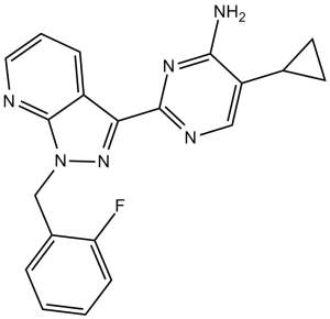
BAY 41-2272 是一种吡唑并吡啶类化合物,其结构为 1H-吡唑并[3,4-b]吡啶,在 1 位被 2-氟苄基取代,在 3 位被 4-氨基-5-环丙基嘧啶-2-基取代。它是一种可溶性鸟苷酸环化酶激活剂。它具有多种功能,包括作为可溶性鸟苷酸环化酶激活剂、血小板聚集抑制剂、血管扩张剂和抗高血压剂。它属于吡唑并吡啶类化合物,属于单氟苯类、氨基嘧啶类和环丙烷类化合物。
总之,我们利用光亲和标记法,确定了 sGC α1 亚基中半胱氨酸 238 和 243 的区域是 NO 依赖性 sGC 刺激剂的作用靶点。然而,该区域作为调控单元的相关性仍需通过突变分析和共结晶研究来证实。[1] 总之,BAY 41-2272对sGC的影响以及光亲和标记研究表明,sGC α1亚基Cys 238和Cys 243区域存在一个新的不依赖于NO的调控位点,该位点调节催化速率和对血红素配体的反应性。我们的数据不仅为理解sGC的调控提供了一种方法,而且还发现了一种强效的新型sGC刺激剂BAY 41-2272,它能诱导血管舒张而不产生硝酸盐耐受性,具有抗血小板活性,并最终降低死亡率。[2] 一氧化氮(NO)是一种广泛存在且作用强大的生物介质,具有许多生理和病理生理功能。一氧化氮(NO)领域的研究似乎遵循着一条清晰的路径,研究成果也稳步推进:NO和环磷酸鸟苷(cGMP)参与血管舒张;硝酸甘油通过生物转化为NO来舒张血管平滑肌;哺乳动物细胞能够合成NO;最后,NO通过刺激可溶性鸟苷酸环化酶(sGC)介导血管舒张,sGC是一种异二聚体(α/β)血红素蛋白,可将GTP转化为cGMP2-4。本文报道了sGC上一个调控位点的发现。利用光亲和标记技术,我们鉴定出sGC α1亚基中的半胱氨酸238和半胱氨酸243区域是新型sGC刺激剂的靶点。此外,我们还介绍了一种吡唑并吡啶类化合物BAY 41-2272,它能够通过该位点有效刺激sGC,且其作用机制不依赖于NO。这导致抗血小板活性,显著降低血压,并提高低NO高血压大鼠模型的生存率,因此可能为治疗心血管疾病提供一种新方法。[2] 目的:在海绵体平滑肌中,一氧化氮(NO)激活可溶性鸟苷酸环化酶,后者催化环磷酸鸟苷(cGMP)的合成,从而导致平滑肌松弛、血流量增加和阴茎勃起。吡唑并吡啶衍生物BAY 41-2272(5-环丙基-2-[1-(2-氟苄基)-1H-吡唑并[3,4-b]吡啶-3-基]嘧啶-4-胺)被发现能够以不依赖于NO的方式刺激可溶性鸟苷酸环化酶。我们研究了BAY41-2272对人和兔阴茎海绵体的影响。 [3]在体内感染模型中,BAY 41-2272对真菌的杀灭效果优于细菌。因此,我们研究了腹膜巨噬细胞对相同病原体的体外和离体杀菌活性。结果表明,体外处理增强了腹膜巨噬细胞对白色念珠菌和金黄色葡萄球菌的杀菌活性,且离体处理后增强效果更为显著。这些结果证实了BAY 41-2272在治疗真菌感染(特别是白色念珠菌感染)方面的潜力。我们还发现,该疗法能有效促进金黄色葡萄球菌的杀灭。这些数据支持我们的假设,即体内金黄色葡萄球菌感染的明显不消退与病原体产生的炎症持续存在有关,而BAY 41-2272可增强这种炎症。 这种杀菌活性的增强可能与氧化爆发、活性氮的产生和吞噬作用有关。然而,我们不能排除其他过程的可能参与,例如吞噬体pH酸化和溶酶体/颗粒酶的释放(Sokolovska等人,2012),以及其他细胞的参与。重要的是,体外反应的广泛性表明,生理环境中存在的化学介质和细胞与吞噬细胞反应的激活和调节密切相关。BAY 41-2272对其他免疫细胞的作用可能创造了一个具有更多刺激巨噬细胞激活的环境。考虑到复杂的生理系统,这些数据为BAY 41-2272或其通路(sGC-cGMP)可用于治疗某些感染(尤其适用于免疫功能低下患者)的观点提供了新的证据。值得强调的是,BAY 41-2272的心血管效应(Thorsen等人,2010;Joshi等人,2011)并未限制其体内应用。 我们得出结论,BAY 41-2272具有促炎作用,可激活单核吞噬细胞(腹腔巨噬细胞)。此外,BAY 41-2272治疗可显著增强小鼠对白色念珠菌(体内和体外)和金黄色葡萄球菌(体外)的免疫反应,提高腹腔巨噬细胞对这些病原体的杀菌活性。我们团队正积极研究BAY 41-2272的药理学特性,旨在阐明其信号通路并揭示其对单核吞噬细胞的影响。基于这些信息,我们计划开发新的治疗方法,以提高易感染患者(尤其是原发性免疫缺陷病患者)的生活质量。 [4] BAY 41-2272(5-环丙基-2-[1-(2-氟苄基)-1H-吡唑并[3,4-b]吡啶-3-基]嘧啶-4-胺)是一种可溶性鸟苷酸环化酶(sGC)刺激剂,已被证实具有抗增殖和血管舒张作用(Evgenov等人,2006),并能增强勃起反应(Bischoff等人,2003),以及放松人类和动物的阴茎海绵体(Baracat等人,2003;Kalsi等人,2003;Claudino等人,2011)。该化合物被认为具有高效力且无磷酸二酯酶(PDE)抑制活性(Stasch等人,2001)。在 NO 缺乏的大鼠模型中,长期口服 BAY 41-2272 可改善受损的海绵体松弛(Claudino 等人,2011)。在之前对 BAY 41-2272 对小鼠 CC 的影响的研究中,我们课题组发现该化合物通过降低其亚基 gp91phox 和 p22phox 的蛋白表达,逆转了 NADPH 氧化酶依赖性超氧化物生成增加(Teixeira 等,2007)。 BAY 41-2272(而非 PDE-5 抑制剂)增强了肛尾肌和阴茎缩肌的硝能舒张反应(Kalsi 等,2004)(这些组织是研究硝能神经传递的理想组织),而链脲佐菌素诱导的糖尿病大鼠的这些反应受损(Cheah 等,2002)。这些数据表明,糖尿病患者体内来自硝基能神经的内源性NO减少,并且显示sGC刺激剂在治疗糖尿病引起的ED方面比PDE-5抑制剂更有效。 据我们所知,此前尚无研究探讨BAY 41-2272对糖尿病阴茎海绵体的作用。此外,尽管db/db−/−小鼠表现出与海绵体松弛受损和阴茎静脉闭塞性疾病一致的血管反应性改变,但利用db/db−/−小鼠研究ED的研究却很少。db/db−/−小鼠缺乏瘦素受体,这种缺陷会导致糖尿病和肥胖的发生。因此,这些小鼠被广泛认为是2型糖尿病的合适模型,并已被用于研究2型糖尿病相关的ED(Luttrell等人,2008)。此外,db/db−/−小鼠会出现高血糖和高胰岛素血症,后者会提高静息交感神经输出,并导致海绵体舒张功能受损(Anderson等,1991)。 在本研究中,我们探讨了BAY 41-2272对db/db−/−肥胖DM2小鼠及其瘦型对照db/+小鼠在血管舒张激动剂刺激下海绵体舒张功能的影响,以及该药物对这些动物氧化应激标志物的影响。 我们的数据显示,在糖尿病肥胖(db/db−/−)小鼠中,BAY 41-2272通过提高细胞内cGMP浓度、抑制NADPH氧化酶亚基表达升高以及减少超氧化物生成,改善了内皮和硝基能海绵体舒张功能障碍。虽然糖尿病患者发生勃起功能障碍的病因是多因素的,但血管功能障碍是导致糖尿病男性勃起功能障碍高发的主要原因(Chu 和 Edelman,2002)。[5] |
| 分子式 |
C20H17FN6
|
|
|---|---|---|
| 分子量 |
360.39
|
|
| 精确质量 |
360.149
|
|
| 元素分析 |
C, 66.65; H, 4.75; F, 5.27; N, 23.32
|
|
| CAS号 |
256376-24-6
|
|
| 相关CAS号 |
|
|
| PubChem CID |
9798973
|
|
| 外观&性状 |
White to off-white solid powder
|
|
| 密度 |
1.5±0.1 g/cm3
|
|
| 沸点 |
496.1±45.0 °C at 760 mmHg
|
|
| 闪点 |
253.8±28.7 °C
|
|
| 蒸汽压 |
0.0±1.3 mmHg at 25°C
|
|
| 折射率 |
1.767
|
|
| LogP |
1.99
|
|
| tPSA |
83.24
|
|
| 氢键供体(HBD)数目 |
1
|
|
| 氢键受体(HBA)数目 |
6
|
|
| 可旋转键数目(RBC) |
4
|
|
| 重原子数目 |
27
|
|
| 分子复杂度/Complexity |
517
|
|
| 定义原子立体中心数目 |
0
|
|
| SMILES |
FC1=C([H])C([H])=C([H])C([H])=C1C([H])([H])N1C2=C(C([H])=C([H])C([H])=N2)C(C2=NC([H])=C(C(N([H])[H])=N2)C2([H])C([H])([H])C2([H])[H])=N1
|
|
| InChi Key |
ATOAHNRJAXSBOR-UHFFFAOYSA-N
|
|
| InChi Code |
InChI=1S/C20H17FN6/c21-16-6-2-1-4-13(16)11-27-20-14(5-3-9-23-20)17(26-27)19-24-10-15(12-7-8-12)18(22)25-19/h1-6,9-10,12H,7-8,11H2,(H2,22,24,25)
|
|
| 化学名 |
5-cyclopropyl-2-[1-[(2-fluorophenyl)methyl]pyrazolo[3,4-b]pyridin-3-yl]pyrimidin-4-amine
|
|
| 别名 |
|
|
| HS Tariff Code |
2934.99.9001
|
|
| 存储方式 |
Powder -20°C 3 years 4°C 2 years In solvent -80°C 6 months -20°C 1 month |
|
| 运输条件 |
Room temperature (This product is stable at ambient temperature for a few days during ordinary shipping and time spent in Customs)
|
| 溶解度 (体外实验) |
|
|||
|---|---|---|---|---|
| 溶解度 (体内实验) |
配方 1 中的溶解度: ≥ 1.75 mg/mL (4.86 mM) (饱和度未知) in 10% DMSO + 40% PEG300 + 5% Tween80 + 45% Saline (这些助溶剂从左到右依次添加,逐一添加), 澄清溶液。
例如,若需制备1 mL的工作液,可将100 μL 17.5 mg/mL 澄清的 DMSO 储备液加入到400 μL PEG300中,混匀;再向上述溶液中加入50 μL Tween-80,混匀;然后加入450 μL 生理盐水定容至1 mL。 *生理盐水的制备:将 0.9 g 氯化钠溶解在 100 mL ddH₂O中,得到澄清溶液。 配方 2 中的溶解度: ≥ 1.75 mg/mL (4.86 mM) (饱和度未知) in 10% DMSO + 90% (20% SBE-β-CD in Saline) (这些助溶剂从左到右依次添加,逐一添加), 澄清溶液。 例如,若需制备1 mL的工作液,可将 100 μL 17.5mg/mL澄清的DMSO储备液加入到900μL 20%SBE-β-CD生理盐水中,混匀。 *20% SBE-β-CD 生理盐水溶液的制备(4°C,1 周):将 2 g SBE-β-CD 溶解于 10 mL 生理盐水中,得到澄清溶液。 View More
配方 3 中的溶解度: ≥ 1.75 mg/mL (4.86 mM) (饱和度未知) in 10% DMSO + 90% Corn Oil (这些助溶剂从左到右依次添加,逐一添加), 澄清溶液。 1、请先配制澄清的储备液(如:用DMSO配置50 或 100 mg/mL母液(储备液)); 2、取适量母液,按从左到右的顺序依次添加助溶剂,澄清后再加入下一助溶剂。以 下列配方为例说明 (注意此配方只用于说明,并不一定代表此产品 的实际溶解配方): 10% DMSO → 40% PEG300 → 5% Tween-80 → 45% ddH2O (或 saline); 假设最终工作液的体积为 1 mL, 浓度为5 mg/mL: 取 100 μL 50 mg/mL 的澄清 DMSO 储备液加到 400 μL PEG300 中,混合均匀/澄清;向上述体系中加入50 μL Tween-80,混合均匀/澄清;然后继续加入450 μL ddH2O (或 saline)定容至 1 mL; 3、溶剂前显示的百分比是指该溶剂在最终溶液/工作液中的体积所占比例; 4、 如产品在配制过程中出现沉淀/析出,可通过加热(≤50℃)或超声的方式助溶; 5、为保证最佳实验结果,工作液请现配现用! 6、如不确定怎么将母液配置成体内动物实验的工作液,请查看说明书或联系我们; 7、 以上所有助溶剂都可在 Invivochem.cn网站购买。 |
| 制备储备液 | 1 mg | 5 mg | 10 mg | |
| 1 mM | 2.7748 mL | 13.8739 mL | 27.7477 mL | |
| 5 mM | 0.5550 mL | 2.7748 mL | 5.5495 mL | |
| 10 mM | 0.2775 mL | 1.3874 mL | 2.7748 mL |
1、根据实验需要选择合适的溶剂配制储备液 (母液):对于大多数产品,InvivoChem推荐用DMSO配置母液 (比如:5、10、20mM或者10、20、50 mg/mL浓度),个别水溶性高的产品可直接溶于水。产品在DMSO 、水或其他溶剂中的具体溶解度详见上”溶解度 (体外)”部分;
2、如果您找不到您想要的溶解度信息,或者很难将产品溶解在溶液中,请联系我们;
3、建议使用下列计算器进行相关计算(摩尔浓度计算器、稀释计算器、分子量计算器、重组计算器等);
4、母液配好之后,将其分装到常规用量,并储存在-20°C或-80°C,尽量减少反复冻融循环。
计算结果:
工作液浓度: mg/mL;
DMSO母液配制方法: mg 药物溶于 μL DMSO溶液(母液浓度 mg/mL)。如该浓度超过该批次药物DMSO溶解度,请首先与我们联系。
体内配方配制方法:取 μL DMSO母液,加入 μL PEG300,混匀澄清后加入μL Tween 80,混匀澄清后加入 μL ddH2O,混匀澄清。
(1) 请确保溶液澄清之后,再加入下一种溶剂 (助溶剂) 。可利用涡旋、超声或水浴加热等方法助溶;
(2) 一定要按顺序加入溶剂 (助溶剂) 。
 sGC/cGMP/PKG signaling pathway in HNSCC cell lines.Cancer Lett. 2016 Jan 28;370(2):279-85. |
|---|
 NO donors, sGC stimulators, and PDE5 inhibitors decrease the viability of HNSCC cells.Cancer Lett. 2016 Jan 28;370(2):279-85. |
 Tadalafil suppressed the growth of CAL27 xenograftsin vivo.Cancer Lett. 2016 Jan 28;370(2):279-85. |
 BAY 41-2272 (BAY) and Tadalafil (Tad) decrease cell proliferation and induce apoptosis in HNSCC cells.Cancer Lett. 2016 Jan 28;370(2):279-85. |
|---|
 BAY 41-2272 (BAY) and Tadalafil (Tad) decrease clonogenic survival of HNSCC cells. CAL27 (A), UM6 (B) and UM47 (C) cells were plated at 150 cells/well in 6 well plates and treated before first doubling. After 72 h, treatments were replaced with growth media and colonies were allowed to form for 2 weeks. Colonies were stained with crystal violet and counted.Cancer Lett. 2016 Jan 28;370(2):279-85. |
 Effect of sGC or PKG inhibitors on apoptosis induced by BAY or Tadalafil.Cancer Lett. 2016 Jan 28;370(2):279-85. |
 SGC stimulator 1
 A-350619
 RIG 200
 S-3448
InvivoChem的所有产品仅用于作科学研究,不面向患者销售
Copyright 2020 InvivoChem LLC | All Rights Reserved 粤ICP备20063088号-1


 COA
COA
 463611831
