| 规格 | 价格 | 库存 | 数量 |
|---|---|---|---|
| 5mg |
|
||
| 10mg |
|
||
| Other Sizes |
|

| 靶点 |
LXRβ/liver X receptor β
|
|---|---|
| 体外研究 (In Vitro) |
在反式激活实验中,与全泛激动剂相比,XL041 (BMS-852927) 表现出 20% LXRα 和 88% LXRβ 活性。 XL041 在体外人全血内源性靶基因激活测定 (WBA) 中的 EC50 为 9 nM,活性为 26%,是一种非常强大的药物。据报道,BMS-852927 对 LXRα 和 LXRβ(分别为 19 和 12 nM)具有相当的结合亲和力 [1]。
当氟R3取代与2,6-二氯A环(15/BMS-852927)结合时,观察到非常强的hWBA活性,EC50值为9 nM(26%的疗效)。尽管15/BMS-852927具有相似的LXRα和LXRβ结合Ki值(分别为19和12 nM),但在激动剂试验中,与全泛激动剂相比,该化合物对LXRβ的疗效为88%,对LXRα的疗效仅为20%。在拮抗剂模式下测试时,15是一种强效的LXRα拮抗剂,IC50值为69 nM(抑制率为83%);而在高达10000 nM的LXRβ测定中没有观察到拮抗作用(支持信息)。通过2-Cl、6-F R1置换模式的进一步SAR研究,16个样本的hWBA效力为41 nM(33%)。在17中引入R3氯原子导致所有四种激动剂测定的疗效降低,hWBA EC50值为5 nM,略高于测定检测限值(16%)。R2处有氢的类似物18具有与宝石二甲基类似物12一致的体外活性。单甲基类似物19被鉴定为15的代谢产物,合成后的特征表明它也是一种强效的部分LXR激动剂[2]。 |
| 体内研究 (In Vivo) |
在有效剂量下,XL041 (BMS-852927) 在食蟹猴和小鼠中表现出非常积极的特征。给予C57BL/6J小鼠XL041 7天后,该系统的胆固醇流出以剂量依赖性方式得到有效刺激。在剂量为 3 mg/kg/天的组中观察到最高的外排率,比媒介物高 70%。 LDLR 敲除 (KO) 的小鼠也有类似的结果。在另一项为期 12 周的试验中,XL041 阻止了缺乏 LDLR 基因的小鼠动脉粥样硬化的进展。值得注意的是,增加巨噬细胞逆转胆固醇转移(RCT)(0.03-3 mg/kg/天)和抑制动脉粥样硬化(0.1-3 mg/kg/天)的剂量反应相当。是 LXR 激动剂影响疾病的关键潜在过程 [1]。
|
| 酶活实验 |
体外检测方法:[2]
配体结合分析。使用编码人RXRα和人LXRα或人LXRβ的杆状病毒共感染Sf9细胞。制备受感染的细胞裂解物,并将含有可溶性RXRα-LXR或RXRα-RXR¦异二聚体的上清液用于闪烁邻近配体结合分析,其中测试化合物与50 nM 3H-24,25-环氧胆固醇(NEN生命科学产品/Perkin Elmer)竞争结合的能力。1确定的Ki代表至少两个独立剂量反应实验的平均值。使用单位点竞争公式通过非线性回归分析确定每种化合物的结合亲和力,以确定IC50,其中:Y=底部+(顶部-底部)/(1+10X-logIC50)。然后计算Ki:Ki=IC50/(1+[配体浓度]/Kd配体)。对于该测定,通常配体浓度=50 nM,受体配体的Kd为200 nM,通过饱和结合测定。 瞬时转染报告检测。[2] LXR和LXR¦LXRE测定(同种型特异性)。对于激动剂试验,CV-1细胞与pCXM hLXRα或pCMX hLXRβ和LXREx1 tk-luc质粒共转染,并在含有5µL培养基和0.5%DMSO或试验激动剂的384孔板中以8000个细胞/孔的密度重新接种。以相同的方式进行2次LXR拮抗剂测定,但存在60 nM的激动剂Pan agonist A。基于在没有激动剂的情况下获得的值计算100%的抑制率。每个板的前两列含有泛激动剂A,以确定0%的抑制水平。将细胞孵育18-20小时,裂解并使用北极星HTS工作站(Applied BioSystems)测定萤光素酶活性。剂量反应曲线由浓度相差½对数单位的10点曲线生成。每个点代表来自32 384孔板的4孔数据的平均值。将该测定的数据拟合到以下方程中,可以求解EC50值:Y=底部+(顶部-底部)/(1+10((logEC50-X)*HillSlope))。EC50被定义为激动剂引发反应的浓度,该反应位于顶部(最大值)和底部(基线)值之间的一半。所表示的EC50值是至少2个独立实验的平均值。通过与泛激动剂A达到的最大反应进行比较来确定激动剂的相对疗效或%疗效(支持信息图1),该反应在每个剂量反应实验中单独测量。 LXR激动剂对巨噬细胞依赖性中性粒细胞减少途径影响的体外研究[1] 将巯基乙酸诱导的C57BL/6J小鼠腹腔巨噬细胞在DMEM-FBS中培养,并在无血清DMEM中与LXR激动剂或载体一起孵育20小时,然后在激动剂或赋形剂的持续存在下用20ng/mL LPS处理5小时。如补充实验程序的RNA制备和分析部分所述,确定了治疗对IL-23α和Mertk mRNA的影响。 |
| 细胞实验 |
用PBS洗涤每个烧瓶中的细胞,加入2 mL胰蛋白酶-EDTA(0.25%),在37℃、5%CO2下孵育5分钟。然后用力敲击烧瓶以破碎细胞聚集体。在加入8ml含有5%木炭/葡聚糖处理的FBS的DMEM后,将整个混合物转移到锥形管中。然后将细胞以1000rpm离心5分钟。将细胞沉淀重新悬浮在冷冻培养基(含20%血清和10%DMSO的DMEM)中,最终计数约为7 x 106个细胞/ml。将细胞悬浮液等分到15ml聚丙烯管中,每管5ml。将细胞放入-80℃的聚苯乙烯泡沫绝缘容器中过夜,缓慢冷冻。24小时后,将小瓶转移到超冷(-140℃)冰箱中长期储存。将冷冻保存的细胞小瓶在温水浴中快速解冻五分钟。将细胞合并,并在50ml锥形瓶中稀释至50ml。将解冻的细胞以1500rpm离心5分钟以收集细胞并丢弃上清液。然后将细胞重新悬浮在新鲜培养基II(DMEM,含有5%木炭/葡聚糖处理的FBS,1%青霉素/链霉素,100M非必需氨基酸、1 mM丙酮酸钠和2 mM L-谷氨酰胺),使用番石榴细胞计数器计数,并在相同的培养基中稀释至1.6 x 10⁵个细胞/ml。将50微升细胞混合物加入到经白色组织培养处理的384孔板第1-23列的孔中,该板含有溶解在100%DMSO中的0.25l试验化合物。将50微升培养基II加入柱24中的孔中。将平板在37℃(5%CO2)下孵育24小时,然后向每个孔中加入5l Alamar Blue试剂。然后将板在37℃、5%CO2下再孵育两小时,然后在室温下孵育一小时。荧光读数为Ex525/Em598。在测量荧光之后,向每个孔中加入25l萤光素酶底物。将平板在室温下孵育15分钟,然后在PheraStar平板读数器上读取发光情况[1]。
|
| 动物实验 |
食蟹猴研究[1]
所有研究均在雄性动物中进行。在药效学研究中,动物被随机分为六个治疗组(每组n=3),每日一次分别给予赋形剂、10 mg/kg/天的T0901317以及0.1、0.3、1或3 mg/kg/天的BMS-852927,持续14天。在药效学研究中,于基线以及给药后的第1、4、7和14天测定血液RNA和血浆脂质;在肝脏甘油三酯磁共振波谱(MTS)研究中(见下文),于给药后的第1和7天测定血液RNA和血浆脂质。在一项作为更大规模毒理学研究一部分的食蟹猴肝脏mRNA研究中,动物被随机分为四组(每组n=5),并连续28天每日分别给予赋形剂以及0.3、3或30 mg/kg/天的BMS-852927。一项类似的研究使用了BMS-779788,动物连续14天分别接受赋形剂以及1、10或30 mg/kg/天的BMS-779788治疗。两项研究中均在最后一次给药后24小时采集肝脏样本,用于测定化合物浓度和mRNA水平。所有血液和肝脏mRNA的定量分析均按照补充实验步骤中RNA制备和分析部分详细描述的方法进行。 为了探究BMS-852927对多种组织转录组的整体影响,我们分别用载体或15 mg/kg/天的BMS-852927灌胃给予三只动物,连续7天。末次给药后5-6小时,对动物进行镇静并处死。采集包括肝脏、脾脏、主动脉、右侧颈总动脉、骨髓等在内的多种组织,并使用Affymetrix芯片进行转录组分析,具体方法见补充实验步骤。 使用Bruker 4.7T/40cm磁共振成像系统和Bruker Topspin软件进行肝脏甘油三酯(TG)的体内磁共振波谱测量,具体方法见补充实验步骤。在每只灵长类动物口服给予赋形剂或BMS-852927 7天后,对肝脏中央相同感兴趣区域进行脂质分析,并与先前测量的相同肝脏感兴趣区域的基线值进行比较。 为了确定BMS-852927对甾醇排泄的影响,在上述14天药效学研究中,于基线和治疗结束时收集粪便。将干燥的样品和添加的标准品溶解于NaOH-乙醇-水中,并用石油醚提取物通过超高效液相色谱法分析胆固醇。 小鼠研究[1] 为了研究LXR激动剂对中性粒细胞的影响,将预先适应口服给药的C57BL/6小鼠(每组n = 8)随机分配到赋形剂组;分别给予小鼠 0.03、0.1、1 或 3 mg/kg/天的 BMS-852927;以及 0.3 或 3 mg/kg/天的 GW3965,并口服给药 3 天。异氟烷麻醉后,通过眼眶后静脉丛取血,并使用 Advia 血液分析仪,采用过氧化物酶染色法分析中性粒细胞水平。 在一项动脉粥样硬化预防研究中,喂食高脂饮食的 8 至 10 周龄 LDL 受体敲除小鼠,每日灌胃给予赋形剂、BMS-852927(0.1、1 或 3 mg/kg/天)或 10 mg/kg/天的 T0901317,持续 12 周。治疗结束后,处死小鼠,并通过图像分析对油红O染色的主动脉进行表面定量分析,以评估动脉粥样硬化程度。病变面积以占主动脉总面积的百分比表示。 为了确定BMS-852927对体内巨噬细胞胆固醇外流的影响,将C57Bl/6J小鼠(12-24周龄,每组n=3-4)分别灌胃给予载体或0.03、0.1或3 mg/kg/天的BMS-852927,每日一次,连续7天。末次给药5小时后,麻醉动物,并通过非禁食动物的眼眶静脉丛注射牛血清白蛋白:[3H]-胆固醇颗粒复合物。注射后 5 至 150 分钟采集尾静脉血样,离心分离后,采用液体闪烁计数法测定血浆放射性。绘制血浆放射性占注射标记物的百分比随时间变化的曲线,并利用快速初始清除后甾醇再出现阶段的线性部分,通过线性回归计算初始巨噬细胞胆固醇外流率。 第一阶段单次、CV201-001 和多次、CV201-002 递增剂量研究[1] 纳入 18-45 岁、BMI 为 18-30 kg/m2 的健康男性和女性,进行随机、双盲、安慰剂对照的单次和多次递增剂量 (SAD 和 MAD) 研究。在每个剂量组中,8 名受试者按 3:1 的比例随机分组,分别接受 BMS-852927(n = 6)或匹配的安慰剂(n = 2)。在单次给药(SAD)研究中,受试者(平均年龄 24.2 岁,BMI 24.1 kg/m2)被分配到六个连续的剂量组,分别接受单次剂量的 BMS-852927(0.4、2、8、20、40 和 80 mg)或安慰剂。SAD 研究中未发生死亡或严重不良事件(SAE);最常见的治疗期间出现的不良事件是高甘油三酯血症,4 名(7.1%)受试者报告了该不良事件,所有这些受试者均接受了 BMS-852927 治疗,研究者认为该不良事件与研究药物相关。在 MAD 研究中,受试者被随机分配到五个连续剂量组,每日一次接受 BMS-852927(0.2、1、2.5、5 和 15 mg)或安慰剂,持续 14 天。MAD 研究的排除标准包括 MRI 检查禁忌症,例如体内有金属植入物或假体。 1b 期研究,CV201008 [1] 纳入年龄在 18 至 75 岁之间、患有原发性高胆固醇血症且 BMI ≤40 kg/m2 的男性和女性患者。受试者需已接受稳定剂量的他汀类药物治疗 ≥6 周,且筛选时血清甘油三酯水平 <400 mg/dL。 97名受试者被随机分组并接受研究药物,其中90名(92.8%)受试者完成了研究,7名(7.2%)受试者中止研究,6名(6.2%)受试者因不良事件中止研究,1名(1.0%)受试者因其他原因中止研究。受试者每日一次服用0.25、1或2.5 mg的BMS-852927或安慰剂,疗程为28天。研究期间,受试者继续服用其研究前的他汀类药物治疗方案。安全性评估基于对不良事件报告的医学审查、心电图结果以及临床实验室检查(包括空腹血脂)的审查。有关研究设计和实施的更多详细信息,请参阅补充实验步骤。 采用经验证的液相色谱-串联质谱 (LC-MS/MS) 法测定人血浆样本中 BMS-852927 的浓度,具体方法详见补充实验步骤。体内方法 [2] 小鼠体内药代动力学 [2] 在雄性 C57BL6 小鼠(21.6-36 g)中进行了药代动力学表征。小鼠经口灌胃给予化合物(隔夜禁食),化合物以 0.75% CMC/0.1% Tween 80 水溶液的悬浮液形式给药。分别于给药后 0.25、0.5、1、3、6、8 和 24 小时,从每组 3 只小鼠采集血样(约 0.2 mL),从而获得综合药代动力学曲线(每只小鼠采集 3 个血样)。血样静置凝固后,于 4 °C 下以 1500-2000×g 离心,分离血清。血清样本储存于 -20 °C,直至进行 LC/MS/MS 分析。 小鼠脑内分布 [2] 对 C57BL6 小鼠(N = 12)口服给药(10 mg/kg)后,研究了化合物 15 在脑内的分布。分别于给药后 1、4、8 和 24 小时采集脑组织和血液样本(每组 3 只小鼠)。脑组织经吸干、称重后,用50%乙腈/水(1:6 v/v)匀浆,并在4℃下以10,000×g离心10分钟。血浆样本通过在4℃下以1500-2000×g离心获得。所有样本均储存于-20℃,直至进行LC/MS/MS分析。 食蟹猴单剂量药代动力学[2] 为了评估食蟹猴的清除率,药代动力学研究(表2)采用两种方法:一是静脉注射单一化合物(例如BMS-852927),二是静脉注射包含5种不同分子量化合物的混合制剂(0.2 mg/kg)。静脉输注在10分钟内完成(溶剂:10%乙醇;50%聚乙二醇400;40%生理盐水),两只猴子均采用如下所述的类似操作。药代动力学在雄性食蟹猴中采用交叉设计进行评估(见补充信息表1)。禁食过夜后,3只动物(4.5至5.7 kg)分别通过股静脉静脉输注(1 mg/kg,10分钟)和灌胃(3 mg/kg)给药,两次给药之间间隔1周的洗脱期。在给药前以及给药后0.17小时(仅限静脉注射)、0.25小时、0.5小时、0.75小时、1小时、2小时、4小时、6小时、8小时、24小时、48小时和72小时,从股动脉采集系列血样(约0.3 mL),并在4℃下以1500-2000×g离心以获得血浆。血浆样本储存在-20℃直至进行LC/MS/MS分析。 食蟹猴药效学研究。[2] 雄性食蟹猴购自Bioculture Group,成对饲养3周以适应环境,随后在研究开始时将动物转移到标准非人灵长类动物笼中进行单笼饲养。提供充足的水和标准灵长类动物饲料。每日提供120克食物。如需禁食,则于下午3点停止喂食,次日上午9点(给药后2小时)重新喂食。在为期14天的药效学研究中,动物被随机分为治疗组(每组3只;体重3-6公斤),并于每日上午7点通过灌胃法接受以下治疗,持续14天:赋形剂(0.5%羧甲基纤维素和2%吐温80)、10 mg/kg/d T0901317、0.1、0.3、1或3 mg/kg/d BMS-852927 或 0.3、1、3、10、30 mg/kg/d BMS-832878。在给药前一天(第-1天)采集初始静脉血样本,用于基线RNA和脂质测定。随后在给药后的第1、4、7和14天采集血样,用于药效学(PD)研究。用于测定RNA和化合物暴露量的血样在给药后5-6小时采集,用于测定血浆脂质的血样在给药后24小时采集。血浆甘油三酯采用罗氏诊断公司的甘油三酯试剂盒测定。报告的数值均为平均值。统计分析采用方差分析(ANOVA),并使用Dunnett事后检验。文中报告的ABCG1诱导水平与基线水平相比具有统计学意义(ANOVA,并使用Dunnett事后检验,p < 0.05或更低)。 |
| 药代性质 (ADME/PK) |
BMS-852927 的口服生物利用度为 42%(小鼠)和 84%(猴),半衰期分别为 12.4 小时(小鼠)和 9.9 小时(猴)。[2] 激动剂 13 和 15/BMS-852927 被选为进一步研究对象,因为这两种化合物均具有强大的 LXRβ 激动剂活性,而 LXRα 激动剂活性较低(<20%),预计这将有助于更好地将所需疗效与 TG 和 LDL-C 的影响区分开来。此外,15/BMS-852927 在 hWBA 中表现出非常强的活性。类似物 13 和 15/BMS-852927 在 16 项核激素受体激动剂试验中均无活性(>10 μM),但对 PXR 受体有活性,其 EC50 值分别为 3 μM(完全激动作用的 85%)和 1 μM(完全激动作用的 108%)。在小鼠中以 10 mg/kg 的剂量给药时,其 Cmax 覆盖率高于 hWBA 效价(见补充信息)。鉴于 LXR 激动剂可能对大脑产生有害影响(如 LXR-623 所示),我们检测了脑内药物浓度,发现其浓度较低,其中化合物 15 的脑/血浆比值 <0.05。在食蟹猴中,化合物 13 和 15 显示出良好的生物利用度、中等的清除率和 10-12 小时的血浆半衰期[2]。
由于化合物 15/BMS-852927 具有卓越的 hWBA 效力和较低的 LXRα 效力(表 3),因此被认为是先导化合物。为了研究 ABCG1 对脂质作用的剂量反应,并与化合物 1 进行比较,研究人员在食蟹猴中对化合物 13 和 15/BMS-852927 进行了为期 14 天的研究。激动剂在根据食蟹猴 WBA 效力预测的药物浓度下,在血浆中表现出对 RCT 靶基因 ABCG1 的强效诱导作用(化合物 1 的食蟹猴 WBA EC50 = 310 nM (100%);化合物 13 的食蟹猴 WBA EC50 = 52 nM (29%);化合物 15 的食蟹猴 WBA EC50 = 5 nM (32%))。在多项食蟹猴研究中,ABCA1 显示出不同的载体效应,因此不适用于作为药效学生物标志物。化合物 13 和 15 的甘油三酯 (TG) 水平较化合物 1 有所改善。化合物 1 以 10 mg/kg 的剂量给药 14 天(给药后 5 小时血浆浓度为 200 nM),可使血细胞中 ABCG1 的表达诱导 6 倍,TG 水平较基线值升高 140%(p < 0.05,ANOVA)。14 天后,化合物 13 以 1 mg/kg 和 3 mg/kg 的剂量给药,分别使血细胞中 ABCG1 的表达诱导 4 倍和 10 倍,血浆浓度分别为 85 nM 和 310 nM。这些剂量使 TG 水平较基线值分别升高 2% 和 58%(差异无统计学意义)。相比之下,化合物 15 的 0.1、0.3 和 1 mg/kg 剂量组在第 14 天分别使血浆药物浓度在 5 小时内达到 7.5、22 和 57 nM,并诱导 ABCG1 表达分别提高 4.7 倍、15 倍和 11 倍。甘油三酯 (TG) 水平较基线分别升高了 20%、8% 和 10%,但差异无统计学意义。正如预期,化合物 15 在极低的血浆药物浓度下即可显著诱导 ABCG1 表达,且对血浆 TG 水平的影响甚微。该食蟹猴研究 15 的完整数据已在其他文献中报道 [2]。 |
| 参考文献 | |
| 其他信息 |
LXR激动剂在冠状动脉疾病治疗中的应用一直受到动物模型中不良特性的挑战。本文研究了一种LXR激动剂对人体脂质和脂蛋白代谢以及中性粒细胞的影响。新型LXRβ选择性化合物BMS-852927在动物模型中表现出良好的特性,在食蟹猴和小鼠中具有较宽的治疗指数。在健康受试者和高胆固醇血症患者中,逆向胆固醇转运途径的诱导与动物模型相似。然而,血浆和肝脏甘油三酯(TG)、血浆低密度脂蛋白胆固醇(LDL-C)、载脂蛋白B(apoB)、载脂蛋白E(apoE)和胆固醇酯转移蛋白(CETP)水平升高,以及循环中性粒细胞减少也十分明显。此外,在正常胆固醇血症受试者和接受他汀类药物治疗的患者中也观察到了类似的LDL-C升高。灵长类动物模型显著低估了人体的脂肪生成反应,并且无法预测人体中性粒细胞的变化。这些研究展示了LXR激动剂的临床疗效,既有有益的也有不良的,并强调了在该领域开展进一步转化研究的重要性。[1] 在一系列联苯咪唑类肝X受体(LXR)激动剂中引入独特的取代苯砜基团,显著提高了其在人全血中诱导ATP结合盒转运蛋白ABCA1和ABCG1的效力。该系列激动剂在细胞实验中表现出强大的LXRβ活性(>70%)和较低的LXRα部分激动剂活性(<25%),为在食蟹猴中诱导所需的血细胞ABCG1基因表达与激动剂15/BMS-852927引起的血浆甘油三酯水平适度升高之间提供了一个平衡窗口。苯砜基团极性的增加也降低了其与血浆蛋白人α-1-酸性糖蛋白的结合。激动剂 15/BMS-852927 因其体外特性、优异的药代动力学参数和良好的脂质谱而被选中进行临床开发。[2]
|
| 分子式 |
C29H28CL2F2N2O4S
|
|---|---|
| 分子量 |
609.511431694031
|
| 精确质量 |
608.111
|
| 元素分析 |
C, 57.15; H, 4.63; Cl, 11.63; F, 6.23; N, 4.60; O, 10.50; S, 5.26
|
| CAS号 |
1256918-39-4
|
| PubChem CID |
49787490
|
| 外观&性状 |
White to off-white solid powder
|
| LogP |
5.7
|
| tPSA |
101
|
| 氢键供体(HBD)数目 |
2
|
| 氢键受体(HBA)数目 |
7
|
| 可旋转键数目(RBC) |
7
|
| 重原子数目 |
40
|
| 分子复杂度/Complexity |
974
|
| 定义原子立体中心数目 |
0
|
| SMILES |
C(C1C(=CC=CC=1Cl)Cl)(C1=NC(C(O)(C)C)=CN1C1C=CC(C2C=C(F)C(CO)=C(S(=O)(=O)C)C=2)=CC=1F)(C)C
|
| InChi Key |
HNAJDMYOTDNOBK-UHFFFAOYSA-N
|
| InChi Code |
InChI=1S/C29H28Cl2F2N2O4S/c1-28(2,26-19(30)7-6-8-20(26)31)27-34-25(29(3,4)37)14-35(27)23-10-9-16(11-22(23)33)17-12-21(32)18(15-36)24(13-17)40(5,38)39/h6-14,36-37H,15H2,1-5H3
|
| 化学名 |
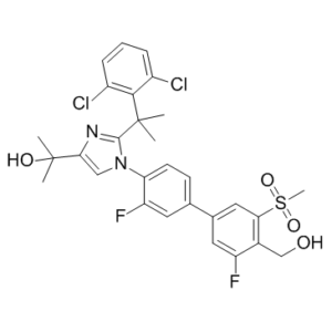
2-(2-(2-(2,6-dichlorophenyl)propan-2-yl)-1-(3,3′-difluoro-4′-(hydroxymethyl)-5′-(methylsulfonyl)biphenyl-4-yl)-1H-imidazol-4-yl)propan-2-ol
|
| 别名 |
BMS-852927; BMS 852927; BMS852927; XL041; 1256918-39-4; BMS-852,927; XL-652; EXEL-04541041; H9649L8MZN; UNII-H9649L8MZN;XL-041; XL 041.
|
| HS Tariff Code |
2934.99.9001
|
| 存储方式 |
Powder -20°C 3 years 4°C 2 years In solvent -80°C 6 months -20°C 1 month |
| 运输条件 |
Room temperature (This product is stable at ambient temperature for a few days during ordinary shipping and time spent in Customs)
|
| 溶解度 (体外实验) |
DMSO : ~100 mg/mL (~164.07 mM)
|
|---|---|
| 溶解度 (体内实验) |
配方 1 中的溶解度: ≥ 2.5 mg/mL (4.10 mM) (饱和度未知) in 10% DMSO + 40% PEG300 + 5% Tween80 + 45% Saline (这些助溶剂从左到右依次添加,逐一添加), 澄清溶液。
例如,若需制备1 mL的工作液,可将100 μL 25.0 mg/mL澄清DMSO储备液加入到400 μL PEG300中,混匀;然后向上述溶液中加入50 μL Tween-80,混匀;加入450 μL生理盐水定容至1 mL。 *生理盐水的制备:将 0.9 g 氯化钠溶解在 100 mL ddH₂O中,得到澄清溶液。 配方 2 中的溶解度: ≥ 2.5 mg/mL (4.10 mM) (饱和度未知) in 10% DMSO + 90% (20% SBE-β-CD in Saline) (这些助溶剂从左到右依次添加,逐一添加), 澄清溶液。 例如,若需制备1 mL的工作液,可将 100 μL 25.0 mg/mL澄清DMSO储备液加入900 μL 20% SBE-β-CD生理盐水溶液中,混匀。 *20% SBE-β-CD 生理盐水溶液的制备(4°C,1 周):将 2 g SBE-β-CD 溶解于 10 mL 生理盐水中,得到澄清溶液。 View More
配方 3 中的溶解度: ≥ 2.5 mg/mL (4.10 mM) (饱和度未知) in 10% DMSO + 90% Corn Oil (这些助溶剂从左到右依次添加,逐一添加), 澄清溶液。 1、请先配制澄清的储备液(如:用DMSO配置50 或 100 mg/mL母液(储备液)); 2、取适量母液,按从左到右的顺序依次添加助溶剂,澄清后再加入下一助溶剂。以 下列配方为例说明 (注意此配方只用于说明,并不一定代表此产品 的实际溶解配方): 10% DMSO → 40% PEG300 → 5% Tween-80 → 45% ddH2O (或 saline); 假设最终工作液的体积为 1 mL, 浓度为5 mg/mL: 取 100 μL 50 mg/mL 的澄清 DMSO 储备液加到 400 μL PEG300 中,混合均匀/澄清;向上述体系中加入50 μL Tween-80,混合均匀/澄清;然后继续加入450 μL ddH2O (或 saline)定容至 1 mL; 3、溶剂前显示的百分比是指该溶剂在最终溶液/工作液中的体积所占比例; 4、 如产品在配制过程中出现沉淀/析出,可通过加热(≤50℃)或超声的方式助溶; 5、为保证最佳实验结果,工作液请现配现用! 6、如不确定怎么将母液配置成体内动物实验的工作液,请查看说明书或联系我们; 7、 以上所有助溶剂都可在 Invivochem.cn网站购买。 |
| 制备储备液 | 1 mg | 5 mg | 10 mg | |
| 1 mM | 1.6407 mL | 8.2033 mL | 16.4066 mL | |
| 5 mM | 0.3281 mL | 1.6407 mL | 3.2813 mL | |
| 10 mM | 0.1641 mL | 0.8203 mL | 1.6407 mL |
1、根据实验需要选择合适的溶剂配制储备液 (母液):对于大多数产品,InvivoChem推荐用DMSO配置母液 (比如:5、10、20mM或者10、20、50 mg/mL浓度),个别水溶性高的产品可直接溶于水。产品在DMSO 、水或其他溶剂中的具体溶解度详见上”溶解度 (体外)”部分;
2、如果您找不到您想要的溶解度信息,或者很难将产品溶解在溶液中,请联系我们;
3、建议使用下列计算器进行相关计算(摩尔浓度计算器、稀释计算器、分子量计算器、重组计算器等);
4、母液配好之后,将其分装到常规用量,并储存在-20°C或-80°C,尽量减少反复冻融循环。
计算结果:
工作液浓度: mg/mL;
DMSO母液配制方法: mg 药物溶于 μL DMSO溶液(母液浓度 mg/mL)。如该浓度超过该批次药物DMSO溶解度,请首先与我们联系。
体内配方配制方法:取 μL DMSO母液,加入 μL PEG300,混匀澄清后加入μL Tween 80,混匀澄清后加入 μL ddH2O,混匀澄清。
(1) 请确保溶液澄清之后,再加入下一种溶剂 (助溶剂) 。可利用涡旋、超声或水浴加热等方法助溶;
(2) 一定要按顺序加入溶剂 (助溶剂) 。
InvivoChem的所有产品仅用于作科学研究,不面向患者销售
Copyright 2020 InvivoChem LLC | All Rights Reserved 粤ICP备20063088号-1


 COA
COA
 463611831
