| 规格 | 价格 | 库存 | 数量 |
|---|---|---|---|
| 10mg |
|
||
| 25mg |
|
||
| 50mg |
|
||
| 100mg |
|
||
| 250mg |
|
||
| Other Sizes |
|

| 靶点 |
Bax (IC50 = 250 nM); Bax (IC50 = 144 nM)
BTSA1: High-affinity and selective activator of BAX (BCL-2-associated X protein), binding to the N-terminal activation site (trigger site) of BAX; [1] |
|---|---|
| 体外研究 (In Vitro) |
BTSA1 没有能力直接激活促凋亡同系物 BAK。通过 BTSA1 处理,重组可溶性 BAX 被有效且剂量响应地易位至线粒体膜,随后诱导 BAX 寡聚化。当 BAX 被 BTSA1 激活时,癌细胞更有可能发生凋亡。 BTSA1 的 IC50 值范围在 1 至 4 μM 之间,以剂量依赖性方式降低所有 AML 细胞系的活力,并在治疗 24 小时内产生完全效果。所有五种 AML 细胞系均表现出剂量依赖性 caspase-3/7 激活[1]。
BAX激活及构象变化:BTSA1 高亲和力特异性结合BAX的N端触发位点(α1、α6区域)。竞争性荧光偏振结合实验显示,它可与FITC-BIM SAHB A2竞争结合BAX;直接荧光偏振结合实验证实其与BAX直接相互作用。对¹⁵N标记BAX的HSQC谱分析表明,BTSA1滴定后,BAX触发位点的残基出现显著主链酰胺化学位移变化,提示诱导BAX构象改变。对接研究显示,BTSA1与BAX形成疏水相互作用,其吡唑酮基团与BAX的K21残基形成关键氢键,模拟BIM BH3螺旋与BAX的相互作用模式[1] - 白血病细胞中BAX介导的凋亡:BTSA1 可诱导BAX激活通路的全步骤,包括BAX向膜转运、寡聚化及线粒体 outer membrane permeabilization(MOMP)。在ANTS/DPX脂质体实验中,BTSA1(100–400 nM)可促进BAX介导的膜通透,而BAX K21E突变体中该效应受损。在分离的小鼠肝脏线粒体中,BTSA1呈剂量依赖性诱导BAX线粒体转运、寡聚化及细胞色素c释放。它能有效诱导人AML细胞系(如NB4、OCI-AML3、THP-1、MOLM-13)和小鼠AML细胞系WEHI凋亡,表现为细胞活力下降、caspase 3/7活性升高、线粒体去极化(TMRE实验检测)及细胞色素c释放。Western blot证实BTSA1可诱导NB4细胞中BAX从胞质向线粒体转运[1] - 选择性及敏感性决定因素:竞争性结合实验显示,BTSA1 与抗凋亡BCL-2家族蛋白(BCL-XL、MCL-1、BFL-1)无显著结合,对非癌细胞系和正常造血祖细胞无明显毒性。BAX表达水平和胞质单体构象决定对BTSA1的敏感性:BAX mRNA/蛋白水平较高且胞质中存在单体BAX的AML细胞(如OCI-AML3、HPB-ALL)更敏感,而BAX敲除(KO)MEF细胞对其耐受。在BAX KO MEF细胞中重构野生型BAX可恢复敏感性,但触发位点功能受损的BAX突变体则不能。BTSA1 与Venetoclax(BCL-2抑制剂)在AML细胞系(THP-1、OCI-AML3)中具有协同作用,可增强caspase 3/7活性并降低细胞活力[1] - 患者AML样本中的药效:BTSA1 可诱导原发性人AML母细胞(n=4)和CD34⁺CD38⁻ AML干细胞富集群体(n=4)凋亡(Annexin V结合实验检测),且不影响健康CD34⁺CD38⁻造血干祖细胞(n=2)。AML患者细胞(n=542)的BAX mRNA表达水平显著高于健康对照细胞(n=74)[1] |
| 体内研究 (In Vivo) |
BTSA1 可有效抑制人类急性髓系白血病 (AML) 异种移植物并提高宿主存活率,且无毒性。健康的富含干细胞 (LSK) 细胞、常见骨髓祖细胞、粒细胞-单核细胞祖细胞和巨核细胞-红细胞祖细胞在小鼠中具有良好的耐受性,并且没有毒性作用。 10 mg/kg 剂量的 BTSA1 达到足够的水平 (~15 μM),引起白血病细胞中的 BAX 激活和凋亡,同时在小鼠血浆中也具有显着的半衰期 (T1/2 = 15 小时) 和口服生物利用度 (%F = 51)。因此,BTSA1 具有优异的药代动力学,可口服生物利用,通过诱导细胞凋亡显着抑制白血病异种移植物中的肿瘤生长,并且在治疗有效剂量下,在造血系统或其他组织中未表现出可检测到的毒性[1]。
AML异种移植模型中的抗肿瘤疗效:在THP-1人AML异种移植NSG小鼠中,BTSA1 治疗可显著抑制肝脏肿瘤负荷(减少人CD45⁺和CD15⁺细胞浸润)并延长宿主生存期(Kaplan-Meier生存曲线,n=7)。在MOLM-13人AML异种移植模型中,BTSA1 可降低骨髓和外周血中人CD45⁺CD15⁺细胞浸润(n=5)。骨髓切片免疫组织化学(IHC)染色显示,BTSA1 处理组的cleaved caspase-3阳性细胞和TUNEL阳性细胞增多,提示凋亡增强。对骨髓来源的人白血病细胞进行TMRE染色,证实线粒体去极化,与BAX介导的MOMP一致[1] - 体内安全性和耐受性:NSG小鼠接受BTSA1(15 mg/kg体重)治疗30天,体重与溶媒对照组相比无显著变化;外周血计数(红细胞、白细胞、血小板)维持在正常范围。主要器官(肝、脾、肾、肺、心、脑、骨髓)的苏木精-伊红(H&E)染色未显示组织病理学异常,表明无全身毒性[1] |
| 酶活实验 |
BAX相互作用的荧光偏振结合实验:
1. 竞争性结合实验:制备含FITC-BIM SAHB A2(与BAX结合)、系列浓度BTSA1 及实验缓冲液的反应体系,室温孵育特定时间后,检测荧光偏振值,评估BTSA1与FITC-BIM SAHB A2竞争结合BAX的能力;同时对BCL-XL、MCL-1、BFL-1进行平行实验以评估选择性。 2. 直接结合实验:使用荧光标记的BTSA1(F-BTSA1)与系列浓度BAX混合孵育,检测荧光偏振值以确定BTSA1与BAX的直接结合亲和力[1] - NMR-based BAX构象变化实验: 1. 制备¹⁵N标记的非活性BAX蛋白,溶解于适宜的NMR缓冲液中。 2. 向¹⁵N标记BAX溶液中滴定BTSA1 至摩尔比1:1,每步滴定后收集¹H-¹⁵N HSQC谱。 3. 分析BAX主链酰胺残基的化学位移变化,将显著变化映射至BAX结构,确定结合位点(触发位点)[1] - 脂质体膜通透实验: 1. 制备负载ANTS/DPX的脂质体,模拟线粒体外膜。 2. 将脂质体与200 nM BAX(或BAX K21E突变体)及系列浓度BTSA1(100–400 nM)共同孵育,以60 nM tBID作为阳性对照。 3. 实时检测脂质体释放的ANTS荧光强度,评估BTSA1诱导的BAX介导膜通透作用[1] |
| 细胞实验 |
将 AML 细胞(以 2 × 10⁴ 个细胞/孔接种)与 BTSA1 或 BTSA2 或载体 (0.15% DMSO) 在无 FBS 培养基中的连续稀释液一起孵育 2.5 小时,然后将 10% FBS 替代物添加至最终体积 100 μl。 24小时后,评估细胞活力。
细胞活力及凋亡实验: 1. 细胞培养:在适宜培养基中培养AML细胞系(NB4、OCI-AML3、THP-1、MOLM-13)、小鼠AML细胞系WEHI、非癌细胞系、MEF细胞(野生型、BAX KO、BAK KO)及重构MEF细胞;从患者样本和健康供体中分离原发性人AML母细胞、CD34⁺CD38⁻ AML细胞及健康造血祖细胞。 2. 药物处理:将细胞以适宜密度接种于96孔板,用系列浓度BTSA1(单独或与Venetoclax联合)处理6–24小时,设置溶媒对照和阳性对照组。 3. 活力检测:使用细胞活力检测试剂盒测量吸光度或发光值,计算细胞活力并确定IC50值。 4. 凋亡检测:对原代细胞进行Annexin V结合实验,对细胞系进行caspase 3/7活性实验,通过荧光或发光检测定量凋亡;采用TMRE染色结合流式细胞术评估线粒体去极化[1] - Western blot及免疫沉淀实验: 1. 蛋白提取:裂解处理后的细胞或分离胞质和线粒体组分,使用含蛋白酶抑制剂的裂解液提取总蛋白。 2. Western blot:通过SDS-PAGE分离蛋白,转移至膜上,封闭后加入针对BAX、细胞色素c、BCL-2、BCL-XL、MCL-1、Actin(胞质内参)、VDAC(线粒体内参)的抗体孵育,化学发光法检测信号。 3. 免疫沉淀:将细胞裂解液与BAX抗体或生物素标记的BTSA1 孵育,用蛋白A/G珠或链霉亲和素珠捕获免疫复合物,洗涤后进行Western blot检测BAX相互作用蛋白[1] - BAX单体检测的凝胶过滤层析实验: 1. 制备MEF、OCI-AML3、HPB-ALL细胞的胞质提取物。 2. 将提取物上样至Superdex 200 HR 10/30柱,用缓冲液洗脱,收集对应不同分子量的组分。 3. 对收集的组分进行BAX抗体Western blot,鉴定单体BAX(低分子量组分)[1] |
| 动物实验 |
配制于 1% DMSO、30% PEG-400、65% D5W(5% 葡萄糖溶液)、4% Tween-80 的溶液中;剂量为 10 mg/kg;口服和静脉注射。
NOD-SCID IL2Rg 基因敲除 (NSG) 小鼠/ICR (CD-1) 雄性小鼠,6-8 周龄。 AML 异种移植小鼠模型: 1. 动物准备:使用 6-8 周龄的 NSG 小鼠,实验前将其适应实验室环境 1 周。 2. 肿瘤细胞接种:通过尾静脉或腹腔注射将人 AML 细胞(THP-1 或 MOLM-13)注射到小鼠体内,建立异种移植模型。 3. 药物配制和给药:将 BTSA1 溶解于合适的溶剂(例如,DMSO/Cremophor/生理盐水)中,配制成适合注射的浓度。按照预定的给药方案(例如,每日一次,持续30天),通过腹腔注射向小鼠施用BTSA1,剂量为15 mg/kg体重。载体处理的小鼠作为对照。 4. 生存率和肿瘤负荷评估:对于THP-1异种移植瘤,每日监测小鼠的生存情况并绘制Kaplan-Meier生存曲线。对于MOLM-13和THP-1异种移植瘤,在指定时间点处死小鼠,并收集肝脏、骨髓和外周血样本。 5. 流式细胞术分析:用小鼠CD45、人CD45和人CD15抗体对样本进行染色,并通过流式细胞术分析以定量人AML细胞浸润。 6. IHC和TMRE检测:制备骨髓切片进行cleaved caspase-3和TUNEL IHC染色以检测细胞凋亡。对骨髓来源的人类白血病细胞进行TMRE染色,以评估线粒体去极化[1] - 体内安全性评估: 1. 动物处理:每日腹腔注射BTSA1(15 mg/kg体重)或载体,连续30天,给予NSG小鼠。 2. 生理监测:每周测量小鼠体重。治疗结束时,采集外周血进行全血细胞计数(红细胞、白细胞、血小板)。 3. 组织病理学分析:采集主要器官(肝脏、脾脏、肾脏、肺脏、心脏、脑、骨髓),用福尔马林固定,石蜡包埋,切片,并进行H&E染色。检查切片是否存在组织病理学异常[1] |
| 毒性/毒理 (Toxicokinetics/TK) |
体外毒性:BTSA1 对非癌细胞系和健康的人造血祖细胞毒性极低,在对 AML 细胞有效的浓度下,细胞活力未见显著降低 [1]
- 体内毒性:BTSA1(15 mg/kg 体重,治疗 30 天)不会引起 NSG 小鼠体重减轻、血液学异常或主要器官的组织病理学损伤。BAX KO MEF 细胞对 BTSA1 具有耐药性,证实其细胞毒性依赖于 BAX,并且对表达 BAX 的癌细胞具有选择性 [1] |
| 参考文献 | |
| 其他信息 |
作用机制:BTSA1 是一种经药理学优化的 BAX 激活剂,它与 BAX 的 N 端触发位点结合,诱导构象变化(膜靶向结构域暴露、寡聚化),从而导致 BAX 转位至线粒体、线粒体外膜通透性 (MOMP)、细胞色素 c 释放以及 caspase 依赖性细胞凋亡。其结合模式模拟 BIM BH3 螺旋,通过疏水相互作用并与 BAX K21 形成关键氢键 [1]。
- 治疗潜力:BTSA1 通过直接激活 BAX(细胞凋亡的关键介质)来克服急性髓系白血病 (AML) 中的凋亡抵抗。 BTSA1 在 AML 细胞系、患者样本(包括富集干细胞的细胞群)和异种移植模型中的疗效,以及其对癌细胞的高选择性和无全身毒性,为直接激活 BAX 作为一种治疗急性髓系白血病的新型策略提供了概念验证 [1] - 与维奈托克(Venetoclax)的协同作用:BTSA1 可与维奈托克(BCL-2 抑制剂)产生协同作用,通过释放 BAX 并直接激活 BAX,增强 AML 细胞的凋亡反应。这种联合疗法可能改善 AML 患者的治疗效果,特别是那些 BCL-2 过表达的患者 [1] |
| 分子式 |
C21H14N6OS2
|
|
|---|---|---|
| 分子量 |
430.51
|
|
| 精确质量 |
430.067
|
|
| 元素分析 |
C, 58.59; H, 3.28; N, 19.52; O, 3.72; S, 14.89
|
|
| CAS号 |
314761-14-3
|
|
| 相关CAS号 |
|
|
| PubChem CID |
3857348
|
|
| 外观&性状 |
Brown to reddish brown solid powder
|
|
| 密度 |
1.5±0.1 g/cm3
|
|
| 沸点 |
625.2±48.0 °C at 760 mmHg
|
|
| 闪点 |
331.9±29.6 °C
|
|
| 蒸汽压 |
0.0±1.8 mmHg at 25°C
|
|
| 折射率 |
1.791
|
|
| LogP |
3.68
|
|
| 氢键供体(HBD)数目 |
1
|
|
| 氢键受体(HBA)数目 |
8
|
|
| 可旋转键数目(RBC) |
5
|
|
| 重原子数目 |
30
|
|
| 分子复杂度/Complexity |
689
|
|
| 定义原子立体中心数目 |
0
|
|
| SMILES |
S1C([H])=C(C2C([H])=C([H])C([H])=C([H])C=2[H])N=C1N1C(C(=C(C2C([H])=C([H])C([H])=C([H])C=2[H])N1[H])/N=N/C1=NC([H])=C([H])S1)=O
|
|
| InChi Key |
CTRCXGFSYFTJIW-UHFFFAOYSA-N
|
|
| InChi Code |
InChI=1S/C21H14N6OS2/c28-19-18(24-25-20-22-11-12-29-20)17(15-9-5-2-6-10-15)26-27(19)21-23-16(13-30-21)14-7-3-1-4-8-14/h1-13,26H
|
|
| 化学名 |
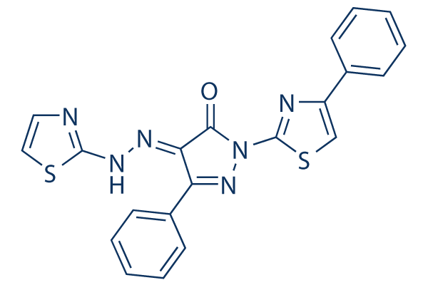
5-phenyl-2-(4-phenyl-1,3-thiazol-2-yl)-4-(1,3-thiazol-2-yldiazenyl)-1H-pyrazol-3-one
|
|
| 别名 |
|
|
| HS Tariff Code |
2934.99.9001
|
|
| 存储方式 |
Powder -20°C 3 years 4°C 2 years In solvent -80°C 6 months -20°C 1 month |
|
| 运输条件 |
Room temperature (This product is stable at ambient temperature for a few days during ordinary shipping and time spent in Customs)
|
| 溶解度 (体外实验) |
|
|---|
| 制备储备液 | 1 mg | 5 mg | 10 mg | |
| 1 mM | 2.3228 mL | 11.6141 mL | 23.2283 mL | |
| 5 mM | 0.4646 mL | 2.3228 mL | 4.6457 mL | |
| 10 mM | 0.2323 mL | 1.1614 mL | 2.3228 mL |
1、根据实验需要选择合适的溶剂配制储备液 (母液):对于大多数产品,InvivoChem推荐用DMSO配置母液 (比如:5、10、20mM或者10、20、50 mg/mL浓度),个别水溶性高的产品可直接溶于水。产品在DMSO 、水或其他溶剂中的具体溶解度详见上”溶解度 (体外)”部分;
2、如果您找不到您想要的溶解度信息,或者很难将产品溶解在溶液中,请联系我们;
3、建议使用下列计算器进行相关计算(摩尔浓度计算器、稀释计算器、分子量计算器、重组计算器等);
4、母液配好之后,将其分装到常规用量,并储存在-20°C或-80°C,尽量减少反复冻融循环。
计算结果:
工作液浓度: mg/mL;
DMSO母液配制方法: mg 药物溶于 μL DMSO溶液(母液浓度 mg/mL)。如该浓度超过该批次药物DMSO溶解度,请首先与我们联系。
体内配方配制方法:取 μL DMSO母液,加入 μL PEG300,混匀澄清后加入μL Tween 80,混匀澄清后加入 μL ddH2O,混匀澄清。
(1) 请确保溶液澄清之后,再加入下一种溶剂 (助溶剂) 。可利用涡旋、超声或水浴加热等方法助溶;
(2) 一定要按顺序加入溶剂 (助溶剂) 。
Cancer Cell.2017 Oct 9;32(4):490-505.e10. |
|---|
BTSA1 Is a High-Affinity and Selective BAX Trigger Site Activator.Cancer Cell.2017 Oct 9;32(4):490-505.e10. |
BTSA1 Induces All Steps of the BAX Activation Pathway.Cancer Cell.2017 Oct 9;32(4):490-505.e10. |
BTSA1 Induces Robust and Rapid BAX-Mediated Apoptosis.Cancer Cell.2017 Oct 9;32(4):490-505.e10. |
|---|
Specificity of BTSA1 for Cellular BAX, Cytosolic BAX Monomer, and the BAX Trigger Site.Cancer Cell.2017 Oct 9;32(4):490-505.e10. |
BTSA1 Is Effective against Patient AML Blasts and Pre-leukemic Stem Cells without Affecting Normal Hematopoietic Progenitor Cells and Demonstrates Significant Synergy with Venetoclax.Cancer Cell.2017 Oct 9;32(4):490-505.e10. |
BTSA1 Demonstrates Potent Efficacy in Killing Human AML In Vivo.Cancer Cell.2017 Oct 9;32(4):490-505.e10. |
|---|
BTSA1 Is Well Tolerated without Toxicity to Normal Cells In Vivo.Cancer Cell.2017 Oct 9;32(4):490-505.e10. |
 PROTAC Bcl-xL degrader-4
 BCL-XL-IN-4
 BAD (103-127) (human), FAM-labeled TFA
 PROTAC BCL-xL/BCL-w Degrader 1
InvivoChem的所有产品仅用于作科学研究,不面向患者销售
Copyright 2020 InvivoChem LLC | All Rights Reserved 粤ICP备20063088号-1


 COA
COA


 463611831
