| 规格 | 价格 | 库存 | 数量 |
|---|---|---|---|
| 5mg |
|
||
| 10mg |
|
||
| 25mg |
|
||
| 50mg |
|
||
| 100mg |
|
||
| 250mg |
|
||
| Other Sizes |
|
| 靶点 |
MIP-1α-CCR1 ( Ki = 1 nM ); RANTES-CCR1 ( Ki = 2.8 nM ); MCP-3-CCR1 ( Ki = 5.5 nM )
CC chemokine receptor 1 (CCR1) (Ki = 1.1 nM for human CCR1; IC₅₀ = 1.8 nM for inhibiting CCL3 binding to human CCR1; IC₅₀ = 2.5 nM for inhibiting CCL5 binding to human CCR1; IC₅₀ = 3.7 nM for inhibiting CCR1-mediated calcium mobilization; IC₅₀ = 5.2 nM for inhibiting CCR1-dependent chemotaxis) [1] |
|
|---|---|---|
| 体外研究 (In Vitro) |
体外活性:BX471(也称为 ZK-811752)是一种新型口服非肽 CCR1(CC 趋化因子受体-1)拮抗剂,对人 CCR1 的 Ki 为 1 nM,可用于治疗慢性炎症性疾病。 BX471 对 CCR1 的选择性是 CCR2、CCR5 和 CXCR4 的 250 倍。 CCR1是治疗自身免疫性疾病的主要治疗靶点。与 28 G 蛋白偶联受体相比,BX 471 对 CCR1 的选择性高出 10,000 倍以上。竞争结合研究表明,BX 471 能够以高亲和力(K(i) 范围为 1 nm)取代 CCR1 配体巨噬细胞炎症蛋白-1α (MIP-1α)、RANTES 和单核细胞趋化蛋白-3 (MCP-3)至 5.5 nm)。 BX 471 是一种有效的功能拮抗剂,因为它能够抑制许多 CCR1 介导的效应,包括 Ca(2+) 动员、细胞外酸化率增加、CD11b 表达和白细胞迁移。此外,BX 471 能有效减少多发性硬化症大鼠实验性过敏性脑脊髓炎模型中的疾病。激酶测定:BX471(也称为 ZK-811752)是一种新型口服非肽 CCR1(CC 趋化因子受体-1)拮抗剂,对人 CCR1 的 Ki 为 1 nM,可能可用于治疗慢性炎症性疾病。 BX471 对 CCR1 的选择性是 CCR2、CCR5 和 CXCR4 的 250 倍。 CCR1是治疗自身免疫性疾病的主要治疗靶点。细胞测定:BX471 (0.1-10 μM) 在分离的血液单核细胞的剪切流中显示出对 RANTES 介导的和对 IL-1β 激活的微血管内皮的抗剪切粘附的剂量依赖性抑制。 BX471 还抑制 RANTES 介导的 T 淋巴细胞与活化内皮细胞的粘附。BX471 还能够以浓度依赖性方式取代 125I-MIP-1α/CCL3 与小鼠 CCR1 的结合,Ki 为 215±46 nM。增加 BX471 浓度可抑制人和小鼠 CCR1 中 MIP-1α/CCL3 诱导的 Ca2+ 瞬变,IC50 分别为 5.8±1 nM 和 198±7 nM。BX 471 是一种有效的功能拮抗剂,因为它能够抑制CCR1 介导的效应包括 Ca2+ 动员、细胞外酸化率增加、CD11b 表达和白细胞迁移。与 28 个 G 蛋白偶联受体相比,BX 471 对 CCR1 的选择性高出 10,000 倍以上。
受体结合抑制:BX-471 HCl以剂量依赖性方式竞争性抑制放射性标记的CCL3(MIP-1α)和CCL5(RANTES)与人CCR1表达细胞的结合,IC₅₀值分别为1.8 nM和2.5 nM。对其他趋化因子受体(CCR2、CCR3、CCR4、CXCR1、CXCR2)的选择性>1000倍,所有受体的IC₅₀均>10 μM [1] - 功能活性抑制:BX-471 HCl抑制CCL3、CCL5和CCL9(MIP-1γ)诱导的CCR1介导的钙流,IC₅₀值分别为3.7 nM、4.1 nM和5.0 nM。它还可抑制人外周血单核细胞向CCL3的CCR1依赖趋化,10 nM浓度下迁移减少85%,100 nM浓度下达到最大抑制(92%)[1] - 无脱靶活性:在浓度高达10 μM时,BX-471 HCl不影响所测试的其他趋化因子受体、G蛋白偶联受体(GPCR)或离子通道的结合或功能[1] |
|
| 体内研究 (In Vivo) |
BX471(20 mg/kg,皮下注射)在约 30 分钟内达到峰值血浆水平 9 μM,并在 2 小时后迅速下降至约 0.4 μM。 4 至 8 小时后,血浆药物水平降至 0.1 μM 或更低。用 20 mg/kg BX471 治疗 10 天的小鼠显示间质 CD45 阳性白细胞减少约 55%。 BX471 对外周血中 CCR5 阳性 CD8 细胞的数量具有临界显着影响。与载体对照相比,BX471 使 UUO 肾脏中 FSP1 阳性细胞的数量减少 65%。用 BX471 预处理可减少缺血再灌注损伤后肾脏中巨噬细胞和中性粒细胞的积累。BX 471(4 mg/kg,口服或静脉注射)是口服有效,狗体内生物利用度为 60%。此外,BX 471 能有效减少多发性硬化症大鼠实验性过敏性脑脊髓炎模型中的疾病。
大鼠单侧输尿管结扎(UUO)诱导的肾纤维化模型:UUO术后24小时开始,口服给予BX-471 HCl(10 mg/kg,每日一次,持续14天),与溶媒对照组相比,显著减轻肾间质纤维化,α-平滑肌肌动蛋白(α-SMA)表达降低45%,胶原沉积(Masson三色染色)减少52%。同时减少CD45+白细胞(减少40%)和CD68+巨噬细胞(减少55%)的间质浸润[2] - 大鼠肾缺血再灌注(I/R)损伤模型:缺血前1小时及再灌注后每日腹腔注射BX-471 HCl(10 mg/kg),持续3天,可减轻肾损伤。中性粒细胞浸润(MPO活性降低60%)和单核细胞/巨噬细胞聚集(CD68+细胞减少50%),血清肌酐水平降低35%,血尿素氮(BUN)水平降低40%[3] - 大鼠心脏移植排斥模型:移植当天开始,口服给予BX-471 HCl(10 mg/kg,每日两次),可将心脏移植物存活时间从溶媒组的7±1天延长至21±3天。减少间质单核细胞浸润(减少65%),并抑制移植物组织中促炎细胞因子(TNF-α、IFN-γ)的表达[4] |
|
| 酶活实验 |
趋化因子结合研究[1]
如前所述,通过过滤进行结合分析。使用终浓度约为0.1-0.2 nm的放射性标记趋化因子作为配体。使用每个测定点8000或300000个细胞表达人CCR1的HEK293细胞作为受体来源。在存在100nm未标记趋化因子的情况下测定非特异性结合。结合数据用计算机程序IGOR进行曲线拟合,以确定亲和力和位点数量。 胞浆Ca2+测量[1] 将表达人CCR1的HEK293细胞以80000个细胞/孔的速度铺在聚-d-赖氨酸涂覆的黑壁96孔板上,并培养过夜。然后,在Hanks的平衡盐溶液中,在37°C下用4μm Fluo-3(一种钙敏感的荧光染料)装载细胞60分钟,该溶液含有20 mm Hepes、3.2 mm氯化钙、1%胎牛血清、2.5 mm丙磺舒和0.04%普朗尼克酸。使用Denley洗涤器用测定缓冲液(Hanks平衡盐溶液,含有20mmHepes、2.5mm丙磺舒和0.1%牛血清白蛋白)轻轻洗涤细胞4次,去除多余的染料。在37°C下加入激动剂后,立即用FLIPR测量细胞内游离Ca2+浓度的变化。为了检测BX471的拮抗活性,在加入激动剂之前,用该化合物对细胞进行15分钟的预处理。根据方程式Ca2+=K D(F−F min)/(F max−F)计算细胞内Ca2+浓度(nm)(7)。K D是Fluo-3和Ca2+复合物的离解常数(Fluo-3为390 nm)。F是测量的荧光强度。F max是在0.1%triton X-100存在的情况下测定的最大荧光强度。F min是在0.1%曲拉通X-100加5 mmEGTA存在下测定的最小荧光强度。 BX471(也称为 ZK-811752)是一种新型口服非肽 CCR1(CC 趋化因子受体-1)拮抗剂,对人 CCR1 的 Ki 为 1 nM。它可能有助于治疗慢性炎症。与 CCR2、CCR5 和 CXCR4 相比,BX471 对 CCR1 的偏好程度高出 250 倍。在治疗自身免疫性疾病时,CCR1 是首要治疗靶点。 CCR1放射性配体结合实验:将表达人CCR1的CHO细胞制备细胞膜并悬浮于结合缓冲液(三羟甲基氨基甲烷-盐酸、氯化镁、牛血清白蛋白)中。将系列稀释(0.001–1000 nM)的BX-471 HCl与细胞膜及氚标记的CCL3或CCL5混合,25°C孵育90分钟后,通过玻璃纤维滤膜过滤分离结合态与游离态配体。闪烁计数器测量放射性强度,通过置换曲线计算Ki/IC₅₀值[1] - CCR1介导的钙流检测实验:表达人CCR1的CHO细胞用钙敏感荧光染料负载30分钟(37°C)。BX-471 HCl(0.01–100 nM)与细胞预孵育15分钟后,加入CCL3(10 nM)刺激。实时测量荧光强度评估钙流,从剂量-反应曲线推导IC₅₀值[1] - 趋化因子诱导的趋化实验:分离人外周血单核细胞并悬浮于趋化缓冲液中。BX-471 HCl(0.1–100 nM)与单核细胞混合后加入Transwell上室,下室加入CCL3(10 nM),37°C孵育2小时。计数下室中的迁移细胞,计算相对于溶媒对照组的抑制率[1] |
|
| 细胞实验 |
总之,在培养皿中培养至汇合的真皮微血管内皮细胞用 IL-1β (10 ng/mL) 刺激持续 12 小时,并在检测前立即与 RANTES (10 nM) 预孵育37°C 30 分钟。将板安装在带有×20和×40相衬物镜的Olympus IMT-2倒置显微镜的载物台上,并将它们组装为平行壁流室的下壁。将分离的人血单核细胞以 5×10⁵ 细胞/mL 的密度重悬于含有 0.5% 人血清白蛋白、10 mM HEPES、pH 7.4 的测定缓冲液 (HBSS) 中。在测定前不久添加 1 mM Mg2+ 和 1 mM Ca2+。将细胞悬浮液以 1.5 dyn/cm2 的速率灌注到流动室中五分钟,同时将其保持在 37°C 的加热块中进行测定。进行抑制实验的单核细胞首先与不同浓度 (0.1–10 μM) 的 Me2SO 对照或 BX471 在 37°C 下预孵育 10 分钟。以细胞/mm2 表示,5 分钟后,通过使用 JVC SR L 900 E 录像机和长镜头进行图像分析,在多个视野(每个实验至少 5 个)中量化牢固贴壁细胞的数量。集成JVC 3CCD 摄像机。原发性粘附,或单核细胞和内皮细胞之间的直接相互作用,是唯一被检查的粘附类型。
BX471 (0.1–10 μM) 以剂量依赖性方式抑制分离血液单核细胞剪切流中 IL-1β 激活的微血管内皮的抗剪切和 RANTES 介导的粘附。此外,BX471 还可抑制 T 细胞 RANTES 介导的与活化内皮细胞的粘附。 BX471 的 Ki 为 215±46 nM,还可以以浓度依赖性方式取代 125I-MIP-1α/CCL3 与小鼠 CCR1 的结合。 BX471 在人和小鼠 CCR1 中抑制 MIP-1α/CCL3 诱导的 Ca2+ 瞬变,随着化合物浓度的增加,IC50 值分别为 5.8±1 nM 和 198±7 nM。 BX 471 能够阻断多种 CCR1 介导的过程,例如白细胞迁移、细胞外酸化率增加、Ca2+ 动员和 CD11b 表达,使其成为强大的功能拮抗剂。 BX 471 对 CCR1 的选择性比 28 G 蛋白偶联受体高 10,000 倍以上。 外周血单个核细胞CD11b的表达[1] 如上所述测量全血测定中外周血单核细胞上表达的CD11b。简而言之,通过静脉穿刺将人全血收集到含有EDTA的2.5ml Vacutainer管中。血液保持在室温下,抽血后立即使用。全血样本(200μl)在37°C下用或不用1μmBX471预处理15分钟,然后用或不用100 nm MIP-1α再处理15分钟。通过加入1 ml冷磷酸盐缓冲盐溶液洗涤液终止反应。试管离心(200×g,4°C下7分钟),通过抽吸去除上清液。将细胞沉淀重新悬浮在冷磷酸盐缓冲盐溶液中,加入10μl 1mg/ml热聚集IgG,并在4°C下孵育试管10分钟。将抗体CD11b FITC(5μl)和CD14 PE(20μl)加入每个检测管中,在4°C下孵育20分钟。最后,加入1ml冰冷的磷酸盐缓冲盐溶液,如上所述将细胞造粒,并通过FACScan进行分析。 CCR1结合特异性实验:将表达人CCR1、CCR2、CCR3、CXCR1或CXCR2的CHO细胞接种到96孔板,孵育过夜。加入系列稀释的BX-471 HCl(0.001–1000 nM)及氚标记的CCL3(针对CCR1)或受体特异性放射性配体(针对其他受体),25°C孵育90分钟。洗涤后测量结合放射性,评估结合亲和力和选择性[1] - 单核细胞趋化抑制实验:分离的人单核细胞与BX-471 HCl(0.1–100 nM)在37°C下预孵育15分钟。将细胞加入Transwell插入物(5 μm孔径),置于含CCL3(10 nM)的孔上方。孵育2小时后移除插入物,用血细胞计数板计数下孔中的迁移细胞,计算相对于未处理细胞的抑制百分比[1] - 钙流特异性实验:表达不同GPCR(肾上腺素能、毒蕈碱、血清素受体)的CHO细胞负载荧光钙染料,用BX-471 HCl(10 μM)预孵育15分钟后,加入受体特异性激动剂刺激。测量荧光强度以确认对钙信号无脱靶效应[1] |
|
| 动物实验 |
|
|
| 药代性质 (ADME/PK) |
BX 471 在犬体内的药代动力学 [1]
本研究考察了 BX 471 在清醒犬体内的口服生物利用度。将 BX 471 以 4 mg/kg 的剂量溶于 40% 环糊精生理盐水中,通过头静脉推注或灌胃的方式给予空腹雄性比格犬。制备血浆样本,并采用高效液相色谱-质谱联用(HPLC-MS)法测定血浆中化合物的浓度。如图 9 所示,BX 471 在口服给药后约 2 小时达到血浆峰浓度,并维持在可测量的浓度长达 6 小时。BX 471 的分布容积(0.5 L/kg)接近体液容积(0.6 L/kg),表明该化合物主要分布于体液中(表 III)。犬体内清除率较低,为 2 ml/min/kg(不足肝脏总血流量的 10%),导致其终末半衰期中等,为 3 小时(图 9 和表 III)。对于口服给药的犬,BX 471 的半衰期约为 3 小时。使用 TOPFIT 软件分析获得的曲线下面积测量值计算口服生物利用度百分比表明,BX 471 在空腹犬中是一种口服吸收的药物,口服生物利用度约为 60%(图 9 和表 III)。 在大鼠中:口服 BX-471 HCl (10 mg/kg) 导致血浆峰浓度 (Cₘₐₓ) 为 1.2 μg/mL,达峰时间 (Tₘₐₓ) 为 1 小时,末端半衰期 (t₁/₂) 为 4.5 小时,分布容积 (Vd) 为 2.3 L/kg。口服生物利用度为 40% [1] - 体外代谢:人肝微粒体研究表明,BX-471 HCl 的代谢极少,固有清除率 (CLint) 为 12 μL/min/mg 蛋白 [1] - 组织分布:大鼠口服给药后,BX-471 HCl 分布于炎症组织(肾脏、心脏),给药后 2 小时,肾脏的组织/血浆比为 2.1,心脏的组织/血浆比为 1.8 [1] |
|
| 毒性/毒理 (Toxicokinetics/TK) |
BX 471 对全身毒性的影响 [1]
为证明 BX 471 对 CCR1 的拮抗作用并非由该化合物的细胞毒性引起,我们用浓度高达 10 μM 的 BX 471 处理 THP-1 或 CCR1 转染的 HEK293 细胞 24 小时,并通过 WST-1 染色监测细胞毒性。未观察到明显的毒性(数据未显示)。我们进一步在体内检测了 BX 471 的毒性,方法是对连续 30 天以 20 mg/kg/天剂量给予 BX 471 的兔子进行一系列血清诊断试验,包括肝肾功能试验和血电解质检测。所有检测结果均在正常范围内(数据未显示)。结果表明,BX 471 对 CCR1 活化的抑制并非由于细胞毒性所致,且该药物的长期治疗对动物的正常生理功能无不良影响。 急性毒性:在大鼠中,BX-471 HCl 的口服 LD₅₀ >200 mg/kg,在剂量高达 100 mg/kg 时未观察到明显的毒性(惊厥、呼吸抑制、体重减轻)[1] 亚慢性毒性:在一项为期 14 天的大鼠重复给药研究(10 mg/kg/天,口服)中,BX-471 HCl 未引起体重、食物摄入量、血液学参数或肝肾功能的显著变化。主要器官(肝、肾、心、肺、脾)未发现组织病理学异常[2][3] - 血浆蛋白结合率:BX-471 HCl在人血浆中的血浆蛋白结合率为94%,在大鼠血浆中的血浆蛋白结合率为92%(超滤法测定)[1] - 药物相互作用:体外研究表明,浓度高达10 μM时,未观察到对细胞色素P450酶(CYP1A2、CYP2C9、CYP2D6、CYP3A4)的抑制作用[1] |
|
| 参考文献 |
|
|
| 其他信息 |
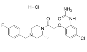
CC趋化因子受体1 (CCR1) 是治疗自身免疫性疾病的主要靶点。通过高通量筛选和化学优化,我们鉴定出一种新型非肽类CCR1拮抗剂,即RN-[5-氯-2-[2-[4-[(4-氟苯基)甲基]-2-甲基-1-哌嗪基]-2-氧代乙氧基]苯基]脲盐酸盐 (BX 471)。竞争性结合实验表明,BX 471能够以高亲和力(Ki范围为1 nM至5.5 nM)置换CCR1配体巨噬细胞炎症蛋白-1α (MIP-1α)、RANTES和单核细胞趋化蛋白-3 (MCP-3)。 BX 471 是一种强效的功能性拮抗剂,其抑制多种 CCR1 介导的效应,包括 Ca²⁺ 动员、细胞外酸化率增加、CD11b 表达和白细胞迁移。与 28 种 G 蛋白偶联受体相比,BX 471 对 CCR1 的选择性高出 10,000 倍以上。药代动力学研究表明,BX 471 具有口服活性,在犬体内的生物利用度为 60%。此外,BX 471 能有效减轻大鼠实验性变应性脑脊髓炎(多发性硬化症)模型中的疾病症状。本研究首次证实,非肽类趋化因子受体拮抗剂在自身免疫性疾病动物模型中具有疗效。总之,我们发现了一种强效、选择性高且口服有效的 CCR1 拮抗剂,该药物可能有助于治疗慢性炎症性疾病。 [1]趋化因子及其受体的表达被认为与单侧输尿管梗阻(UUO)后的白细胞浸润和进行性肾纤维化有关。我们假设使用非肽类拮抗剂BX471阻断趋化因子受体CCR1可以减少UUO后的白细胞浸润和肾纤维化。经BX471治疗的小鼠(第0-10天和第6-10天)的UUO肾脏显示,与对照组相比,间质巨噬细胞和淋巴细胞浸润减少了40-60%。治疗组小鼠的CCR1和CCR5 mRNA水平也显著降低,FACS分析显示CD8+/CCR5+ T细胞数量也相应减少。与载体对照组相比,BX471治疗显著降低了肾纤维化标志物,例如间质成纤维细胞、间质体积以及I型胶原蛋白的mRNA和蛋白表达。相反,仅在第0至5天给药则无效。总之,阻断CCR1可显著减少UUO后的细胞浸润和肾纤维化。最重要的是,延迟给药也有效。因此,我们得出结论,阻断CCR1可能代表一种新的治疗策略,用于减少细胞浸润和肾纤维化,而细胞浸润和肾纤维化是导致终末期肾衰竭进展的主要因素。[2]
中性粒细胞和巨噬细胞在肾脏缺血再灌注损伤后迅速浸润肾脏,但这些细胞类型的具体分子募集机制尚未完全阐明。本文通过遗传学和药理学证据,证实趋化因子受体CCR1在7天小鼠肾脏缺血再灌注损伤模型中对巨噬细胞和中性粒细胞浸润起积极作用。到第7天,CCR1缺失小鼠受损肾脏中的中性粒细胞和巨噬细胞数量分别比野生型对照小鼠减少35%和45%。用特异性CCR1拮抗剂BX471预处理野生型小鼠也能抑制该模型中的中性粒细胞和巨噬细胞浸润。与野生型对照小鼠相比,CCR1缺失小鼠受损肾脏中CCR1配体CCL3(MIP-1α)和CCL5(RANTES)的含量也降低,提示这些炎症趋化因子来源于白细胞,并且该模型中存在CCR1依赖的白细胞浸润正反馈环路。损伤后检测到局部白细胞增殖和凋亡,但这些过程并不依赖于CCR1。此外,野生型小鼠和CCR1缺陷型小鼠受损肾脏的坏死和纤维化损伤程度以及肾功能下降情况相似。因此,在肾脏缺血再灌注损伤的小鼠模型中,CCR1似乎调节巨噬细胞和中性粒细胞向肾脏的迁移,但这种活性似乎并不影响组织损伤。[3] 趋化因子如RANTES似乎在器官移植排斥反应中发挥作用。由于RANTES是趋化因子受体CCR1的强效激动剂,我们研究了CCR1受体拮抗剂BX471在大鼠异位心脏移植排斥模型中的疗效。 BX471联合亚治疗剂量环孢素(2.5 mg/kg)治疗动物,虽然单独使用环孢素或BX471治疗并不能有效延长移植排斥反应,但其疗效远优于单独使用环孢素或BX471治疗动物。我们利用体外微血管内皮细胞流动实验研究了CCR1拮抗剂的作用机制,发现该拮抗剂能够阻断RANTES诱导的单核细胞在炎症内皮细胞上的牢固黏附。综上所述,这些数据表明CCR1在同种异体移植排斥反应中发挥着重要作用。 [4] BX-471 HCl是一种强效、选择性、口服有效的非肽类CC趋化因子受体1 (CCR1)拮抗剂,旨在阻断促炎趋化因子(CCL3、CCL5、CCL9)与CCR1的结合[1] - 其核心作用机制是抑制CCR1介导的炎症细胞(单核细胞、中性粒细胞、T细胞)的募集和活化,从而减轻炎症和组织纤维化[1][2][3][4] - 临床前数据支持其在炎症和纤维化疾病(肾纤维化)、器官移植排斥反应和缺血再灌注损伤中的潜在治疗用途[2][3][4] - BX-471 HCl对CCR1的选择性远高于其他趋化因子受体和G蛋白偶联受体(GPCR),从而最大限度地减少了脱靶副作用[1] - 口服BX-471 HCl 具有良好的生物利用度和药代动力学特性,使其适用于临床长期口服给药[1] |
| 分子式 |
C21H25CL2FN4O3
|
|
|---|---|---|
| 分子量 |
471.35
|
|
| 精确质量 |
470.129
|
|
| 元素分析 |
C, 53.51; H, 5.35; Cl, 15.04; F, 4.03; N, 11.89; O, 10.18
|
|
| CAS号 |
288262-96-4
|
|
| 相关CAS号 |
BX471; 217645-70-0
|
|
| PubChem CID |
5311124
|
|
| 外观&性状 |
White to yellow solid powder
|
|
| LogP |
4.346
|
|
| tPSA |
88.89
|
|
| 氢键供体(HBD)数目 |
3
|
|
| 氢键受体(HBA)数目 |
5
|
|
| 可旋转键数目(RBC) |
6
|
|
| 重原子数目 |
31
|
|
| 分子复杂度/Complexity |
591
|
|
| 定义原子立体中心数目 |
1
|
|
| SMILES |
O=C(N)NC1=CC(Cl)=CC=C1OCC(N2[C@H](C)CN(CC3=CC=C(F)C=C3)CC2)=O.[H]Cl
|
|
| InChi Key |
FRUCNQBAWUHKLS-PFEQFJNWSA-N
|
|
| InChi Code |
InChI=1S/C21H24ClFN4O3.ClH/c1-14-11-26(12-15-2-5-17(23)6-3-15)8-9-27(14)20(28)13-30-19-7-4-16(22)10-18(19)25-21(24)29;/h2-7,10,14H,8-9,11-13H2,1H3,(H3,24,25,29);1H/t14-;/m1./s1
|
|
| 化学名 |
[5-chloro-2-[2-[(2R)-4-[(4-fluorophenyl)methyl]-2-methylpiperazin-1-yl]-2-oxoethoxy]phenyl]urea;hydrochloride
|
|
| 别名 |
|
|
| HS Tariff Code |
2934.99.9001
|
|
| 存储方式 |
Powder -20°C 3 years 4°C 2 years In solvent -80°C 6 months -20°C 1 month 注意: 请将本产品存放在密封且受保护的环境中,避免吸湿/受潮。 |
|
| 运输条件 |
Room temperature (This product is stable at ambient temperature for a few days during ordinary shipping and time spent in Customs)
|
| 溶解度 (体外实验) |
|
|||
|---|---|---|---|---|
| 溶解度 (体内实验) |
配方 1 中的溶解度: ≥ 3 mg/mL (6.36 mM) (饱和度未知) in 10% DMSO + 40% PEG300 + 5% Tween80 + 45% Saline (这些助溶剂从左到右依次添加,逐一添加), 澄清溶液。
例如,若需制备1 mL的工作液,可将100 μL 30.0 mg/mL 澄清的 DMSO 储备液加入到400 μL PEG300中,混匀;再向上述溶液中加入50 μL Tween-80,混匀;然后加入450 μL 生理盐水定容至1 mL。 *生理盐水的制备:将 0.9 g 氯化钠溶解在 100 mL ddH₂O中,得到澄清溶液。 配方 2 中的溶解度: ≥ 3 mg/mL (6.36 mM) (饱和度未知) in 10% DMSO + 90% (20% SBE-β-CD in Saline) (这些助溶剂从左到右依次添加,逐一添加), 澄清溶液。 例如,若需制备1 mL的工作液,可将 100 μL 30.0 mg/mL澄清DMSO储备液加入900 μL 20% SBE-β-CD生理盐水溶液中,混匀。 *20% SBE-β-CD 生理盐水溶液的制备(4°C,1 周):将 2 g SBE-β-CD 溶解于 10 mL 生理盐水中,得到澄清溶液。 View More
配方 3 中的溶解度: ≥ 3 mg/mL (6.36 mM) (饱和度未知) in 10% DMSO + 90% Corn Oil (这些助溶剂从左到右依次添加,逐一添加), 澄清溶液。 1、请先配制澄清的储备液(如:用DMSO配置50 或 100 mg/mL母液(储备液)); 2、取适量母液,按从左到右的顺序依次添加助溶剂,澄清后再加入下一助溶剂。以 下列配方为例说明 (注意此配方只用于说明,并不一定代表此产品 的实际溶解配方): 10% DMSO → 40% PEG300 → 5% Tween-80 → 45% ddH2O (或 saline); 假设最终工作液的体积为 1 mL, 浓度为5 mg/mL: 取 100 μL 50 mg/mL 的澄清 DMSO 储备液加到 400 μL PEG300 中,混合均匀/澄清;向上述体系中加入50 μL Tween-80,混合均匀/澄清;然后继续加入450 μL ddH2O (或 saline)定容至 1 mL; 3、溶剂前显示的百分比是指该溶剂在最终溶液/工作液中的体积所占比例; 4、 如产品在配制过程中出现沉淀/析出,可通过加热(≤50℃)或超声的方式助溶; 5、为保证最佳实验结果,工作液请现配现用! 6、如不确定怎么将母液配置成体内动物实验的工作液,请查看说明书或联系我们; 7、 以上所有助溶剂都可在 Invivochem.cn网站购买。 |
| 制备储备液 | 1 mg | 5 mg | 10 mg | |
| 1 mM | 2.1216 mL | 10.6078 mL | 21.2157 mL | |
| 5 mM | 0.4243 mL | 2.1216 mL | 4.2431 mL | |
| 10 mM | 0.2122 mL | 1.0608 mL | 2.1216 mL |
1、根据实验需要选择合适的溶剂配制储备液 (母液):对于大多数产品,InvivoChem推荐用DMSO配置母液 (比如:5、10、20mM或者10、20、50 mg/mL浓度),个别水溶性高的产品可直接溶于水。产品在DMSO 、水或其他溶剂中的具体溶解度详见上”溶解度 (体外)”部分;
2、如果您找不到您想要的溶解度信息,或者很难将产品溶解在溶液中,请联系我们;
3、建议使用下列计算器进行相关计算(摩尔浓度计算器、稀释计算器、分子量计算器、重组计算器等);
4、母液配好之后,将其分装到常规用量,并储存在-20°C或-80°C,尽量减少反复冻融循环。
计算结果:
工作液浓度: mg/mL;
DMSO母液配制方法: mg 药物溶于 μL DMSO溶液(母液浓度 mg/mL)。如该浓度超过该批次药物DMSO溶解度,请首先与我们联系。
体内配方配制方法:取 μL DMSO母液,加入 μL PEG300,混匀澄清后加入μL Tween 80,混匀澄清后加入 μL ddH2O,混匀澄清。
(1) 请确保溶液澄清之后,再加入下一种溶剂 (助溶剂) 。可利用涡旋、超声或水浴加热等方法助溶;
(2) 一定要按顺序加入溶剂 (助溶剂) 。
| NCT Number | Recruitment | interventions | Conditions | Sponsor/Collaborators | Start Date | Phases |
| NCT00185341 | Completed | Drug: Placebo Drug: CCR1-Antagonist (BAY86-5047, ZK811752) |
Endometriosis | Bayer | February 2005 | Phase 2 |
 |
|---|
 |
 |
 HGS004
 CCR3 antagonist 2
 LMD-902
 RAP-103
InvivoChem的所有产品仅用于作科学研究,不面向患者销售
Copyright 2020 InvivoChem LLC | All Rights Reserved 粤ICP备20063088号-1


 COA
COA
 463611831
