| 规格 | 价格 | 库存 | 数量 |
|---|---|---|---|
| 10mg |
|
||
| 25mg |
|
||
| 50mg |
|
||
| 100mg |
|
||
| 250mg |
|
||
| 500mg |
|
||
| Other Sizes |
|

| 靶点 |
COX-2 (IC50 = 1 nM); COX-1 (IC50 >500 μM)
|
|---|---|
| 体外研究 (In Vitro) |
CAY10404(化合物 7)不会抑制 COX-1 (IC50>500 µM)[1]。 CAY10404(10-100 µM;持续 3 天)的平均 50% 抑制浓度 (IC50) 为 60-100 µM,以浓度依赖性方式抑制 NSCLC 细胞系的发育 [3]。三天内,CAY10404 (20–100 µM) 会导致 NSCLC 细胞发生凋亡 [3]。 CAY10404 (80 µM) 在三天内诱导 pAkt、pGSK-3β 和抗凋亡蛋白(Bcl-2 和 Bcl-XL)水平呈浓度依赖性降低 [3]。 CAY10404(20、50、80 和 100 µM;持续 14 天)以浓度依赖性方式抑制 H460 细胞在不依赖贴壁的生长中形成集落的能力 [3]。
在10-100微M范围内用CAY10404治疗可引起剂量依赖性生长抑制,平均50%抑制浓度(IC(50))为60-100微mol/L,具体取决于细胞系。CAY10404处理的细胞的蛋白质印迹分析显示聚ADP核糖聚合酶(PARP)和前天冬氨酸蛋白酶-3的切割,表明胱天蛋白酶活性和凋亡细胞死亡。CAY10404处理抑制了H460和H358细胞中Akt、糖原合酶激酶-3β和细胞外信号调节激酶1/2的磷酸化。 结论:这些结果表明,CAY10404是NSCLC细胞凋亡的强效诱导剂,可能通过抑制多种蛋白激酶B/Akt和丝裂原活化蛋白激酶途径发挥作用[3]。 |
| 体内研究 (In Vivo) |
在 HTV 小鼠中,腹腔注射 50 mg/kg/天的 CAY10404 可改善肺部炎症和呼吸机引起的肺损伤 [2]。
抑制COX-2可减轻呼吸机诱导的肺损伤[2] 在机械通气开始前4天,用COX-2特异性抑制剂CAY10404(50mg/kg/天,腹腔注射)治疗小鼠。使用COX-2特异性抑制剂CAY10404(50mg/kg/天,持续4天)治疗可减弱环氧化酶活性,显著降低BAL PGE2和6-酮PGF1α(图3)。同样,与未经治疗的HTV小鼠相比,全身COX-2抑制使血浆PGE2降低了66%(P<0.05)。CAY10404对COX-2的药理学抑制显著降低了HTV机械通气引起的肺泡毛细血管渗漏(图1,深色条;P<0.05)。CAY10404治疗对对照组或LTV小鼠的组织EBD或BAL蛋白没有显著影响。同样,与未经治疗的HTV小鼠相比,抑制COX-2可减少HTV小鼠的肺部炎症(图2A-2D,深色条;P<0.05),降低BAL白细胞、组织PMN、组织MPO和BAL IL-6。用CAY10404治疗对对照组或LTV小鼠的BAL细胞计数、PMN评分或IL-6没有显著影响,尽管在接受COX-2抑制的LTV小鼠中,肺MPO降低的趋势并不显著。COX-2抑制对白细胞粘附分子产生不同的影响,在两个通气组中显著降低ICAM-1的表达,但在两个通风组中增加VCAM-1(图2E,底部)。值得注意的是,用CAY10404治疗对照组小鼠增加了基础VCAM-1表达。 在存在或不存在3-(4-甲基磺酰基苯基)-4-苯基-5-三氟甲基异恶唑(CAY10404)对环氧合酶-2-特异性药物抑制的情况下,在低潮气量和高潮气量下对小鼠进行机械通气。使用肺泡毛细血管渗漏和肺部炎症标志物评估肺损伤。通过蛋白质印迹、实时PCR和肺/血浆前列腺素分析测量环氧化酶-2的表达和活性,并通过免疫组织化学分析组织切片中环氧化酶-2的染色。高潮气量通气引起肺损伤,与对照和低潮气量通气相比,肺漏和肺部炎症显著增加。与对照组小鼠和低潮气量小鼠相比,高潮气量机械通气显著诱导了肺和全身环氧合酶-2的表达和活性。肺切片的免疫组织化学分析将环氧化酶-2的表达定位在肺泡中的单核细胞和巨噬细胞上。CAY10404对环氧合酶-2的药理学抑制显著降低了高潮气量通气小鼠的环氧合酶活性,减轻了肺损伤,减轻了屏障破坏、组织炎症和炎性细胞信号传导。本研究证明了机械通气诱导环氧化酶-2,并表明环氧化酶-2的治疗性抑制可能会减轻呼吸机诱导的急性肺损伤[2]。 |
| 酶活实验 |
环氧合酶抑制研究。[1]
使用COX-(绵羊)抑制剂筛选试剂盒测试了本文所述的所有化合物抑制COX-1和COX-2的能力。简而言之,环氧化酶催化花生四烯酸(AA)生物合成为PGH2的第一步。PGF2α由PGH2经氯化亚锡还原产生,通过酶免疫测定法进行测量。该检测基于PG和PG-乙酰胆碱酯酶结合物(PG示踪剂)之间对有限量PG抗血清的竞争。能够结合PG抗血清的PG示踪剂的量与孔中PG的浓度成反比,因为PG示踪剂的浓度保持恒定,而PG的浓度变化。这种抗体-PG复合物与之前附着在孔上的小鼠抗abbit单克隆抗体结合。清洗平板以去除任何未结合的试剂,然后将含有乙酰胆碱酯酶底物的Ellman试剂加入孔中。这种酶反应的产物产生明显的黄色,在405nm处吸收。通过分光光度法测定的这种颜色的强度与结合到孔上的PG示踪剂的量成正比,而结合到孔中的PG示踪剂与孵育过程中孔中存在的PG的量成反比:吸光度├[结合的PG示踪剂]├1/PG。通过比较经不同对照培养处理的化合物来计算抑制百分比。根据浓度-抑制反应曲线(重复测定)计算引起50%抑制的试验化合物的浓度(IC50,μM)。 COX-2抑制[2] CAY10404(3-(4-甲基磺酰基苯基)-4-苯基-5-三氟甲基异恶唑)50mg/kg/天,在开始通气前每天腹腔注射3天加1小时。根据我们的初步剂量范围研究和之前发表的研究,选择该剂量是为了在疗效和毒性之间达到最佳平衡。 |
| 细胞实验 |
细胞活力测定 [3]
细胞类型: 非小细胞肺癌 (NSCLC) 细胞(H1703、H358、H460) 测试浓度: 10 -100 µM 孵育时间: 3 天 实验结果: 以一定的浓度依赖性方式抑制 NSCLC 细胞系的生长。 细胞凋亡分析[3] 细胞类型: H460 细胞 测试浓度: 20、50、100 µM 孵育时间:3天 实验结果:诱导细胞凋亡。 蛋白质印迹分析 [3] 细胞类型: NSCLC 细胞(H358、H460) 测试浓度: 80 µM 孵育时间: 3 天 实验结果:诱导抗凋亡蛋白(Bcl-2 和 Bcl-XL)和 pAkt 水平随浓度依赖性降低pGSK-3β 不改变促凋亡蛋白 (Bax) 的水平以及总 Akt 和 GSK-3β 蛋白水平。 细胞增殖试验[3] 为了测量CAY10404对NSCLC细胞增殖的影响,将3×10³个细胞/孔(H-1703、H-358、H-460)镀在96孔板上,并在37°C下粘附过夜。第二天,将细胞转移到含有10%血清和DMSO中浓度范围为CAY-10404的新鲜培养基中(终浓度为0.1%)。对照细胞用0.1%DMSO处理。在潜伏期结束时,使用3-(4,5-二甲基噻唑-2-基)-2,5-二苯基溴化四唑(MTT)法测量细胞增殖。每次分析使用六个重复孔。生长抑制百分比由以下方程式计算:生长抑制百分比=(1-At/Ac)×100,其中At和Ac分别表示处理和对照培养物的吸光度值。通过剂量-反应曲线插值确定引起50%细胞生长抑制的药物浓度(IC50)。至少进行了三次独立实验。 锚地独立生长试验[3] 将NSCLC细胞与低温熔融琼脂糖(0.5%)混合,并以2000个细胞/孔的速度放置在六孔板中的凝固琼脂糖(1%)上。琼脂糖下层和上层均含有不同浓度的0.1%DMSO(作为对照)或CAY10404。将含有上层琼脂糖层的细胞在4°C下固化,然后在37°C下在95%空气/5%二氧化碳的加湿气氛中孵育14天。3天后,将RPMI 1640加10%FBS(含或不含CAY10404;0.5 mL)放置在琼脂糖上,此后每3天更换一次。实验结束时,在倒置显微镜(×40)下计数直径>125µm的菌落。 细胞凋亡检测[3] 将NSCLC细胞暴露于不同浓度的CAY10404或0.1%DMSO中,然后在含有10%血清的培养基中生长3天。如前所述,使用APO BrdU染色试剂盒(一种改良的末端脱氧核苷酸转移酶dUTP缺口末端标记(TUNEL)测定法)测量细胞凋亡。22简而言之,收集漂浮和贴壁细胞,用1%多聚甲醛和70%乙醇固定。通过末端脱氧尿苷转移酶诱导BrdU三磷酸(Br-dUTP)掺入DNA链的3'-OH末端来检测DNA断裂。在配备488nm氩激光和CellQuest软件的FACScan流式细胞仪上分析细胞。DNA含量(线性红色荧光)和Br-dUTP掺入(FITC-PRB-1)的双重显示用于确定群体中凋亡细胞的百分比。凋亡细胞被鉴定为FITC阳性细胞在10000个门控细胞总数中的比例。 |
| 动物实验 |
动物/疾病模型:成年雄性C57Bl/6J小鼠,体重24-30克[2]
剂量:50毫克/千克 给药途径:腹腔注射;每日一次;持续4天 实验结果:环氧合酶活性降低,支气管肺泡灌洗液中PGE2和6-酮PGF1α显著降低。降低HTV小鼠的肺部炎症(潮气量峰值;20 ml/kg;持续时间4小时)和呼吸机相关性肺损伤。 急性肺损伤/呼吸机相关性肺损伤的体内模型[2] 体重24-30 g的成年雄性C57Bl/6J小鼠用氯胺酮/甲苯噻嗪麻醉,气管插管,并以低潮气量(LTV,7 ml/kg)或高潮气量(HTV,20 ml/kg)通气4小时,呼吸频率为160次/分钟,呼气末正压为3 cm H2O。对照组动物麻醉后自主呼吸。HTV小鼠设置外部死腔。所有接受机械通气的动物在开始机械通气时均静脉注射 0.5 ml 无菌乳酸林格氏液,以防止低血压。 |
| 参考文献 |
|
| 其他信息 |
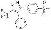
异噁唑,3-[4-(甲磺酰基)苯基]-4-苯基-5-(三氟甲基)-是一种磺酸衍生物。
合成了在其中一个苯环的对位具有不同取代基(H、F、MeS、MeSO2)的4,5-二苯基-4-异噁唑啉类化合物(13a-k),并评估其作为镇痛剂和选择性环氧合酶-2 (COX-2) 抑制剂的抗炎活性。虽然不含C-3位甲基取代基的4,5-苯基-4-异噁唑啉类化合物(13a-d、f)表现出较强的镇痛和抗炎活性,但所评估的化合物(13a、13b、13h和13k)并非COX-2的选择性抑制剂。相比之下,2,3-二甲基-5-(4-甲基磺酰基苯基)-4-苯基-4-异噁唑啉 (13j) 表现出优异的镇痛和抗精神病活性,并且是一种强效且选择性的 COX-2 抑制剂(COX-1,IC50 = 258 μM;COX-2,IC50 = 0.004 μM)。在 4-苯环对位具有氟取代基的相关化合物 13k 也是一种选择性 COX-2 抑制剂(选择性指数 SI = 3162),但其效力(IC50 = 0.0316 μM)低于 13j。对化合物 13j 的分子建模(分子对接研究)表明,MeSO2 取代基的 S 原子位于 COX-2 二级口袋 (Val(523)) 入口内约 6.46 Å 处,并且 C-3 Me (13j, 13k) 中心异噁唑啉环取代基对于此类化合物选择性抑制 COX-2 至关重要。[1] 背景和目的:肺癌是全球男性和女性癌症死亡的最常见原因。本研究评估了高选择性环氧合酶-2抑制剂CAY10404在三种非小细胞肺癌(NSCLC)细胞系(H460、H358和H1703)中诱导细胞死亡的机制。 方法:为检测CAY10404对NSCLC细胞增殖的影响,将3×10³个细胞/孔接种于96孔板中,并在37℃下培养过夜使其贴壁。用CAY10404处理3天后,采用MTT法检测细胞增殖。在H460 NSCLC细胞中,采用TUNEL法和Western blot分析检测细胞凋亡。 [3] H460 和 H358 细胞对 CAY10404 敏感的原因尚不完全清楚。然而,这可能归因于多种促凋亡效应的诱导。既往研究表明,法尼基转移酶抑制剂 SCH66336 和特异性 COX-2 抑制剂 NS-398 分别降低了 SqCC/Y1 头颈部鳞状细胞癌细胞和鼠 B 淋巴瘤 A20 细胞中 Bcl-2/Bcl-XL 的水平。然而,地瓜素并未改变 H322 非小细胞肺癌细胞中 Bcl-2 蛋白的水平。32 在本研究中,CAY10404 处理降低了抗凋亡蛋白 Bcl-2 和 Bcl-XL 的水平,而未影响促凋亡蛋白 Bax 的水平,导致抗凋亡蛋白与促凋亡蛋白的比值下降。这种差异可能归因于细胞类型或COX-2抑制剂的不同。然而,目前的结果或许可以解释CAY10404的促凋亡作用。 作为持续研究CAY10404对非小细胞肺癌(NSCLC)细胞作用机制的一部分,我们研究了PKB/Akt和MAPK通路的作用。MAPK和Akt酶在调节细胞凋亡和增殖中发挥着重要作用。已有研究表明,PKB/Akt和MAPK对细胞凋亡具有显著的抑制作用33。Akt的磷酸化最近被认为与COX-2介导的肺癌和肝细胞癌细胞的存活有关。在本研究中,用CAY10404处理H460和H358细胞3天后,Akt活性降低,导致磷酸化Akt底物(包括pGSK-3β)水平降低,这些变化也可能导致细胞凋亡增加。此外,本研究表明,CAY10404以浓度依赖的方式降低了H460和H358细胞中pAkt和pGSK-3β的水平,而总Akt和GSK-3β蛋白水平未发生改变。在分子水平上探究MAPK通路的作用时,CAY10404处理3天后,磷酸化Erk1/2的表达略有下降,而总Erk1/2蛋白水平未发生改变。这些结果提示CAY10404优先影响PKB/Akt信号通路,该通路在调控细胞凋亡和增殖中发挥重要作用。这是首次报道CAY10404通过抑制参与细胞增殖和存活的信号转导机制诱导非小细胞肺癌(NSCLC)细胞凋亡。其他报告表明,胰岛素样生长因子(IGF)结合蛋白-3抑制了非小细胞肺癌(NSCLC)细胞中IGF-I诱导的PI3K/Akt/PKB和MAPK通路激活,而PI3K调节亚基dnp85α的过表达诱导细胞凋亡,LY294002或磷酸酶和张力蛋白同源物(PTEN)的过表达则诱导H1299 NSCLC细胞增殖停滞。37在癌前HBE细胞中,地瓜素抑制PI3K活性并降低pAkt水平和活性,但对MAPK通路的影响甚微。38因此,我们评估了CAY10404是否抑制了NSCLC细胞中抑制PI3K和MAPK的信号转导通路。 许多实验和临床研究已证实,特异性COX-2抑制剂可能有助于预防和治疗多种恶性肿瘤。然而,近期研究表明,长期使用高浓度COX-2抑制剂会显著增加心脏毒性的风险。克服这一局限性的一个方法是使用较低剂量的COX-2抑制剂与其他已上市药物联合使用。两种药物同时使用的协同作用可能使特异性COX-2抑制剂CAY10404能够以更低、更安全的浓度使用,并为更有效治疗人类非小细胞肺癌铺平道路。未来的研究应探讨COX-2抑制剂与其他已上市药物联合使用以改善癌症控制的效果。[2] |
| 分子式 |
C17H12NO3F3S
|
|---|---|
| 分子量 |
367.342
|
| 精确质量 |
367.049
|
| 元素分析 |
C, 55.58; H, 3.29; F, 15.52; N, 3.81; O, 13.07; S, 8.73
|
| CAS号 |
340267-36-9
|
| PubChem CID |
10429020
|
| 外观&性状 |
White to off-white solid powder
|
| 密度 |
1.4±0.1 g/cm3
|
| 沸点 |
498.6±45.0 °C at 760 mmHg
|
| 熔点 |
196.47 °C(Predicted)
|
| 闪点 |
255.3±28.7 °C
|
| 蒸汽压 |
0.0±1.2 mmHg at 25°C
|
| 折射率 |
1.538
|
| LogP |
2.92
|
| tPSA |
68.55
|
| 氢键供体(HBD)数目 |
0
|
| 氢键受体(HBA)数目 |
7
|
| 可旋转键数目(RBC) |
3
|
| 重原子数目 |
25
|
| 分子复杂度/Complexity |
547
|
| 定义原子立体中心数目 |
0
|
| SMILES |
CS(=O)(=O)C1=CC=C(C=C1)C2=NOC(=C2C3=CC=CC=C3)C(F)(F)F
|
| InChi Key |
KKBWWVXRKULXHF-UHFFFAOYSA-N
|
| InChi Code |
InChI=1S/C17H12F3NO3S/c1-25(22,23)13-9-7-12(8-10-13)15-14(11-5-3-2-4-6-11)16(24-21-15)17(18,19)20/h2-10H,1H3
|
| 化学名 |
3-[4-(Methylsulfonyl)phenyl]-4-phenyl-5-(trifluoromethyl)-isoxazole
|
| 别名 |
CAY 10404; 340267-36-9; CAY10,404; CAY 10,404; 3-(4-methylsulphonylphenyl)-4-phenyl-5-trifluoromethylisoxazole; 3-(4-methylsulfonylphenyl)-4-phenyl-5-trifluoromethylisoxazole; 3-(4-methylsulfonylphenyl)-4-phenyl-5-(trifluoromethyl)-1,2-oxazole; CHEMBL97943; 3-[4-(Methylsulfonyl)phenyl]-4-phenyl-5-(trifluoromethyl)-isoxazole; CAY-10404; CAY10404
|
| HS Tariff Code |
2934.99.9001
|
| 存储方式 |
Powder -20°C 3 years 4°C 2 years In solvent -80°C 6 months -20°C 1 month 注意: 请将本产品存放在密封且受保护的环境中(例如氮气保护),避免吸湿/受潮。 |
| 运输条件 |
Room temperature (This product is stable at ambient temperature for a few days during ordinary shipping and time spent in Customs)
|
| 溶解度 (体外实验) |
DMSO : ~100 mg/mL (~272.23 mM)
|
|---|---|
| 溶解度 (体内实验) |
配方 1 中的溶解度: 2.5 mg/mL (6.81 mM) in 10% DMSO + 40% PEG300 + 5% Tween80 + 45% Saline (这些助溶剂从左到右依次添加,逐一添加), 悬浮液;超声助溶。
例如,若需制备1 mL的工作液,可将100 μL 25.0 mg/mL澄清DMSO储备液加入到400 μL PEG300中,混匀;然后向上述溶液中加入50 μL Tween-80,混匀;加入450 μL生理盐水定容至1 mL。 *生理盐水的制备:将 0.9 g 氯化钠溶解在 100 mL ddH₂O中,得到澄清溶液。 配方 2 中的溶解度: 2.5 mg/mL (6.81 mM) in 10% DMSO + 90% (20% SBE-β-CD in Saline) (这些助溶剂从左到右依次添加,逐一添加), 悬浊液; 超声助溶。 例如,若需制备1 mL的工作液,可将 100 μL 25.0 mg/mL澄清DMSO储备液加入900 μL 20% SBE-β-CD生理盐水溶液中,混匀。 *20% SBE-β-CD 生理盐水溶液的制备(4°C,1 周):将 2 g SBE-β-CD 溶解于 10 mL 生理盐水中,得到澄清溶液。 View More
配方 3 中的溶解度: ≥ 2.5 mg/mL (6.81 mM) (饱和度未知) in 10% DMSO + 90% Corn Oil (这些助溶剂从左到右依次添加,逐一添加), 澄清溶液。 1、请先配制澄清的储备液(如:用DMSO配置50 或 100 mg/mL母液(储备液)); 2、取适量母液,按从左到右的顺序依次添加助溶剂,澄清后再加入下一助溶剂。以 下列配方为例说明 (注意此配方只用于说明,并不一定代表此产品 的实际溶解配方): 10% DMSO → 40% PEG300 → 5% Tween-80 → 45% ddH2O (或 saline); 假设最终工作液的体积为 1 mL, 浓度为5 mg/mL: 取 100 μL 50 mg/mL 的澄清 DMSO 储备液加到 400 μL PEG300 中,混合均匀/澄清;向上述体系中加入50 μL Tween-80,混合均匀/澄清;然后继续加入450 μL ddH2O (或 saline)定容至 1 mL; 3、溶剂前显示的百分比是指该溶剂在最终溶液/工作液中的体积所占比例; 4、 如产品在配制过程中出现沉淀/析出,可通过加热(≤50℃)或超声的方式助溶; 5、为保证最佳实验结果,工作液请现配现用! 6、如不确定怎么将母液配置成体内动物实验的工作液,请查看说明书或联系我们; 7、 以上所有助溶剂都可在 Invivochem.cn网站购买。 |
| 制备储备液 | 1 mg | 5 mg | 10 mg | |
| 1 mM | 2.7223 mL | 13.6114 mL | 27.2227 mL | |
| 5 mM | 0.5445 mL | 2.7223 mL | 5.4445 mL | |
| 10 mM | 0.2722 mL | 1.3611 mL | 2.7223 mL |
1、根据实验需要选择合适的溶剂配制储备液 (母液):对于大多数产品,InvivoChem推荐用DMSO配置母液 (比如:5、10、20mM或者10、20、50 mg/mL浓度),个别水溶性高的产品可直接溶于水。产品在DMSO 、水或其他溶剂中的具体溶解度详见上”溶解度 (体外)”部分;
2、如果您找不到您想要的溶解度信息,或者很难将产品溶解在溶液中,请联系我们;
3、建议使用下列计算器进行相关计算(摩尔浓度计算器、稀释计算器、分子量计算器、重组计算器等);
4、母液配好之后,将其分装到常规用量,并储存在-20°C或-80°C,尽量减少反复冻融循环。
计算结果:
工作液浓度: mg/mL;
DMSO母液配制方法: mg 药物溶于 μL DMSO溶液(母液浓度 mg/mL)。如该浓度超过该批次药物DMSO溶解度,请首先与我们联系。
体内配方配制方法:取 μL DMSO母液,加入 μL PEG300,混匀澄清后加入μL Tween 80,混匀澄清后加入 μL ddH2O,混匀澄清。
(1) 请确保溶液澄清之后,再加入下一种溶剂 (助溶剂) 。可利用涡旋、超声或水浴加热等方法助溶;
(2) 一定要按顺序加入溶剂 (助溶剂) 。
 COX-2/15-LOX-IN-4
 Diclofenac-13C6 sodium heminonahydrate (Diclofenac sodium 13C6 (1/2 water))
 COX-2-IN-31
 Oxaprozin-d4 (Oxaprozin D4; Wy-21743-d4)
InvivoChem的所有产品仅用于作科学研究,不面向患者销售
Copyright 2020 InvivoChem LLC | All Rights Reserved 粤ICP备20063088号-1


 COA
COA
 463611831
