| 规格 | 价格 | 库存 | 数量 |
|---|---|---|---|
| 10 mM * 1 mL in DMSO |
|
||
| 1mg |
|
||
| 5mg |
|
||
| 10mg |
|
||
| 25mg |
|
||
| 50mg |
|
||
| 100mg |
|
||
| 250mg |
|
||
| Other Sizes |
|

| 靶点 |
MNK1 (IC50 = 2.2 μM)
Mitogen-activated protein kinase-interacting kinase 1 (MNK1) (IC50=300 nM) [1][4] - Mitogen-activated protein kinase-interacting kinase 2 (MNK2) (IC50=280 nM) [1][4] |
|---|---|
| 体外研究 (In Vitro) |
CGP57380 在细胞测定中抑制 eIF4E 磷酸化,IC50 约为 3 μM。使用 CGP57380 去磷酸化 eIF4E 会导致 293 细胞表达更多帽依赖性报告基因。 [1] CGP57380 以剂量依赖性方式抑制蛋白质合成、VSMC 肥大和 Ang II 刺激的 eIF4E 磷酸化。 [2]在小鼠胚胎成纤维细胞 (MEF) 中,CGP57380 使野生型细胞更容易受到血清撤除引起的细胞凋亡的影响。 [3] CGP57380 阻止 BC 祖细胞重复复制。[4]
重组酶实验中,CGP 57380 可浓度依赖性抑制MNK1和MNK2的激酶活性,1 μM浓度时对两者的抑制率均超过80%,且对其他相关激酶(如ERK1、p38α)无明显抑制作用,靶点选择性良好[1] - HEK293细胞中,CGP 57380 处理(10 μM,24小时)可显著抑制eIF4E的磷酸化(Ser209位点),抑制率达75%,同时减少下游Cyclin D1、c-Myc等蛋白的翻译合成,不影响其mRNA表达水平[1] - 血管平滑肌细胞(VSMC)中,血管紧张素II(Ang II)诱导的蛋白合成可被CGP 57380 剂量依赖性抑制,10 μM浓度时蛋白合成速率降低60%,且该效应与抑制MNK1-eIF4E通路相关,不影响Ang II介导的ERK1/2磷酸化[2] - 小鼠胚胎成纤维细胞(MEF)中,血清饥饿条件下,CGP 57380 处理(5 μM,48小时)可显著增加凋亡细胞比例,从对照组的12%升至38%,同时上调促凋亡蛋白Bax表达,下调抗凋亡蛋白Bcl-2水平[3] - 急变期慢性髓系白血病(BC-CML)细胞系(K562、KU812)中,CGP 57380 可浓度依赖性抑制细胞增殖,IC50值分别为2.3 μM和1.8 μM,且显著降低白血病干细胞(LSC)的集落形成能力(集落数减少55%)[4] - BC-CML细胞中,CGP 57380 处理后eIF4E磷酸化水平降低82%,下游促存活蛋白Mcl-1、Bcl-xL的表达减少,同时激活caspase-3/7依赖的凋亡通路,凋亡率较对照组升高4倍[4] |
| 体内研究 (In Vivo) |
CGP57380 (40 mg/kg/d ip) 可有效消除 BC CML 细胞作为 LSC 和连续移植免疫缺陷小鼠的功能。 [4]
在这里,我们如前所述,对BC样本中的GMP进行FACS分选,并将其注射到8至12周龄的雌性NSG小鼠体内(图S8A)。移植后6周,用DMSO、CGP57380或达沙替尼治疗移植小鼠3周(每个治疗组n=5只小鼠)。在治疗期结束时,杀死所有小鼠,并使用免疫磁珠从造血组织中获得人类细胞。我们发现,每个治疗组的外周血或骨髓中CD45+人细胞的百分比没有差异(图6A)。然而,我们观察到达沙替尼和CGP57380对承诺的BC祖细胞具有特异性活性,因为与对照组相比,它们显著减少了BM中检测到的集落形成单位的数量(分别为P≤0.05和P≤0.005),尽管CGP57370的效果更大(图6B)。然后将从原代小鼠获得的人类细胞移植到次级受体中,并在16周内通过流式细胞术监测植入情况。到4周时,我们能够检测到三个治疗组中所有动物的移植物(图6C)。在DMSO或达沙替尼治疗的动物中,在整个16周的实验时间框架内,移植率保持在80%(即五只动物中的四只),但相比之下,CGP57380治疗的小鼠均无法保持长期移植(图6C)。16周时,对小鼠实施安乐死,并检查BM中是否存在BCR-ABL1。在用DMSO或达沙替尼治疗的每只动物中都检测到BCR-ABL1转录本(即每个治疗组五只动物中有四只),而在CGP57380治疗组的四只动物中只有一只检测到非常微弱的条带(图6D)。使用来自不同个体的CD34+BC细胞重复该实验,得到了类似的结果(图S8 B-J)。综上所述,我们的研究结果表明,体内MNK抑制可以有效地消除BC CML细胞连续移植免疫缺陷小鼠的能力,并作为LSCs发挥作用[4]。 免疫缺陷小鼠BC-CML异种移植模型中,CGP 57380 以50 mg/kg剂量腹腔注射,每日一次,连续14天,可显著抑制肿瘤生长,肿瘤体积较对照组缩小62%,肿瘤重量减轻58%[4] - 白血病小鼠模型中,给药组骨髓和脾脏中白血病细胞浸润程度显著降低,LSC数量减少65%,且外周血中白血病细胞比例从对照组的78%降至32%[4] - 实验期间,给药组小鼠体重无明显下降(体重变化率≤4%),未观察到明显的器官毒性,肝肾功能相关血清指标(ALT、AST、肌酐)与对照组无显著差异[4] |
| 酶活实验 |
CGP 57380 是 MNK1 的有效抑制剂,IC50 值为 2.2 μM。
MNK1和PRAK通过与活化的p38预孵育而磷酸化,p38是通过与重组MKK6b孵育而产生的(E)。制备重组激酶和eIF4E,并如前所述进行体外激酶反应。使用Oligotex Direct mRNA试剂盒从293细胞中纯化Poly(A)+mRNA。对于体外翻译,根据制造商的说明,在每毫升3或10μg激酶、[35S]蛋氨酸(0.6 mCi/ml)、1.5 mM乙酸镁、75 mM KCl、2 mM DTT和100μM ATP的存在下,用每毫升10μg mRNA对兔网织红细胞裂解物(Promega)进行编程。注意确保所有测定中的缓冲条件相同。翻译反应在30°C下孵育90分钟,并测量掺入TCA可沉淀材料的放射性。[1] 重组p38异构体在以下条件下被Mkk6(E)激活:p38(100 ng/mL)、Mkk6。Mnk1活性的典型测定反应包含激酶缓冲液中的Mnk1(2ng/mL)、HA-eIF4E(10ng/mL)、ATP(300mM)。通过加入活化的p38(0.03-3ng/mL)开始反应,并在30°C下30分钟后通过加入SDS负载缓冲液停止反应。在相同的测定条件下鉴定Mnk1抑制剂,除了在暴露于底物和抑制剂之前使用活性p38a预激活Mnk1[1]。 MNK1/MNK2激酶活性测定:重组人MNK1或MNK2蛋白与eIF4E重组蛋白、ATP在反应缓冲液中孵育,加入梯度浓度(0.01-10 μM)的CGP 57380,30℃反应60分钟后,通过Western blot检测eIF4E的磷酸化水平,计算激酶活性抑制率及IC50值[1] - 激酶选择性检测:采用相同实验体系,分别以ERK1、p38α、JNK2等为靶点激酶,加入10 μM CGP 57380 后检测激酶活性,对比其对不同激酶的抑制效果,验证靶点特异性[1] - 蛋白合成速率检测:VSMC细胞经CGP 57380 预处理1小时后,加入放射性标记的亮氨酸和Ang II,培养4小时后,检测细胞内放射性掺入量,计算蛋白合成速率[2] |
| 细胞实验 |
重组 p38 同种型在以下条件下被 Mkk6(E) 激活:p38 (100 ng/mL)、Mkk6(E) (30 ng/mL)、ATP (100 mM) 在激酶缓冲液(25 mM Hepes、25 mM b-甘油磷酸盐、0.1 mM 原钒酸钠、25 mM MgCl2、2.5 mM DTT,pH 7.4)并在 30°C 下孵育 30 分钟。 Mnk1 (2 ng/mL)、HA-eIF4E (10 ng/mL) 和 ATP (300 mM) 是 Mnk1 活性的典型检测反应的主要成分。添加活化的 p38 (0.03–3 ng/mL) 启动反应,30 分钟后在 30°C 下添加 SDS 上样缓冲液终止反应。使用相同的测定条件来鉴定 Mnk1 抑制剂,但在暴露于底物和抑制剂之前,首先使用活性 p38a 预激活 Mnk1。
细胞增殖与凋亡检测:BC-CML细胞系接种于96孔板,加入梯度浓度(0.1-20 μM)的CGP 57380,培养72小时后,MTT法检测细胞活力并计算IC50;Annexin V/PI双染法通过流式细胞仪检测凋亡率[4] - eIF4E磷酸化及下游蛋白检测:不同细胞(HEK293、VSMC、BC-CML细胞)经CGP 57380 处理后,提取总蛋白,Western blot检测p-eIF4E(Ser209)、eIF4E、Cyclin D1、Mcl-1等蛋白表达水平;RT-PCR检测对应mRNA表达,验证药物对蛋白翻译的特异性抑制[1][2][4] - 成纤维细胞凋亡实验:MEF细胞接种后,血清饥饿处理12小时,加入5 μM CGP 57380 继续培养48小时,Hoechst 33342染色观察细胞核形态变化,流式细胞仪定量凋亡率,Western blot检测Bax、Bcl-2蛋白表达[3] - 白血病干细胞集落形成实验:从BC-CML患者骨髓中分离CD34+CD38- LSC,加入梯度浓度(0.5-10 μM)的CGP 57380,接种于半固体培养基,培养14天后计数集落形成数量,评估LSC自我更新能力[4] |
| 动物实验 |
将 CD34+ 细胞 (5×10⁵) 或 GMP (1×10⁵) 重悬于 25 μL 1% FBS/PBS 溶液中,并注射到 8~10 周龄经亚致死剂量 (200 cGy) 照射的雌性小鼠(每组 n=5 只)的右侧股骨中。每个实验中,注射 1% FBS/PBS 溶液的小鼠作为假手术对照。移植后每 4 周,使用流式细胞术检测小鼠,以观察人源细胞是否成功植入。移植成功后,在移植后 6 周,对小鼠进行以下处理:腹腔注射 CGP57380 (40 mg/kg/d),灌胃给予达沙替尼 (5 mg/kg/d),或仅给予载体(每组 n = 5 只)。治疗结束后,将小鼠处死,并使用抗人CD45特异性免疫磁珠从骨髓和脾脏中提取CD45+细胞。在集落形成细胞(CFC)测定中,将1×10⁵个人CD45+细胞接种到甲基纤维素中,2周后计数集落。然后将每个初次移植受体小鼠剩余的人类细胞全部经股动脉注射到次级受体小鼠体内,并从第4周开始每两周监测一次人细胞植入情况。所有小鼠在16周后处死。采用RT-PCR检测BCR-ABL1转录本,并采用流式细胞术评估骨髓和血液中的移植情况。
BC-CML异种移植模型实验:将6-8周龄的NOD/SCID小鼠尾静脉注射BC-CML患者骨髓来源的CD34+细胞(5×10^5个细胞/只),建立白血病模型。造血7天后,将小鼠随机分为对照组和治疗组(每组8只)。治疗组腹腔注射CGP 57380(50 mg/kg,溶于5% DMSO + 45% PEG300 + 50%生理盐水),每日一次,连续14天;对照组注射等体积的溶剂[4]。 - 实验监测和样本采集:实验期间,每3天测量小鼠体重和外周血中白血病细胞的比例。给药结束后,处死小鼠,收集骨髓、脾脏和肝脏组织,用于病理切片分析、白血病细胞计数和p-eIF4E表达的Western blot检测[4] |
| 毒性/毒理 (Toxicokinetics/TK) |
在体内实验中,小鼠腹腔注射 CGP 57380 50 mg/kg,连续 14 天,未出现明显的毒性症状,且在肝脏、脾脏和肾脏等主要器官的病理切片中未观察到坏死、炎症或其他损伤[4]
|
| 参考文献 |
|
| 其他信息 |
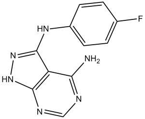
N3-(4-氟苯基)-2H-吡唑并[3,4-d]嘧啶-3,4-二胺是一种吡唑并嘧啶类化合物。
真核起始因子4E (eIF4E) 是翻译机制的关键组成部分,也是细胞生长和增殖的重要调节因子。eIF4E的活性被认为受其与抑制性结合蛋白 (4E-BP) 的相互作用以及丝裂原活化蛋白 (MAP) 激酶相互作用激酶 (MNK) 在丝裂原和细胞应激刺激下对Ser209位点的磷酸化调控。本文证明,MNK1介导的eIF4E磷酸化是通过激活Erk或p38通路实现的。我们进一步发现,在293细胞中表达MNK1和MNK2的活性突变体,会降低帽依赖性翻译相对于帽非依赖性翻译的活性(通过瞬时报告基因检测)。当MNK1被Erk或p38通路激活时,也观察到了对帽依赖性翻译的相同影响。与这些发现一致,在兔网织红细胞裂解液中添加重组活性MNK1导致体外蛋白质合成减少,而MNK2的过表达导致293细胞中蛋白质合成速率降低。我们使用一种新型的MNK1小分子激酶抑制剂CGP57380,证明eIF4E磷酸化对于起始复合物的形成、有丝分裂原刺激的帽依赖性翻译增加以及细胞增殖并非至关重要。我们的结果表明,MAP激酶通路激活MNK并非帽依赖性翻译的正向调控机制。相反,我们提出MNK的激酶活性最终可能通过eIF4E的磷酸化,在生理条件下限制帽依赖性翻译。 [1]血管紧张素II (Ang II) 可刺激血管平滑肌细胞 (VSMC) 中的蛋白质合成,这可能继发于mRNA翻译起始阶段的调控改变。丝裂原活化蛋白激酶 (MAP) 信号整合激酶-1 (Mnk1) 是ERK和p38 MAP激酶的底物,可磷酸化真核起始因子4E (eIF4E),而eIF4E是翻译过程中的一个重要因子。本研究旨在探讨Mnk1在Ang II诱导的蛋白质合成中的作用,并阐明Mnk1和eIF4E在大鼠VSMC中被激活的分子机制。Ang II处理导致Mnk1活性和eIF4E磷酸化水平升高。表达显性失活的Mnk1突变体可消除Ang II诱导的eIF4E磷酸化。 PD98059 或引入激酶失活的 MEK1/MKK1,而非 SB202190 或激酶失活的 p38 MAP 激酶,抑制了 Ang II 诱导的 Mnk1 活化和 eIF4E 磷酸化,提示 ERK 而非 p38 MAP 激酶是 Ang II 诱导的 Mnk1-eIF4E 活化所必需的。此外,Ras 的显性失活构建体(而非 Rho、Rac 或 Cdc42 的显性失活构建体)消除了 Ang II 诱导的 Mnk1 活化。最后,用新型 Mnk1 特异性激酶抑制剂 CGP57380 处理 VSMC 可导致 Ang II 刺激的 eIF4E 磷酸化、蛋白质合成和 VSMC 肥大呈剂量依赖性降低。总之,这些数据表明:(1) 血管平滑肌细胞 (VSMC) 中 Ang II 诱导的 Mnk1 激活是由 Ras-ERK 信号通路介导的;(2) Mnk1 参与 Ang II 介导的蛋白质合成和肥大,推测是通过激活翻译起始实现的。Mnk1-eIF4E 通路可能为血管肥大和其他 Ang II 介导的病理状态的分子机制提供新的见解。[2]MAP 激酶相互作用蛋白激酶 1 和 2 (MNK1、MNK2) 位于 p38 和 ERK MAP 激酶的下游,但我们对 MNK 的调控和功能知之甚少。敲除这两个基因的小鼠表型正常,提示 MNK 在应激反应的适应性通路中发挥作用。本文研究表明,与野生型细胞相比,从 mnk1 (-/-)/mnk2 (-/-) 以及 mnk1 (-/-) 和 mnk2 (-/-) 小鼠中获得的胚胎成纤维细胞 (MEF) 在血清撤离后对 caspase-3 激活更加敏感。所有细胞均发生 caspase-3 裂解,但来自缺失 MNK 基因小鼠的细胞裂解速度最快、强度最高。用 MNK1 和 MNK2 抑制剂 (CGP57380) 处理野生型 MEF 后,野生型细胞对血清撤离诱导的细胞凋亡更加敏感,表明这种敏感性增加是由于 MNK 功能丧失所致,而非继发事件。在双敲除MEF细胞中重新引入野生型MNK1会导致血清撤离敏感性降低,而野生型MNK2或激酶失活变体则未观察到这种现象。我们的研究表明,MNK激酶参与了血清撤离反应中的抗凋亡信号传导。[3]慢性粒细胞白血病在慢性期对靶向致癌融合蛋白BCR-ABL1的治疗反应良好,但在进展至急变期(BC)后则对治疗产生耐药性。BC的特征是粒细胞巨噬细胞祖细胞(GMP)中β-catenin信号通路增强,这使得该细胞群能够发挥白血病干细胞(LSC)的功能,并成为耐药性的储存库。由于正常造血干细胞 (HSC) 和白血病干细胞 (LSC) 的自我更新依赖于 β-catenin 信号通路,因此,针对乳腺癌的特异性治疗策略需要识别能够区分乳腺癌 LSC 和正常 HSC 自我更新的可药物靶向因子。本文研究表明,MAP 激酶相互作用丝氨酸/苏氨酸激酶 (MNK)-真核翻译起始因子 4E (eIF4E) 轴在乳腺癌粒单核细胞 (GMP) 中过表达,但在正常 HSC 中未见过表达;并且 MNK 激酶依赖的 eIF4E 在丝氨酸 209 位点的磷酸化激活了乳腺癌 GMP 中的 β-catenin 信号通路。机制上,eIF4E 的过表达和磷酸化导致 β-catenin 蛋白合成增加,而 MNK 依赖的 eIF4E 磷酸化是 β-catenin 核转位和激活所必需的。因此,我们发现一组小分子MNK激酶抑制剂在体外和体内均能抑制eIF4E磷酸化、β-catenin激活以及BC LSC功能。我们的研究结果表明,MNK-eIF4E轴是BC自我更新的一个特异性且关键的调控因子,并提示MNK激酶的药理学抑制可能对BC慢性粒细胞白血病具有治疗价值。[4] CGP 57380是首个报道的选择性MNK1/MNK2小分子抑制剂。它通过直接结合激酶催化结构域抑制eIF4E的磷酸化,从而阻断帽依赖性蛋白翻译[1] - 该药物的核心作用机制是靶向MNK-eIF4E轴,该轴在调节细胞增殖、存活和凋亡中起关键作用,其异常激活与肿瘤发生发展密切相关[1][4] - CGP 57380对血清饥饿成纤维细胞的促凋亡作用表明,MNK激酶在营养缺乏条件下细胞存活信号通路中发挥重要的调控作用[3] - 在BC-CML中,CGP 57380不仅抑制白血病细胞增殖,还能靶向并杀死白血病干细胞,为治疗耐药性白血病提供了一种潜在策略[4] - CGP 57380不影响上游MAPK通路(如ERK、p38)的激活。但仅阻断 MNK 介导的 eIF4E 磷酸化,表现出明显的信号通路选择性 [1][2] |
| 分子式 |
C11H9FN6
|
|
|---|---|---|
| 分子量 |
244.23
|
|
| 精确质量 |
244.087
|
|
| 元素分析 |
C, 54.10; H, 3.71; F, 7.78; N, 34.41
|
|
| CAS号 |
522629-08-9
|
|
| 相关CAS号 |
|
|
| PubChem CID |
11644425
|
|
| 外观&性状 |
Light brown to brown solid powder
|
|
| 密度 |
1.6±0.1 g/cm3
|
|
| 沸点 |
541.6±50.0 °C at 760 mmHg
|
|
| 闪点 |
281.4±30.1 °C
|
|
| 蒸汽压 |
0.0±1.4 mmHg at 25°C
|
|
| 折射率 |
1.809
|
|
| LogP |
1.28
|
|
| tPSA |
92.51
|
|
| 氢键供体(HBD)数目 |
3
|
|
| 氢键受体(HBA)数目 |
6
|
|
| 可旋转键数目(RBC) |
2
|
|
| 重原子数目 |
18
|
|
| 分子复杂度/Complexity |
283
|
|
| 定义原子立体中心数目 |
0
|
|
| SMILES |
FC1C([H])=C([H])C(=C([H])C=1[H])N([H])C1=C2C(N([H])[H])=NC([H])=NC2=NN1[H]
|
|
| InChi Key |
UQPMANVRZYYQMD-UHFFFAOYSA-N
|
|
| InChi Code |
InChI=1S/C11H9FN6/c12-6-1-3-7(4-2-6)16-11-8-9(13)14-5-15-10(8)17-18-11/h1-5H,(H4,13,14,15,16,17,18)
|
|
| 化学名 |
3-N-(4-fluorophenyl)-2H-pyrazolo[3,4-d]pyrimidine-3,4-diamine
|
|
| 别名 |
|
|
| HS Tariff Code |
2934.99.9001
|
|
| 存储方式 |
Powder -20°C 3 years 4°C 2 years In solvent -80°C 6 months -20°C 1 month |
|
| 运输条件 |
Room temperature (This product is stable at ambient temperature for a few days during ordinary shipping and time spent in Customs)
|
| 溶解度 (体外实验) |
|
|||
|---|---|---|---|---|
| 溶解度 (体内实验) |
配方 1 中的溶解度: ≥ 2.5 mg/mL (10.24 mM) (饱和度未知) in 10% DMSO + 40% PEG300 + 5% Tween80 + 45% Saline (这些助溶剂从左到右依次添加,逐一添加), 澄清溶液。
例如,若需制备1 mL的工作液,可将100 μL 25.0 mg/mL澄清DMSO储备液加入到400 μL PEG300中,混匀;然后向上述溶液中加入50 μL Tween-80,混匀;加入450 μL生理盐水定容至1 mL。 *生理盐水的制备:将 0.9 g 氯化钠溶解在 100 mL ddH₂O中,得到澄清溶液。 配方 2 中的溶解度: ≥ 2.5 mg/mL (10.24 mM) (饱和度未知) in 10% DMSO + 90% (20% SBE-β-CD in Saline) (这些助溶剂从左到右依次添加,逐一添加), 澄清溶液。 例如,若需制备1 mL的工作液,可将 100 μL 25.0 mg/mL澄清DMSO储备液加入900 μL 20% SBE-β-CD生理盐水溶液中,混匀。 *20% SBE-β-CD 生理盐水溶液的制备(4°C,1 周):将 2 g SBE-β-CD 溶解于 10 mL 生理盐水中,得到澄清溶液。 View More
配方 3 中的溶解度: 4% DMSO +30%PEG 300 +ddH2O: 10mg/mL 1、请先配制澄清的储备液(如:用DMSO配置50 或 100 mg/mL母液(储备液)); 2、取适量母液,按从左到右的顺序依次添加助溶剂,澄清后再加入下一助溶剂。以 下列配方为例说明 (注意此配方只用于说明,并不一定代表此产品 的实际溶解配方): 10% DMSO → 40% PEG300 → 5% Tween-80 → 45% ddH2O (或 saline); 假设最终工作液的体积为 1 mL, 浓度为5 mg/mL: 取 100 μL 50 mg/mL 的澄清 DMSO 储备液加到 400 μL PEG300 中,混合均匀/澄清;向上述体系中加入50 μL Tween-80,混合均匀/澄清;然后继续加入450 μL ddH2O (或 saline)定容至 1 mL; 3、溶剂前显示的百分比是指该溶剂在最终溶液/工作液中的体积所占比例; 4、 如产品在配制过程中出现沉淀/析出,可通过加热(≤50℃)或超声的方式助溶; 5、为保证最佳实验结果,工作液请现配现用! 6、如不确定怎么将母液配置成体内动物实验的工作液,请查看说明书或联系我们; 7、 以上所有助溶剂都可在 Invivochem.cn网站购买。 |
| 制备储备液 | 1 mg | 5 mg | 10 mg | |
| 1 mM | 4.0945 mL | 20.4725 mL | 40.9450 mL | |
| 5 mM | 0.8189 mL | 4.0945 mL | 8.1890 mL | |
| 10 mM | 0.4095 mL | 2.0473 mL | 4.0945 mL |
1、根据实验需要选择合适的溶剂配制储备液 (母液):对于大多数产品,InvivoChem推荐用DMSO配置母液 (比如:5、10、20mM或者10、20、50 mg/mL浓度),个别水溶性高的产品可直接溶于水。产品在DMSO 、水或其他溶剂中的具体溶解度详见上”溶解度 (体外)”部分;
2、如果您找不到您想要的溶解度信息,或者很难将产品溶解在溶液中,请联系我们;
3、建议使用下列计算器进行相关计算(摩尔浓度计算器、稀释计算器、分子量计算器、重组计算器等);
4、母液配好之后,将其分装到常规用量,并储存在-20°C或-80°C,尽量减少反复冻融循环。
计算结果:
工作液浓度: mg/mL;
DMSO母液配制方法: mg 药物溶于 μL DMSO溶液(母液浓度 mg/mL)。如该浓度超过该批次药物DMSO溶解度,请首先与我们联系。
体内配方配制方法:取 μL DMSO母液,加入 μL PEG300,混匀澄清后加入μL Tween 80,混匀澄清后加入 μL ddH2O,混匀澄清。
(1) 请确保溶液澄清之后,再加入下一种溶剂 (助溶剂) 。可利用涡旋、超声或水浴加热等方法助溶;
(2) 一定要按顺序加入溶剂 (助溶剂) 。
|
|---|
InvivoChem的所有产品仅用于作科学研究,不面向患者销售
Copyright 2020 InvivoChem LLC | All Rights Reserved 粤ICP备20063088号-1


 COA
COA

 463611831
