| 规格 | 价格 | 库存 | 数量 |
|---|---|---|---|
| 1mg |
|
||
| 5mg |
|
||
| 10mg |
|
||
| 25mg |
|
||
| 50mg |
|
||
| 100mg |
|
||
| 250mg |
|
||
| 500mg |
|
||
| Other Sizes |
|

| 靶点 |
BRD4BD1 (DC50/5h = 500 nM)[1]; DC50: half-maximal degradation concentration, at the half-maximal degradation concentration that degrades 50% of the target protein.
|
|---|---|
| 体外研究 (In Vitro) |
我们发现,dBET6(DC50/5h~10 nM,DC50/5h是指处理5小时后的一半最大降解)、dBET23(DC50/5h~50 nM)和dBET70(DC50/5h~5 nM)对BRD4BD1蛋白水平的影响最为显著,其次是dBET1(8)(DC50/5-500 nM),dBET57(DC50/5k~500 nM)(图3a和补充图4)。对于BRD4BD2,dBET70(DC50/5h~5 nM)的影响最为明显,其次是dBET6(DC50/5-50 nM)、dBET23(DC50/5h>1μM)和dBET1(DC50/5h~1μM)dBET57表现出BRD4BD1的显著退化,对BRD4BD2无效(图3b和补充图4)。因此,细胞活性在很大程度上与观察到的协同因子成正比(补充图3),发现dBET57在生化和细胞测定中对BRD4BD1具有显著的选择性(图2e和图3a,b)。[1]
|
| 酶活实验 |
我们还注意到,具有短连接体的分子,如dBET57 ,将无法在CRBN-dBET23-BRD4BD1结构中观察到的构象中使CRBN和BRD4二聚,因为将E3分子与靶部分桥接需要至少8个碳,而dBET57包含一个2-碳连接体(补充图5c)。因此,我们询问与观察到的结合模式不相容的降解分子,如dBET57或dBET1,是否会以不同的整体构象结合。
为了探索结合的潜在差异,我们进行了突变分析。在CRBN和BRD4BD1中引入了一组单氨基酸点突变,以获得结合的突变特征。除了IMiD结合缺陷(IBD)对照(CRBNP353G W386A)17外,这些CRBN突变之前已被证明与沙利度胺具有相当的亲和力。当比较不同降解剂的突变特征时,我们发现,虽然dBET6和23具有相似的特征(图4a-c和补充图2和5),但dBET1和dBET57的突变特征是不同的(图4d-f和补充图5),这与dBET6/23和dBET五十七的不同结合面是一致的(图4b,e)。这表明,不同的降解分子——取决于接头长度和连接位置——导致CRBN-BRD4复合物形成的不同结合构象。[1]
|
| 参考文献 | |
| 其他信息 |
通过连接酶介导的泛素化诱导蛋白质降解的异双功能小分子降解剂,作为一种新型药物疗法展现出巨大的潜力。然而,我们目前对靶点募集和选择性的分子机制缺乏深入理解,而这对于降解剂的合理设计至关重要。本文利用配体依赖性CRBN/BRD4相互作用的全面表征,证明了非进化形成相互作用的蛋白质之间的结合具有可塑性。多个X射线晶体结构表明,这种可塑性导致了几种不同的低能结合构象,这些构象可被配体选择性结合。我们证明,计算蛋白质-蛋白质对接可以揭示潜在的蛋白质间相互作用,并指导设计一种能够区分高度同源的BET溴结构域的BRD4选择性降解剂。我们发现,蛋白质间的塑料接触赋予配体诱导的蛋白质二聚化选择性,这为异双功能配体的开发提供了一个概念框架。[1]
PROTACs 或异双功能降解分子(以下简称降解剂)通常包含一个 E3 连接酶结合支架(以下简称 E3 部分),通常是沙利度胺类似物或 von Hippel-Lindau 肿瘤抑制蛋白 (VHL) 的配体,并通过连接子连接到另一个小分子(以下简称靶标部分),该靶标部分可结合目标蛋白。该目标蛋白被募集到 E3 泛素连接酶上,从而促进目标蛋白的泛素化和随后的降解。该原理已成功应用于多个靶点,包括溴结构域和末端外结构域 (BET) 家族 (BRD2、BRD3、BRD4)、RIPK2、BCR-ABL、FKBP12、BRD9 和 ERRα,代表了一种有前景的新药理学模式,目前在化学生物学和药物发现领域得到了广泛的探索。 |
| 分子式 |
C34H31CLN8O5S
|
|---|---|
| 分子量 |
699.178544282913
|
| 精确质量 |
698.182
|
| 元素分析 |
C, 58.41; H, 4.47; Cl, 5.07; N, 16.03; O, 11.44; S, 4.59
|
| CAS号 |
1883863-52-2
|
| 相关CAS号 |
1883863-52-2
|
| PubChem CID |
118912822
|
| 外观&性状 |
Typically exists as Light yellow to yellow solids at room temperature
|
| LogP |
3.7
|
| tPSA |
196Ų
|
| 氢键供体(HBD)数目 |
3
|
| 氢键受体(HBA)数目 |
10
|
| 可旋转键数目(RBC) |
8
|
| 重原子数目 |
49
|
| 分子复杂度/Complexity |
1380
|
| 定义原子立体中心数目 |
1
|
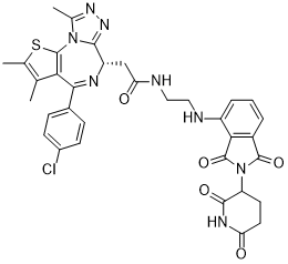
| SMILES |
CC1=C(SC2=C1C(=N[C@H](C3=NN=C(N32)C)CC(=O)NCCNC4=CC=CC5=C4C(=O)N(C5=O)C6CCC(=O)NC6=O)C7=CC=C(C=C7)Cl)C
|
| InChi Key |
CZRLOIDJCMKJHE-UXMRNZNESA-N
|
| InChi Code |
InChI=1S/C34H31ClN8O5S/c1-16-17(2)49-34-27(16)29(19-7-9-20(35)10-8-19)38-23(30-41-40-18(3)42(30)34)15-26(45)37-14-13-36-22-6-4-5-21-28(22)33(48)43(32(21)47)24-11-12-25(44)39-31(24)46/h4-10,23-24,36H,11-15H2,1-3H3,(H,37,45)(H,39,44,46)/t23-,24?/m0/s1
|
| 化学名 |
2-((S)-4-(4-chlorophenyl)-2,3,9-trimethyl-6H-thieno[3,2-f][1,2,4]triazolo[4,3-a][1,4]diazepin-6-yl)-N-(2-((2-(2,6-dioxopiperidin-3-yl)-1,3-dioxoisoindolin-4-yl)amino)ethyl)acetamide
|
| 别名 |
dBET57;dBET-57; dBET57; 1883863-52-2; 2-((S)-4-(4-chlorophenyl)-2,3,9-trimethyl-6H-thieno[3,2-f][1,2,4]triazolo[4,3-a][1,4]diazepin-6-yl)-N-(2-((2-(2,6-dioxopiperidin-3-yl)-1,3-dioxoisoindolin-4-yl)amino)ethyl)acetamide; 2-[(9S)-7-(4-chlorophenyl)-4,5,13-trimethyl-3-thia-1,8,11,12-tetrazatricyclo[8.3.0.02,6]trideca-2(6),4,7,10,12-pentaen-9-yl]-N-[2-[[2-(2,6-dioxopiperidin-3-yl)-1,3-dioxoisoindol-4-yl]amino]ethyl]acetamide; dBET57?; CHEMBL5180012; SCHEMBL17553391; TQP1624; dBET 57
|
| HS Tariff Code |
2934.99.9001
|
| 存储方式 |
Powder -20°C 3 years 4°C 2 years In solvent -80°C 6 months -20°C 1 month 注意: 本产品在运输和储存过程中需避光。 |
| 运输条件 |
Room temperature (This product is stable at ambient temperature for a few days during ordinary shipping and time spent in Customs)
|
| 溶解度 (体外实验) |
DMSO : ~250 mg/mL (~357.56 mM)
|
|---|---|
| 溶解度 (体内实验) |
配方 1 中的溶解度: ≥ 2.08 mg/mL (2.97 mM) (饱和度未知) in 10% DMSO + 40% PEG300 +5% Tween-80 + 45% Saline (这些助溶剂从左到右依次添加,逐一添加), 澄清溶液。
例如,若需制备1 mL的工作液,可将100 μL 20.8 mg/mL澄清DMSO储备液加入400 μL PEG300中,混匀;然后向上述溶液中加入50 μL Tween-80+,混匀;加入450 μL生理盐水定容至1 mL。 *生理盐水的制备:将 0.9 g 氯化钠溶解在 100 mL ddH₂O中,得到澄清溶液。 请根据您的实验动物和给药方式选择适当的溶解配方/方案: 1、请先配制澄清的储备液(如:用DMSO配置50 或 100 mg/mL母液(储备液)); 2、取适量母液,按从左到右的顺序依次添加助溶剂,澄清后再加入下一助溶剂。以 下列配方为例说明 (注意此配方只用于说明,并不一定代表此产品 的实际溶解配方): 10% DMSO → 40% PEG300 → 5% Tween-80 → 45% ddH2O (或 saline); 假设最终工作液的体积为 1 mL, 浓度为5 mg/mL: 取 100 μL 50 mg/mL 的澄清 DMSO 储备液加到 400 μL PEG300 中,混合均匀/澄清;向上述体系中加入50 μL Tween-80,混合均匀/澄清;然后继续加入450 μL ddH2O (或 saline)定容至 1 mL; 3、溶剂前显示的百分比是指该溶剂在最终溶液/工作液中的体积所占比例; 4、 如产品在配制过程中出现沉淀/析出,可通过加热(≤50℃)或超声的方式助溶; 5、为保证最佳实验结果,工作液请现配现用! 6、如不确定怎么将母液配置成体内动物实验的工作液,请查看说明书或联系我们; 7、 以上所有助溶剂都可在 Invivochem.cn网站购买。 |
| 制备储备液 | 1 mg | 5 mg | 10 mg | |
| 1 mM | 1.4302 mL | 7.1512 mL | 14.3025 mL | |
| 5 mM | 0.2860 mL | 1.4302 mL | 2.8605 mL | |
| 10 mM | 0.1430 mL | 0.7151 mL | 1.4302 mL |
1、根据实验需要选择合适的溶剂配制储备液 (母液):对于大多数产品,InvivoChem推荐用DMSO配置母液 (比如:5、10、20mM或者10、20、50 mg/mL浓度),个别水溶性高的产品可直接溶于水。产品在DMSO 、水或其他溶剂中的具体溶解度详见上”溶解度 (体外)”部分;
2、如果您找不到您想要的溶解度信息,或者很难将产品溶解在溶液中,请联系我们;
3、建议使用下列计算器进行相关计算(摩尔浓度计算器、稀释计算器、分子量计算器、重组计算器等);
4、母液配好之后,将其分装到常规用量,并储存在-20°C或-80°C,尽量减少反复冻融循环。
计算结果:
工作液浓度: mg/mL;
DMSO母液配制方法: mg 药物溶于 μL DMSO溶液(母液浓度 mg/mL)。如该浓度超过该批次药物DMSO溶解度,请首先与我们联系。
体内配方配制方法:取 μL DMSO母液,加入 μL PEG300,混匀澄清后加入μL Tween 80,混匀澄清后加入 μL ddH2O,混匀澄清。
(1) 请确保溶液澄清之后,再加入下一种溶剂 (助溶剂) 。可利用涡旋、超声或水浴加热等方法助溶;
(2) 一定要按顺序加入溶剂 (助溶剂) 。
|
|
|
InvivoChem的所有产品仅用于作科学研究,不面向患者销售
Copyright 2020 InvivoChem LLC | All Rights Reserved 粤ICP备20063088号-1


 COA
COA


 463611831
