| 规格 | 价格 | 库存 | 数量 |
|---|---|---|---|
| 1mg |
|
||
| 5mg |
|
||
| 10mg |
|
||
| 25mg |
|
||
| 50mg |
|
||
| 100mg |
|
||
| 250mg |
|
||
| 500mg |
|
||
| Other Sizes |
|

| 靶点 |
DNA; apoptosis
|
|---|---|
| 体外研究 (In Vitro) |
在 HeLa 和 SiHa 细胞中,薯蓣皂苷(1.25–5 μg/mL;6–24 小时)会提高细胞内钙水平并引发细胞凋亡[4]。薯蓣皂苷(1.25–5 μg/mL;6–24 小时):薯蓣皂苷下调 Bcl-2 和 Bcl-xl 水平蛋白,上调 Bak、Bax、Bid、p53、caspase-3 和 caspase-9 蛋白[ 4]。
薯蓣皂苷是一种天然甾体皂苷,来源于多种植物,对多种肿瘤细胞系具有强大的抗癌作用。在本研究中,我们研究了薯蓣皂苷对人LNCaP细胞的抗癌活性,并评估了其抗肿瘤作用的可能机制。研究发现,薯蓣皂苷(1、2和4μmol/L)可以以时间和浓度依赖的方式显著抑制LNCaP细胞的存活率。流式细胞术显示,薯蓣皂苷处理LNCaP细胞24h后细胞凋亡率升高,说明凋亡是薯蓣皂苷抑制癌症的重要机制。Western blot检测LNCaP细胞中caspase-3、Bcl-2和Bax的表达。切割的半胱氨酸天冬氨酸蛋白酶-3的表达显著增加,同时前天冬氨酸酶-3的表达明显降低。抗凋亡蛋白Bcl-2的表达下调,而促凋亡蛋白Bax的表达上调。Bcl-2/Bax比值显著降低。这些结果表明,薯蓣皂苷通过凋亡途径在人LNCaP细胞中具有潜在的抗肿瘤活性,这可能与半胱氨酸天冬氨酸蛋白酶-3和Bcl-2蛋白家族有关。[1] 在这项研究中,我们首次证明薯蓣皂苷在体外以剂量依赖的方式抑制RANKL介导的破骨细胞分化和骨吸收。薯蓣皂苷的抑制作用得到了破骨细胞特异性标志物表达减少的支持。进一步的分子分析表明,薯蓣皂苷消除了AKT磷酸化,从而损害了RANKL诱导的核因子κB(NF-κB)信号通路并抑制了NFATc1的转录活性。[2] 确定薯蓣皂苷是否通过防止凋亡来保护心肌细胞免受缺血/再灌注(I/R)损伤。对心脏H9c2细胞进行模拟I/R。通过甲基噻唑四氮唑(MTT)比色法评估细胞存活率。用二氯二氢荧光素(DCF)检测活性氧(ROS)。用流式细胞术评估细胞凋亡。罗丹明123(Rho123)用于测量线粒体膜电位(ΔΨm)。ELISA用于检测细胞色素c(Cyt-c)从线粒体释放到细胞质中的情况。RT-PCR检测Bax和Bcl-2 mRNA的表达。薯蓣皂苷减少了I/R诱导的细胞凋亡和细胞色素c从线粒体释放到细胞质中的细胞死亡和乳酸脱氢酶(LDH)释放,而薯蓣皂甙可以阻止这一过程。作为支持,薯蓣皂苷降低了Bax,但增加了Bcl-2 mRNA的表达。薯蓣皂苷阻止了I/R诱导的ΔΨm的耗散。最后,薯蓣皂苷增加了超氧化物歧化酶(SOD)的表达,但降低了细胞内ROS和丙二醛(MDA)的水平。薯蓣皂苷通过减轻氧化应激调节线粒体凋亡途径,保护H9c2细胞免受H/R损伤。[3] Dioscin是一种天然产物,具有对抗多形性胶质母细胞瘤、肺癌和癌症的活性。本研究进一步证实了薯蓣皂苷对人宫颈癌HeLa和SiHa细胞的作用,并探讨了可能的机制。透射电子显微镜(TEM)分析和DAPI染色用于检测细胞形态。流式细胞术用于检测细胞凋亡、ROS和Ca(2+)水平。单细胞凝胶电泳和免疫荧光分析用于检测DNA损伤和细胞色素C释放。结果表明,薯蓣皂苷显著抑制HeLa和SiHa细胞的增殖,并导致DNA损伤。机制研究表明,薯蓣皂苷导致细胞色素C从线粒体释放到细胞质中。此外,薯蓣皂苷显著上调Bak、Bax、Bid、p53、caspase-3、caspase-9的蛋白水平,下调Bcl-2和Bcl-xl的蛋白水平。因此,我们的研究表明,薯蓣皂苷通过调节ROS介导的DNA损伤和线粒体信号通路,显著诱导HeLa和SiHa细胞凋亡[4]。 |
| 体内研究 (In Vivo) |
给予薯蓣皂苷/Dioscin(75-300mg/kg,10mL/kg;口服灌胃;90天)后,雌性大鼠无亚慢性毒性,雄性大鼠有轻度亚慢性毒性。在雄性大鼠中,多利辛使胃肠道适度扩张,并在血液学检查中显示出溶血性贫血[5]。薯蓣皂苷可以通过抑制氧化硝化应激、细胞凋亡和炎症来减轻大鼠肝脏缺血再灌注损伤[6]。
薯蓣皂苷是许多中药中的主要活性化合物,但这种天然产物的安全性评估尚未得到研究。因此,本研究旨在评估Dioscin/薯蓣皂苷 对大鼠的90天亚慢性毒性。将大鼠分为四组,分别以0、75、150和300mg/kg/天的剂量口服<强>薯蓣皂苷。根据临床观察、眼科检查、体重、食物和饮水量、尿液分析、血液学、临床生化和病理学评估薯蓣皂苷的毒性。结果表明,薯蓣皂苷对雌性大鼠没有亚慢性毒性,对雄性大鼠有轻微的亚慢性毒性。然而,300mg/kg/天组的雄性大鼠在治疗期间表现出轻微的胃肠道扩张,在血液学评估中表现出溶血性贫血。与对照组相比,雄性大鼠的体重增加显著减少。在雄性和雌性组中,其他显著变化与薯蓣皂苷无关。总之,雌性和雄性大鼠的薯蓣皂苷无观测不良反应水平(NOAEL)和最低观测不良反应程度(LOAEL)估计分别为300mg/kg/天。我们的工作为薯蓣皂苷的进一步研究和新药开发提供了有用的数据。[5] 研究发现,薯蓣皂苷显著降低了血清丙氨酸氨基转移酶和天冬氨酸氨基转移酶的活性,提高了大鼠的存活率,改善了I/R诱导的肝细胞异常。此外,薯蓣皂苷明显提高了SOD、CAT、GSH-Px、GSH的水平,降低了MDA、TNOS、iNOS、NO的水平,并防止了I/R损伤引起的DNA断裂。进一步的研究表明,Dioscin/薯蓣皂苷显著降低了白介素-1β、白介素-6、肿瘤坏死因子-α、细胞间粘附分子-1、MIP-1α、MIP-2、Fas、FasL的基因表达,降低了NF-κB、AP-1、COX-2、HMGB-1、CYP2E1、Bak、caspase-3、p53、PARP、caspase-9的蛋白表达,降低JNK、ERK和p38 MAPK磷酸化水平,上调Bcl-2和Bcl-x水平。 结论:上述结果表明,薯蓣皂苷通过抑制炎症、氧化硝化应激和细胞凋亡对肝脏I/R损伤具有强效作用,应作为未来治疗肝脏I/R伤害的新药开发[6]。 |
| 酶活实验 |
透射电子显微镜(TEM)分析[4]
将HeLa和SiHa细胞(2×10⁵个细胞/mL)铺在6孔板中,用Dioscin/薯蓣皂苷处理,收获,并在4%戊二醛中于4°C下固定过夜。如前所述,对样品进行处理。然后对获得的切片进行染色,并使用透射电子显微镜进行观察。 DAPI染色[4] 将HeLa和SiHa细胞接种在六孔板中并培养过夜,然后分别用Dioscin/薯蓣皂苷(1.25、2.5和5.0μg/mL)处理12小时和24小时。对于DAPI染色,如上所述处理细胞,然后用DAPI(1.0μg/mL)溶液染色。最后,用荧光显微镜拍摄图像。 细胞内ROS积累的检测[4] HeLa和SiHa细胞以1×10⁵个细胞/孔的密度铺在6孔板上,并用Dioscin/薯蓣皂苷(1.25、2.5和5.0μg/mL)处理。收集细胞并重新悬浮在500μL DCFH-DA(10.0μM)中,所有细胞均通过流式细胞术进行分析。 细胞凋亡和细胞内Ca2+释放的检测[4] HeLa和SiHa细胞以1×10⁵个细胞/孔的密度铺在6孔板上,用Dioscin/薯蓣皂苷(1.25、2.5和5.0μg/mL)处理,然后收集并重新悬浮在500μL Fluo-3/AM(2.5μM)中,所有细胞均通过流式细胞术进行分析。 单细胞凝胶电泳分析[4] 癌症细胞用Dioscin(1.25、2.5和5.0μg/mL)处理后,用单细胞凝胶电泳(SCGE)法检测Dioscin诱导的DNA损伤。根据制造商的说明,通过荧光显微镜(奥林巴斯)获得细胞的图像。最终,从三个重复的孔中随机选择50多个细胞,并通过彗星分析软件项目(CASP)1.2.2进行分析。 细胞色素C释放的检测[4] 将HeLa和SiHa细胞接种在六孔板中,用Dioscin/薯蓣皂苷(1.25、2.5和5.0μg/mL)处理,然后与第一抗体一起孵育过夜。之后,将平板与第二抗体在37°C下孵育1小时,并用DAPI(5.0μg/mL)染色5分钟。通过激光扫描共聚焦显微镜获得细胞图像。 |
| 细胞实验 |
MTT法检测细胞存活率[1]
根据文献记载的方法,通过MTT法测定薯蓣皂苷对LNCaP细胞的细胞毒性作用。简而言之,将8×10³个细胞接种到96孔平底板中,每个孔有200μL完全生长培养基。24小时后,用不同浓度的薯蓣皂苷(0-10μmol/L)处理LNCaP细胞12、24和48小时。此后,向每个孔中加入10μL 5mg/mL MTT,并将平板再孵育4小时。实验结束时,从每个孔中取出培养基,加入150μL DMSO终止反应。使用自动ELISA酶标仪 测量570nm波长下的吸光度(A)。抑制率(%)根据以下方程式计算:抑制率(百分比)=[(对照-Atreated)/Acontrol]×100%,其中Acontrol是载体处理孔的A值,Atreated是薯蓣皂苷处理孔的B值。薯蓣皂苷/Dioscin对LNCaP细胞的细胞毒性以IC50值表示(相对于0.1%DMSO处理的细胞,用受试药物处理的细胞对细胞存活率的抑制率为50%,并通过Probit正常法计算)。 细胞凋亡的流式细胞术检测[1] 按照以下说明,使用AnnexinⅤ-FITC/PI凋亡检测试剂盒通过流式细胞术测定细胞凋亡。简而言之,收获2×105个细胞,用冰冷的PBS(pH 7.4)洗涤两次。然后,将细胞悬浮在500μL结合缓冲液中,用5μL膜联蛋白Ⅴ-FITC和5μL碘化丙啶(PI)双重标记后,在室温下黑暗中孵育15分钟。然后,对约1×10⁴个染色的凋亡细胞进行流式细胞术计数,结果如下:左下象限代表活细胞(Annexin V-/PI-);右下象限代表早期凋亡细胞(膜联蛋白V+/PI-);右上象限代表晚期凋亡细胞(膜联蛋白V+/PI+);左上象限代表原发性坏死细胞(膜联蛋白V–/PI+)。 蛋白质印迹[1] Western blot检测caspase-3、Bcl-2和Bax的表达。简而言之,用薯蓣皂苷处理24小时后,收获细胞,置于冰上,用冰冷的PBS洗涤,然后在冰上裂解缓冲液中裂解30分钟。将缓冲液在4°C下以15000 g离心10分钟后,收集所得上清液作为总细胞蛋白。使用BCA蛋白测定试剂盒 检测蛋白质浓度。蛋白质提取物(20μg,20μL)通过10%十二烷基硫酸钠聚丙烯酰胺凝胶电泳(SDS-PAGE)分离,然后通过电印迹转移到聚二氟乙烯(PVDF)膜上。转移的膜在室温下用TBST(1 mol/L Tris缓冲盐水,pH 7.4;0.1%吐温-20)中的5%脱脂奶粉封闭1小时,然后在4°C下用一抗(cas-pase-3、Bcl-2和Bax,1:1000稀释)培养过夜。将膜在TBST缓冲液中洗涤三次,然后在室温下用与辣根过氧化物酶连接的适当二抗(1:2000稀释)孵育90分钟。用TBST洗涤膜4次后,使用增强化学发光试剂在膜上显影印迹中的蛋白质。使用Bio-Rad Quantity One V4.62对上述蛋白质的表达水平进行密度测定。β-actin以与上述相同的方式检测,并用作内部负荷参考。 细胞活力测定[2] 使用细胞计数试剂盒-8评估薯蓣皂苷对BMMs细胞的抗增殖作用。简而言之,处理后,向每个孔中加入10μl CCK-8溶液;孵育4小时后,使用酶标仪在450nm处测量吸光度。Dioscin薯蓣皂苷对细胞存活率的影响以细胞存活率百分比表示,载体处理的对照细胞设置为100%。 体外破骨细胞生成试验[2] 进行体外破骨细胞生成试验,以检查薯蓣皂苷对破骨细胞分化的影响。如前所述制备骨髓巨噬细胞(BMM)细胞。简而言之,从6周龄C57/BL6小鼠的股骨和胫骨中提取的细胞在T-75 cm2烧瓶中的全细胞培养基和30 ng/mL M-CSF中孵育以增殖。更换培养基时,清洗细胞以耗尽残留的基质细胞。达到90%融合后,用磷酸缓冲盐水(PBS)洗涤细胞三次,并用胰蛋白酶处理30分钟以收获BMM。培养皿底部的粘附细胞被归类为BMM;将这些BMM以8×10³个细胞/孔的密度铺在96孔板上,一式三份,在37°C下含5%CO2的加湿培养箱中孵育24小时。然后用不同浓度的Dioscin/薯蓣皂苷(0、1或4μM)加M-CSF(30 ng/mL)和RANKL(50 ng/mL)处理细胞。五天后,固定细胞并对TRAP活性进行染色。TRAP+具有五个以上核的多核细胞被计数为破骨细胞。 吸收坑分析[2] 为了进行如上所述的骨吸收试验,将BMM以8×10³个细胞/孔的密度接种在96孔板的骨切片上,进行三次重复,并用M-CSF(30 ng/mL)加RANKL(50 ng/mL)刺激。三天后,细胞在培养后用指定浓度的Dioscin/薯蓣皂苷处理48小时。然后用2.5%戊二醛固定细胞。使用放大倍数为200倍、电压为10 kV的扫描电子显微镜 对骨切片进行成像。为每个骨切片随机选择三个视场进行进一步分析。使用Image J软件对凹坑区域进行量化。类似的独立实验重复了至少三次。 蛋白质印迹分析[2] 将BMMS细胞以5×10⁵个细胞/孔的速度接种到6孔板中,在RANKL刺激(50 ng/mL)之前,用或不用Dioscin/薯蓣皂苷 4μM)预处理4小时,持续指定时间(0、5、15或30分钟)。将BMMs以5×10⁵个细胞/孔的速度接种到6孔板中,并用或不用薯蓣皂苷(4μM)和RANKL(50 ng/mL)处理指定时间。细胞在含有50 mM Tris-HCl、150 mM NaCl、5 mM EDTA、1%Triton X-100、1 mM氟化钠、1 mM钒酸钠、1%脱氧胆酸盐和蛋白酶抑制剂混合物的RIPA裂解缓冲液中裂解。将裂解物在12000rcf下离心10分钟,收集上清液中的蛋白质。通过BCA测定法测量蛋白质浓度。使用8-10%的凝胶通过十二烷基硫酸钠-聚丙烯酰胺凝胶电泳(SDS-PAGE)分离每种蛋白质裂解物的30微克,然后将蛋白质转移到聚偏二氟乙烯膜上。用5%脱脂乳阻断非特异性相互作用1小时,然后在4°C下用指定的一抗探测膜过夜。用与IRDye 800CW(分子量,1166 Da)偶联的适当二抗孵育膜,并通过在Odyssey红外成像系统(Li-COR)中暴露来检测抗体反应性。 萤光素酶报告基因活性测定[2] 如前所述,使用用NF-κB荧光素酶报告构建体稳定转染的RAW264.7细胞测量了薯蓣皂苷对RANKL诱导的NF-κB活化的影响。简而言之,将细胞接种到48孔板中,并在细胞培养基中保持24小时。然后用或不用指定浓度的Dioscin/薯蓣皂苷预处理细胞1小时,然后加入RANKL(50 ng/mL)8小时。使用Promega萤光素酶测定系统测量萤光素酶活性,并使其标准化为载体对照。同样,薯蓣皂苷对RANKL诱导的AP-1或NFATc1依赖性萤光素酶报告基因检测的影响如前所述进行了测定。 |
| 动物实验 |
根据先前发表的关于薯蓣皂苷急性毒性作用的研究(Liang et al., 2010),本研究分别以0(对照组)、75、150和300 mg/kg/天的剂量给予大鼠薯蓣皂苷。薯蓣皂苷与0.5%羧甲基纤维素钠的蒸馏水溶液混合。连续90天,每天(每周7天)向每只大鼠给予适量的薯蓣皂苷或对照(溶剂)。每只动物均采用经口灌胃给药,使用连接合适注射器的球头灌胃针。每天给药时间均相同(±1小时)。根据体重计算个体剂量,并每日调整剂量以维持所有大鼠的目标剂量水平。所有剂量均按体积给药,并校正稀释度。所有组均以10 mL/kg的恒定剂量体积给药。对照组仅接受与试验组相同体积的赋形剂。本研究依据美国食品药品监督管理局(FDA,2000)和经合组织(OECD)测试指南408《啮齿动物重复剂量90天口服毒性研究》进行。[5]
眼科检查:在实验第一天(给药前)和实验结束的第90天,对所有大鼠(40只雄性,40只雌性)进行眼科检查。分别采用肉眼和间接检眼镜检查每只大鼠双眼的周边和内部结构。 实验前,动物适应环境1周。如图 5(B) 所示,在预防性试验中,连续 7 天,每天一次,以 20、40 和 60 mg/kg 的剂量灌胃给予动物薯蓣皂苷。假手术组和模型组的大鼠则灌胃给予溶剂。第 8 天,按照先前描述的方法建立 70% 部分肝缺血的小鼠模型。简而言之,将大鼠麻醉后,通过正中剖腹术暴露肝脏,然后用血管钳阻断肝脏左侧叶和中叶的血流,保持右侧叶的血流灌注,以防止肠道充血阻塞。肝脏缺血 60 分钟后,移除血管钳,肝脏再灌注 6 小时。手术结束后,对动物进行麻醉以采集血液,然后处死动物并采集肝脏左叶和中叶。如图 5(C) 所示,在治疗试验中,将大鼠随机分为三组,其中假手术组和模型组动物给予溶剂,而薯蓣皂苷组大鼠在缺血/再灌注 (I/R) 前 2 小时灌胃给予 60 mg/kg 的薯蓣皂苷。肝脏缺血 60 分钟后,移除血管钳,并在再灌注后 0.5、1、2、4、6、12、24、48 和 96 小时采集眼眶血样。此外,还检测了动物的存活率。[6] 背景:我们之前的研究表明,薯蓣皂苷对肝损伤具有显著的保护作用;然而,薯蓣皂苷对肝脏缺血再灌注(I/R)损伤的作用机制尚不清楚。本文旨在探讨薯蓣皂苷对肝脏I/R损伤的作用及其可能的机制。方法:在Wistar大鼠中诱导70%肝脏部分缺血60分钟,随后进行再灌注。在预防性试验中,于I/R前连续7天,每日一次,经胃内灌注给予大鼠薯蓣皂苷,剂量分别为20、40和60 mg/kg。在治疗性试验中,于I/R前2小时,经胃内灌注给予大鼠薯蓣皂苷,剂量为60 mg/kg。[6] |
| 毒性/毒理 (Toxicokinetics/TK) |
小鼠皮下注射LD50 >300 mg/kg 日本公会东京公报专利,编号91-271224
|
| 参考文献 |
|
| 其他信息 |
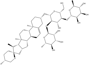
薯蓣皂苷是一种螺甾烷糖苷,由三糖α-L-鼠李糖-(1→4)-[α-L-鼠李糖-(1→2)]-β-D-葡萄糖通过糖苷键连接到薯蓣皂苷元的3位组成。它具有多种活性,包括作为代谢产物、抗真菌剂、抗病毒剂、抗肿瘤剂、抗炎剂、保肝剂、细胞凋亡诱导剂以及酪氨酸酶(EC 1.14.18.1)抑制剂。它是一种螺甾烷糖苷、螺缩酮、六环三萜类化合物和三糖衍生物。其功能与薯蓣皂苷元相关。它来源于螺甾烷的氢化物。
薯蓣皂苷已在薯蓣属植物(如薯蓣、三角叶薯蓣)和其他有相关数据的生物体中被报道。 另见:多穗薯蓣块茎(部分)。 在本研究中,我们首次证实天然化合物薯蓣皂苷能够抑制破骨细胞分化和骨吸收,表明薯蓣皂苷对破骨细胞相关疾病具有额外的保护作用。此外,我们揭示了薯蓣皂苷作用于破骨细胞的分子机制是通过抑制Akt/NF-κB和Akt/NFATc1信号通路。 在破骨细胞中,Akt信号级联是三种破骨细胞表面受体(包括c-fms、αvβ3整合素和RANK)的关键下游信号通路。既往研究表明,M-CSF 和 RANKL 刺激均可激活 Akt 磷酸化,并通过影响 NF-κB 和 NFATc1 的激活,在破骨细胞生成中发挥关键作用。Moon 等人证实,在骨髓单核细胞 (BMM) 中过表达 Akt 可显著诱导 NFATc1 表达,并导致破骨细胞生成增强。此外,NF-κB 的激活可由多种不同的激酶启动,例如 Akt 和 NF-κB 诱导激酶。Gingery 等人证实,AKT/NF-κB 轴在破骨细胞生成和维持成熟破骨细胞存活中至关重要。与这些研究一致,我们证实薯蓣皂苷可抑制 Akt 磷酸化,从而抑制 RANKL 诱导的 NF-κB 和 NFATc1 活性,而这两者对于破骨细胞分化都至关重要。有趣的是,在检测薯蓣皂苷对 MAPK 通路的功能时,并未观察到显著的抑制作用。 总之,薯蓣皂苷能够抑制破骨细胞的形成和功能,表明其对破骨细胞相关疾病具有潜在的治疗价值。此外,本研究还明确揭示了薯蓣皂苷通过抑制 Akt/NF-κB 和 Akt/NFATc1 信号通路来抑制破骨细胞的分子机制。此外,我们的体外实验结果进一步证实了薯蓣皂苷在 LPS 诱导的骨溶解模型中的骨保护作用。然而,仍需进一步研究薯蓣皂苷对骨内其他细胞的作用。[1] 薯蓣皂苷通过诱导 ROS 介导的 DNA 损伤和激活线粒体信号通路来诱导人宫颈癌 HeLa 和 SiHa 细胞凋亡。然而,根据体外实验结果,薯蓣皂苷的分子作用机制和药物靶点仍需进一步研究。[4] 由于薯蓣皂苷具有药用价值和广泛应用,这种天然化合物在未来的医药领域应用和研究中具有巨大的潜力。因此,评估薯蓣皂苷的毒性至关重要。我们为期90天的亚慢性毒性研究结果显示,剂量依赖性与个体体重呈变化趋势。与对照组相比,所有雄性组的体重均显著下降,尤其是在300 mg/kg剂量组的雄性中。这种变化被认为与皂苷的特性有关,例如胃肠道扩张导致的摄食减少(Shen et al., 2008)、肠道蠕动减弱(Klita et al., 1996)以及蛋白质消化率降低(Potter et al., 1993)。由于采食量减少,食物消耗量下降,因此体重减轻。尿液分析显示,75 mg/kg 治疗组的雄性小鼠尿液 pH 值、尿比重和尿蛋白发生改变。300 mg/kg 治疗组未发现类似改变。这些改变是偶然发生的,与剂量无明显相关性。血液学评估显示,300 mg/kg 治疗组的雄性小鼠红细胞计数和血细胞比容发生改变,提示溶血性贫血。甾体皂苷的生物活性成分具有溶血活性(Santos等,1997;Zhang等,1999),已知皂苷对红细胞膜具有溶解作用(Howard和Wallace,1953)。据报道,这种溶解作用是由于皂苷配基与膜甾醇(尤其是胆固醇)具有亲和力,在膜的开放孔道中形成不溶性复合物所致(Bangham等,1962;Cho等,2009)。红细胞膜破裂可导致皂苷释放入血,引起轻微肾毒性。临床生化评估显示,男性患者的血尿素氮(BUN)水平有显著变化,但肾脏未观察到组织病理学病变。其他血液学指标的变化是偶然的,因为未观察到剂量-反应关系。在临床生化评估中,雌性300 mg/kg和雄性300 mg/kg治疗组中观察到的ALT升高被认为与薯蓣皂苷治疗相关,然而,各组的AST、AKP和T-BIL的变化未显示出明显的剂量依赖性。尽管各组ALT的变化提示肝损伤,但未观察到肝脏重量的变化,组织病理学检查也未发现异常。雄性和雌性大鼠BUN的显著变化表明存在轻度肾毒性作用,然而,肾脏未记录到组织病理学损伤。虽然雄性和雌性动物的肌酸(Cr)、甘油三酯(TG)、γ-谷氨酰转移酶(γ-GT)和葡萄糖水平均出现显著波动,但这些变化是偶然的,没有明显的剂量依赖性或毒理学意义。 在器官重量和组织病理学评估中,尽管在肝脏、肾脏、心脏和大脑等器官中观察到绝对重量和相对重量的显著变化,但未观察到组织病理学变化,这些变化可被认为是由于体重下降所致。表5显示了体重减轻和器官相对重量增加的情况,这些变化被认为是由于脂肪增加所致。 结论:药用成分薯蓣皂苷具有显著的开发潜力。然而,基于本研究的结果,也有必要关注薯蓣皂苷的毒性。我们的研究结果为薯蓣皂苷的潜在临床应用和广泛使用提供了一些安全性证据,但在薯蓣皂苷的临床应用之前,还需要进行进一步的评估。总之,本研究确定雌性大鼠每日300 mg/kg剂量的薯蓣皂苷为未观察到不良反应剂量(NOAEL),雄性大鼠每日300 mg/kg剂量为最低观察到不良反应剂量(LOAEL)。[5] 综上所述,薯蓣皂苷通过减轻氧化-硝化应激、炎症和细胞凋亡,对大鼠肝脏缺血/再灌注损伤具有良好的保护作用,未来有望成为治疗缺血/再灌注损伤的新型有效候选药物。当然,薯蓣皂苷的作用机制、药物靶点和临床应用仍需进一步研究。[6] |
| 分子式 |
C45H72O16
|
|
|---|---|---|
| 分子量 |
869.05
|
|
| 精确质量 |
868.482
|
|
| 元素分析 |
C, 62.19; H, 8.35; O, 29.46
|
|
| CAS号 |
19057-60-4
|
|
| 相关CAS号 |
|
|
| PubChem CID |
119245
|
|
| 外观&性状 |
White to off-white solid powder
|
|
| 密度 |
1.4±0.1 g/cm3
|
|
| 熔点 |
294-296 ℃
|
|
| 折射率 |
1.614
|
|
| LogP |
7.24
|
|
| tPSA |
235.68
|
|
| 氢键供体(HBD)数目 |
8
|
|
| 氢键受体(HBA)数目 |
16
|
|
| 可旋转键数目(RBC) |
7
|
|
| 重原子数目 |
61
|
|
| 分子复杂度/Complexity |
1600
|
|
| 定义原子立体中心数目 |
26
|
|
| SMILES |
C[C@@H]1CC[C@@]2([C@H]([C@H]3[C@@H](O2)C[C@@H]4[C@@]3(CC[C@H]5[C@H]4CC=C6[C@@]5(CC[C@@H](C6)O[C@H]7[C@@H]([C@H]([C@@H]([C@H](O7)CO)O[C@H]8[C@@H]([C@@H]([C@H]([C@@H](O8)C)O)O)O)O)O[C@H]9[C@@H]([C@@H]([C@H]([C@@H](O9)C)O)O)O)C)C)C)OC1
|
|
| InChi Key |
VNONINPVFQTJOC-ZGXDEBHDSA-N
|
|
| InChi Code |
InChI=1S/C45H72O16/c1-19-9-14-45(54-18-19)20(2)30-28(61-45)16-27-25-8-7-23-15-24(10-12-43(23,5)26(25)11-13-44(27,30)6)57-42-39(60-41-36(52)34(50)32(48)22(4)56-41)37(53)38(29(17-46)58-42)59-40-35(51)33(49)31(47)21(3)55-40/h7,19-22,24-42,46-53H,8-18H2,1-6H3/t19-,20+,21+,22+,24+,25-,26+,27+,28+,29-,30+,31+,32+,33-,34-,35-,36-,37+,38-,39-,40+,41+,42-,43+,44+,45-/m1/s1
|
|
| 化学名 |
(2S,3R,4R,5R,6S)-2-[(2R,3S,4S,5R,6R)-4-hydroxy-2-(hydroxymethyl)-6-[(1S,2S,4S,5'R,6R,7S,8R,9S,12S,13R,16S)-5',7,9,13-tetramethylspiro[5-oxapentacyclo[10.8.0.02,9.04,8.013,18]icos-18-ene-6,2'-oxane]-16-yl]oxy-5-[(2S,3R,4R,5R,6S)-3,4,5-trihydroxy-6-methyloxan-2-yl]oxyoxan-3-yl]oxy-6-methyloxane-3,4,5-triol
|
|
| 别名 |
|
|
| HS Tariff Code |
2934.99.9001
|
|
| 存储方式 |
Powder -20°C 3 years 4°C 2 years In solvent -80°C 6 months -20°C 1 month |
|
| 运输条件 |
Room temperature (This product is stable at ambient temperature for a few days during ordinary shipping and time spent in Customs)
|
| 溶解度 (体外实验) |
|
|||
|---|---|---|---|---|
| 溶解度 (体内实验) |
配方 1 中的溶解度: ≥ 2.25 mg/mL (2.59 mM) (饱和度未知) in 10% DMSO + 40% PEG300 + 5% Tween80 + 45% Saline (这些助溶剂从左到右依次添加,逐一添加), 澄清溶液。
例如,若需制备1 mL的工作液,可将100 μL 22.5 mg/mL澄清的DMSO储备液加入到400 μL PEG300中,混匀;再向上述溶液中加入50 μL Tween-80,混匀;然后加入450 μL生理盐水定容至1 mL。 *生理盐水的制备:将 0.9 g 氯化钠溶解在 100 mL ddH₂O中,得到澄清溶液。 配方 2 中的溶解度: ≥ 2.25 mg/mL (2.59 mM) (饱和度未知) in 10% DMSO + 90% (20% SBE-β-CD in Saline) (这些助溶剂从左到右依次添加,逐一添加), 澄清溶液。 例如,若需制备1 mL的工作液,可将 100 μL 22.5 mg/mL澄清DMSO储备液加入900 μL 20% SBE-β-CD生理盐水溶液中,混匀。 *20% SBE-β-CD 生理盐水溶液的制备(4°C,1 周):将 2 g SBE-β-CD 溶解于 10 mL 生理盐水中,得到澄清溶液。 View More
配方 3 中的溶解度: ≥ 2.25 mg/mL (2.59 mM) (饱和度未知) in 10% DMSO + 90% Corn Oil (这些助溶剂从左到右依次添加,逐一添加), 澄清溶液。 1、请先配制澄清的储备液(如:用DMSO配置50 或 100 mg/mL母液(储备液)); 2、取适量母液,按从左到右的顺序依次添加助溶剂,澄清后再加入下一助溶剂。以 下列配方为例说明 (注意此配方只用于说明,并不一定代表此产品 的实际溶解配方): 10% DMSO → 40% PEG300 → 5% Tween-80 → 45% ddH2O (或 saline); 假设最终工作液的体积为 1 mL, 浓度为5 mg/mL: 取 100 μL 50 mg/mL 的澄清 DMSO 储备液加到 400 μL PEG300 中,混合均匀/澄清;向上述体系中加入50 μL Tween-80,混合均匀/澄清;然后继续加入450 μL ddH2O (或 saline)定容至 1 mL; 3、溶剂前显示的百分比是指该溶剂在最终溶液/工作液中的体积所占比例; 4、 如产品在配制过程中出现沉淀/析出,可通过加热(≤50℃)或超声的方式助溶; 5、为保证最佳实验结果,工作液请现配现用! 6、如不确定怎么将母液配置成体内动物实验的工作液,请查看说明书或联系我们; 7、 以上所有助溶剂都可在 Invivochem.cn网站购买。 |
| 制备储备液 | 1 mg | 5 mg | 10 mg | |
| 1 mM | 1.1507 mL | 5.7534 mL | 11.5068 mL | |
| 5 mM | 0.2301 mL | 1.1507 mL | 2.3014 mL | |
| 10 mM | 0.1151 mL | 0.5753 mL | 1.1507 mL |
1、根据实验需要选择合适的溶剂配制储备液 (母液):对于大多数产品,InvivoChem推荐用DMSO配置母液 (比如:5、10、20mM或者10、20、50 mg/mL浓度),个别水溶性高的产品可直接溶于水。产品在DMSO 、水或其他溶剂中的具体溶解度详见上”溶解度 (体外)”部分;
2、如果您找不到您想要的溶解度信息,或者很难将产品溶解在溶液中,请联系我们;
3、建议使用下列计算器进行相关计算(摩尔浓度计算器、稀释计算器、分子量计算器、重组计算器等);
4、母液配好之后,将其分装到常规用量,并储存在-20°C或-80°C,尽量减少反复冻融循环。
计算结果:
工作液浓度: mg/mL;
DMSO母液配制方法: mg 药物溶于 μL DMSO溶液(母液浓度 mg/mL)。如该浓度超过该批次药物DMSO溶解度,请首先与我们联系。
体内配方配制方法:取 μL DMSO母液,加入 μL PEG300,混匀澄清后加入μL Tween 80,混匀澄清后加入 μL ddH2O,混匀澄清。
(1) 请确保溶液澄清之后,再加入下一种溶剂 (助溶剂) 。可利用涡旋、超声或水浴加热等方法助溶;
(2) 一定要按顺序加入溶剂 (助溶剂) 。
InvivoChem的所有产品仅用于作科学研究,不面向患者销售
Copyright 2020 InvivoChem LLC | All Rights Reserved 粤ICP备20063088号-1


 COA
COA
 463611831
